
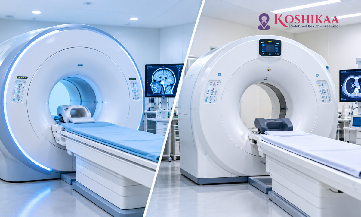
If your doctor has asked you to have a scan, you may wonder why one person is sent for an MRI while another is sent for a CT scan. Both produce detailed images of the inside of the body, and both are widely used in diagnosis, but they work in very different ways and are suited to different medical questions.
Understanding the difference between MRI and CT scans can help you feel more prepared and confident about the test you have been recommended. In simple terms, CT uses X-rays and is fast, while MRI uses magnetic fields and radio waves and is especially good at showing soft tissues. Neither is better in every situation. The right choice depends on the part of the body being examined, the suspected condition, how urgent the situation is and your individual safety considerations.
This guide compares MRI and CT side by side, explains when doctors recommend each and outlines what you should know about preparation, safety and radiation.
What Is the Difference Between MRI and CT Scans?
The main difference lies in the technology. A CT (computed tomography) scan uses X-rays taken from many angles, which a computer combines into cross-sectional images. An MRI (magnetic resonance imaging) scan uses a strong magnetic field and radio waves to create detailed images, without using ionising radiation.
These different technologies mean each scan sees the body in its own way. CT is particularly good at showing bone, bleeding, the lungs and many abdominal problems quickly. MRI provides exceptional detail of soft tissues such as the brain, spinal cord, nerves, muscles, ligaments and cartilage.
How Does a CT Scan Work?
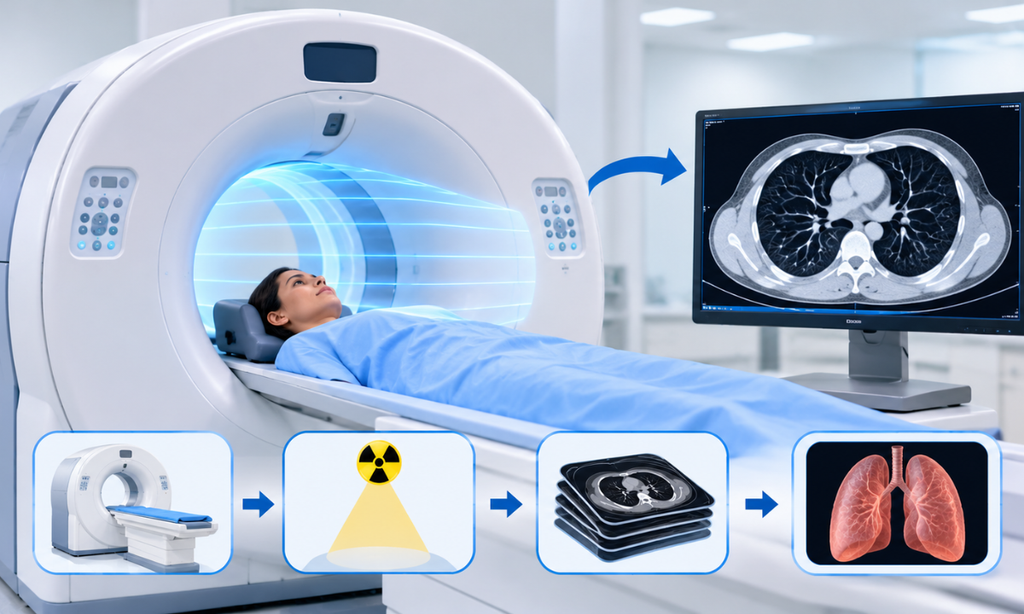
During a CT scan, you lie on a table that moves through a wide, ring-shaped scanner. An X-ray tube rotates around you, taking many images from different angles, and a computer turns these into detailed slices that can be viewed individually or combined into 3D images.
The scanning itself is very quick, often taking only seconds for a single area, and the whole appointment usually takes 15 to 30 minutes including preparation. In some cases, a contrast agent containing iodine is injected into a vein or taken by mouth to make blood vessels and organs stand out more clearly.
Because CT is fast and widely available, it is often the first choice in emergencies. Learn more about our CT scan in Bangalore services.
How Does an MRI Scan Work?
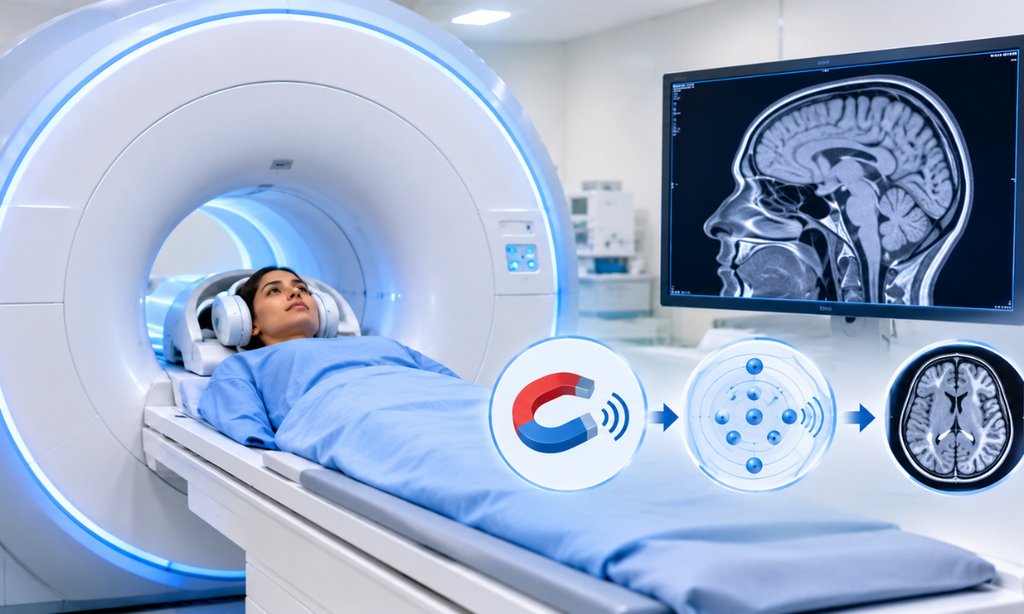
During an MRI scan, you lie on a table that slides into a tunnel-shaped scanner containing a powerful magnet. The magnetic field and radio waves cause tiny particles in the body’s tissues to give off signals, which a computer converts into highly detailed images.
An MRI scan takes longer than a CT scan, often 20 to 60 minutes depending on the area being examined, and you need to stay very still throughout. The scanner makes loud knocking and buzzing sounds, so you will be given earplugs or headphones. Some MRI scans use a contrast agent containing gadolinium, injected into a vein, to highlight certain tissues or abnormalities.
Find out more about our MRI scan in Bangalore services.
MRI vs CT Scan: Main Differences
| Feature | CT Scan | MRI Scan |
| Technology | X-rays and computer processing | Magnetic field and radio waves |
| Ionising radiation | Yes | No |
| Typical scanning time | Often seconds to a few minutes | Often 20 to 60 minutes, depending on the body area |
| Best at showing | Bone, bleeding, lungs, many abdominal conditions, kidney stones | Brain, spinal cord, nerves, ligaments, cartilage, soft tissues |
| Noise | Relatively quiet | Loud knocking and buzzing |
| Space inside the scanner | Open ring, less enclosed | Narrower tunnel, can feel enclosed |
| Contrast agent (if used) | Iodine-based | Gadolinium-based |
| Metal implants | Usually not a problem | Must be checked for safety first |
| Use in emergencies | Very common | Less common |
Imaging Technology
CT relies on X-rays, which pass through the body and are absorbed differently by bone, soft tissue and air. MRI relies on the behaviour of water and fat molecules in a magnetic field, which allows it to distinguish between different soft tissues in great detail.
Radiation Exposure
CT uses ionising radiation, while MRI does not. The dose from a single CT scan is carefully controlled and varies with the body part scanned. A scan of the chest or abdomen, for example, delivers more radiation than a simple X-ray, roughly comparable to the natural background radiation a person receives over months to a few years. The risk from a single scan is small, and doctors recommend CT only when the expected benefit outweighs it. Our article on CT radiation exposure and safety measures explains this in more detail.
Scan Duration and Preparation
CT scans are much quicker, which makes them easier for people who are in pain, unwell or find it difficult to keep still. MRI scans take longer and require you to remain still in a more enclosed space. Preparation for both depends on the type of scan. Some scans need fasting, particularly when contrast is used or when the abdomen is examined, while many need no special preparation.
Metal Implants, Contrast and Safety Considerations
The strong magnet in an MRI scanner can affect certain metal implants and electronic devices, so every patient is screened before entering the scan room. Tell the MRI team about every implanted device, including pacemakers, defibrillators, aneurysm clips, cochlear implants, pumps, stents, ports and metal fragments. Some devices are MRI-safe, some are MRI-conditional and some are not suitable for MRI, so the team must check the device type, manufacturer and model before scanning. Metal is rarely a problem for CT.
Both scans may use contrast agents. People with kidney problems, diabetes or previous reactions to contrast should tell the team in advance, as extra precautions or a different test may be needed.
When Do Doctors Recommend CT Instead of MRI?
Doctors often choose CT when:
•Speed matters: in many emergencies, such as suspected stroke, head injury or serious trauma, CT is often used because it can quickly show bleeding, fractures or internal injuries
•The lungs need to be examined: CT provides excellent detail of the lungs, including infections, nodules and other lung conditions
•Bone detail is needed: CT shows complex fractures and fine bone structure clearly
•Kidney stones are suspected: a CT KUB scan is highly accurate for detecting and measuring stones
•Many abdominal problems need assessment: such as appendicitis, abscesses or bowel problems
•MRI is unsuitable: for example, because of certain implants or severe claustrophobia
When Is MRI Better Than CT?
Doctors often choose MRI when:
•Detailed brain or spinal cord images are needed: for example, to investigate seizures, multiple sclerosis, certain tumours or spinal nerve compression
•Joints and soft tissues need to be assessed: MRI shows ligament tears, cartilage damage and muscle injuries clearly
•Some abdominal and pelvic organs need closer evaluation: including the liver, prostate, uterus and ovaries
•Radiation should be avoided where possible: for example, in children, or during pregnancy, when MRI may be used if imaging is clinically needed
•A finding on another scan needs further characterisation: MRI can help clarify what an abnormality seen on CT or ultrasound is likely to be
Sometimes both tests are needed, as they provide complementary information. For example, a person with a suspected tumour may have a CT scan to look at the chest and abdomen and an MRI to examine a specific organ in more detail.
Can You Choose Between MRI and CT Yourself?
It is natural to have a preference, especially if you are concerned about radiation or feel anxious in enclosed spaces. However, the choice of scan should be made by your doctor and the radiology team based on the medical question being asked.
Choosing the wrong test can mean missing important information or needing a second scan later. If you have concerns, discuss them openly. Tell your doctor if you are pregnant, have metal implants or a medical device, have kidney problems, have reacted to contrast before or experience claustrophobia. These details help the team choose the safest and most useful test for you.
Do MRI and CT Scans Cost the Same?
In general, an MRI scan costs more than a CT scan of the same body part, mainly because MRI equipment is more expensive to run and each scan takes longer. The final cost of either test also depends on the area being scanned, whether contrast is needed and whether more than one region is examined.
Cost should not be the deciding factor when choosing between the two. A cheaper scan that does not answer the medical question may lead to further tests later. Ask the centre for the exact price of the scan your doctor has requested, including any contrast charges, before your appointment.
What Should You Expect on the Day?
For both scans, you will be asked to remove metal items such as jewellery, watches and belts, and you may be given a gown to wear. The radiographer will explain the procedure and stay in contact with you throughout, usually through an intercom.
If you feel anxious about an MRI scan, let the team know in advance. Simple measures such as music, a blanket, an eye mask or having a friend nearby can help, and your doctor may discuss other options if needed. After the scan, a radiologist reviews the images and prepares a report for your doctor. For a practical checklist before your appointment, see our guide on how to prepare for an MRI scan.
Frequently Asked Questions
Which is better, MRI or CT?
Neither is universally better. The right test depends on the body area, symptoms, urgency, previous imaging and the clinical question your doctor needs to answer.
Which is safer, MRI or CT?
Both are safe when used appropriately. MRI does not use radiation but requires careful screening for metal implants and devices. CT involves a small dose of radiation, which is justified when the scan is medically needed.
Is MRI more accurate than CT?
Not in every case. MRI shows soft tissues in greater detail, while CT is often better for bone, lungs, bleeding and kidney stones. The more accurate test depends on what your doctor is looking for.
Why would a doctor order a CT instead of an MRI?
CT is quicker, more widely available and better for certain conditions, such as head injuries, lung problems, complex fractures and kidney stones. It may also be used when MRI is unsuitable.
Can I have an MRI if I have a metal implant?
Many people with implants can safely have an MRI, but this depends on the type of implant or device. Always tell the team about any implants, pacemakers, stents or metal fragments before your scan.
Which scan takes longer, MRI or CT?
MRI scans usually take longer, often 20 to 60 minutes. CT scanning itself takes only seconds to a few minutes, with the whole appointment usually lasting 15 to 30 minutes.
Is MRI or CT better during pregnancy?
MRI does not use ionising radiation and may be used during pregnancy when clinically needed. However, gadolinium-based MRI contrast is generally avoided during pregnancy unless it is expected to provide a significant diagnostic benefit. CT may be used when the clinical benefit outweighs the radiation risk, and the radiology team will use dose-optimisation measures where appropriate. Ultrasound is often considered first where it can answer the question.
Conclusion
MRI and CT are both powerful imaging tools, but they answer different questions. CT is fast and excellent for emergencies, bones, lungs and many abdominal conditions. MRI offers exceptional soft-tissue detail without radiation, making it ideal for the brain, spine and joints. Your doctor will recommend the test that gives the clearest answer for your situation.
If you have been referred for an MRI or CT scan, contact Koshikaa in Banashankari, Bengaluru, on +91 7996666106 to book your appointment and receive preparation instructions.
References