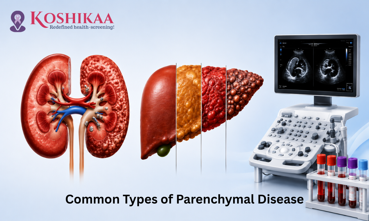
Opening a diagnostic report to find unfamiliar clinical terms can immediately cause a surge of unnecessary worry. If you or a loved one has recently undergone a routine checkup at a specialized Health Screening centre in Bangalore, you might see a note pointing toward structural changes in your organs. One of the most frequent phrases that causes panic is a reference to the different Types of parenchymal disease.
While it sounds incredibly intimidating, understanding this term is your first step toward proactive health management.
To break it down simply, medical professionals divide your vital organs into two core components: the structural scaffolding (the stroma) and the actual functional, working tissue (the parenchyma). The parenchyma is where the real biological magic happens. In your kidneys, it represents the microscopic filters that purify your blood; in your liver, it represents the specialized cells that process nutrients and clear toxins.
Therefore, when a report highlights an abnormality, it simply means that these working cells are experiencing localized stress or cellular inflammation. It is not a single, unmanageable illness, but rather a highly descriptive umbrella term.
As these structural shifts usually develop silently without causing any initial pain, early detection through an advanced Ultrasound scan in Bangalore or a targeted Blood test in Bangalore provides a vital biological window.
In this guide, we will break down the most common Types of parenchymal disease, helping you translate the medical jargon into a clear, empowering blueprint for your long-term wellness.
Medical Disclaimer
The information provided in this article regarding organ parenchyma, renal and hepatic tissue changes, and diagnostic imaging is intended strictly for educational and informational purposes. It does not constitute formal medical advice, a clinical diagnosis, or a personalized treatment protocol. Structural changes in internal organs vary drastically based on individual medical histories, metabolic factors, and genetics. Always consult with a qualified healthcare professional, radiologist, or specialist to interpret your diagnostic scans, ICD-10 codes, and laboratory reports. Never delay seeking professional medical evaluation or alter your diet and medications based solely on the contents of this guide.
Understanding Renal Parenchymal Disease
The kidneys are your body’s sophisticated, continuous filtration systems. They clear toxins, balance essential fluids, and regulate blood pressure every single minute of the day.
When a diagnostic report references renal bilateral parenchymal disease, it means that the functional, working tissue of both kidneys, specifically the outer cortex and inner medulla where filtration occurs, is experiencing cellular stress or structural changes.
Seeing the word bilateral simply means the condition is affecting both kidneys simultaneously. This is very common because the primary drivers of kidney tissue changes are systemic, blood-driven conditions that impact your entire vascular system equally.
The Microscopic Impact of Metabolic Stress
To understand what is happening inside the organs, imagine millions of microscopic, ultra-fine filters called nephrons. These nephrons make up the bulk of the renal parenchyma. When your body experiences chronic health stresses, these delicate filters bear the brunt of the workload:
- Vascular Pressure Overload: Chronic high blood pressure forces blood through these microscopic filters at a damaging velocity, eventually causing the delicate cellular walls to thicken and stiffen.
- Metabolic Stress: Sustained high blood sugar levels chemically alter the structure of the filtering tissue, causing inflammation and reducing the kidneys’ overall efficiency over time.
- Silent Progression: Because you have millions of these filters, your body can adapt seamlessly even when a portion of the tissue is under strain. This is why early tissue modifications do not cause pain or obvious symptoms.
Catching these changes early through a preventive Ultrasound scan in Bangalore or a routine metabolic Blood test in Bangalore gives you a massive health advantage, allowing you to modify your lifestyle and protect your remaining kidney tissue long before any functional decline occurs.
How Kidney Changes are Measured
When you lie down for a diagnostic imaging session, the radiologist is essentially acting as a biological detective. To identify renal parenchymal disease on ultrasound, they rely on high-frequency sound waves that bounce off your internal organs to create a real-time picture on the monitor.
Understanding what they are looking for takes the mystery out of the diagnostic process.
The Secret Language of Ultrasounds: Echogenicity
If you read your report, you might see the word echogenic or hyperechoic. This simply refers to how bright the tissue appears on the screen.
- The Healthy State: A perfectly healthy kidney is relatively soft and less dense than the organs around it. Because of this, it absorbs sound waves and appears relatively dark (hypoechoic) on the monitor, with a sharply defined internal structure.
- The Stressed State: When the microscopic filters become inflamed or scarred due to metabolic stress, the tissue becomes physically denser. Denser tissue reflects more sound waves to the probe, causing the kidney to look unusually bright (hyperechoic) or white on the screen.
Breaking Down the Grading System
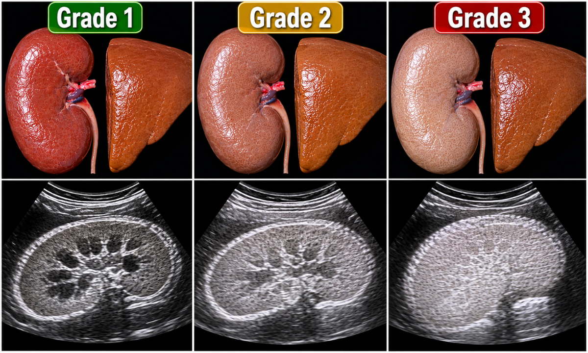
To ensure that every doctor understands exactly how much stress your organ is experiencing, radiologists use a standardized framework known as renal parenchymal disease grading. They compare the brightness of your kidney to the brightness of your neighboring liver or spleen.
Here is exactly what those grades mean for your health:
| The Clinical Grade | What The Radiologist Sees | What It Means For You |
|---|---|---|
| Grade 1 | The kidney tissue is slightly brighter than normal, but the boundaries between the outer and inner sections of the kidney remain perfectly sharp and visible. | The Biological Warning: The tissue is experiencing early stress, but the structure is intact. With prompt lifestyle adjustments, this is highly manageable and often reversible. |
| Grade 2 | The kidney has become just as bright as the liver. The delicate internal architectural lines are beginning to blur and fade. | Active Strain: The tissue is under significant, prolonged stress. This stage requires active medical intervention and medication to protect remaining function. |
| Grade 3 | The kidney is intensely bright (brighter than the liver), and the internal structural landmarks are completely wiped out. The organ may also appear physically smaller. | Advanced Alteration: Significant scarring has occurred. This requires dedicated, specialized nephrology care to manage symptoms and preserve overall health. |
The Empowering Takeaway: If a preventive scan catches your tissue changes at Grade 1, you are in a position of immense power. You have the precise biological data needed to tweak your diet, manage your blood pressure, and completely halt the progression before you ever feel a single physical symptom.
Navigating Hepatic Parenchymal Disease

While your kidneys handle filtration, your liver is your body’s primary metabolic powerhouse. It acts as a massive chemical processing plant, breaking down fats, storing energy, and neutralizing toxins before they can enter your systemic bloodstream.
When a doctor diagnoses liver parenchymal disease, they are observing that the essential, functional cells (hepatocytes) that perform this massive daily workload are becoming inflamed, congested, or scarred.
You may also see the term diffuse liver parenchymal disease on your medical report. The word diffuse simply means that the tissue changes are spread out evenly across the entire organ, rather than being isolated to a single, localized spot (like a cyst or a benign tumor).
The Root Causes of Liver Tissue Stress
Because the liver processes everything you consume, its parenchymal tissue is highly sensitive to metabolic and chemical imbalances. The most frequent drivers of diffuse hepatic changes include:
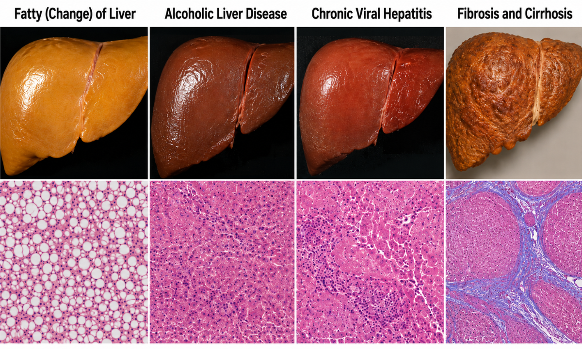
1. Non-Alcoholic Fatty Liver Disease (NAFLD): This is currently the leading cause of early-stage parenchymal stress globally.
Driven by metabolic syndrome, obesity, or unmanaged type 2 diabetes, excess fat slowly accumulates inside the liver cells. The parenchyma literally becomes congested with fat, causing chronic inflammation.
2. Toxin Processing Overload (Alcohol): Prolonged and excessive alcohol consumption directly poisons the hepatocytes.
As the liver struggles to metabolize the alcohol, the parenchymal cells become inflamed (alcoholic hepatitis) and eventually form rigid scar tissue.
3. Viral Infiltration: Chronic viral infections, specifically Hepatitis B and C, invade the parenchymal cells. The body’s own immune system then attacks the infected liver tissue, causing a cycle of smoldering inflammation and gradual scarring.
The Trajectory of Hepatic Changes
Similar to the kidneys, the liver attempts to heal its stressed tissue by laying down collagen, leading to fibrosis (scarring).
If caught in the early inflammatory stages (like a mild fatty liver), the liver possesses an incredible, unique biological superpower: it can regenerate. Simply removing the metabolic stressor, such as adopting a liver-friendly diet, managing cholesterol, or eliminating alcohol, the early-stage diffuse changes can often be completely reversed. However, if the stress continues unabated for years, the flexible parenchyma is permanently replaced by hard, nodular scar tissue, resulting in irreversible cirrhosis.
This is precisely why undergoing a routine diagnostic evaluation is so crucial; it identifies the congestion before the permanent scarring phase begins.
How Doctors Classify the Condition: The Medical Blueprint
When you receive your final diagnostic report, hospital discharge summary, or insurance paperwork, you might notice a string of alphanumeric codes printed next to your diagnosis. For many patients, seeing these clinical codes is highly confusing and can unnecessarily escalate anxiety. However, these codes are simply the healthcare system’s universal language.
To ensure that your care is perfectly standardized and to guarantee that your health insurance claims are processed smoothly, doctors rely on the International Classification of Diseases, 10th Revision (ICD-10). This system provides a specific administrative code for every possible medical observation.
For example, if your report indicates liver tissue stress, a common term you might look up is hepatic parenchymal disease icd 10. While there is not one single catch-all code for the word parenchyma itself, the ICD-10 system requires doctors to code the specific root cause of the tissue change.
To help you decode your medical paperwork, we have built a quick translation guide for the most common liver and kidney classifications.
Decoder Ring 1: Liver (Hepatic) Tissue Classifications
When your liver tissue shows diffuse changes on an ultrasound, your doctor will link that visual finding to a specific metabolic or chemical cause using the following administrative categories:
| The Clinical Diagnosis | Common ICD-10 Category | What It Actually Means For You |
|---|---|---|
| Fatty (Change) of Liver | K76.0 | Non-alcoholic fat accumulation in the liver cells. This is an early-warning signal that is highly responsive to dietary changes. |
| Alcoholic Liver Disease | K70 (Series) | Tissue inflammation is directly linked to toxin processing. It requires immediate lifestyle modification to halt progression. |
| Chronic Viral Hepatitis | B18 (Series) | Smoldering parenchymal inflammation caused by a virus (like Hep B or C), which requires targeted antiviral medication. |
| Fibrosis and Cirrhosis | K74 (Series) | Advanced structural changes where the flexible tissue has begun to scar. This requires specialized hepatology management. |
Decoder Ring 2: Kidney (Renal) Tissue Classifications
Similarly, if your kidneys show increased echogenicity (brightness) on an ultrasound, your physician will assign a code based on the underlying systemic stressor that is driving the kidney strain:
| The Clinical Diagnosis | Common ICD-10 Category | What It Actually Means For You |
|---|---|---|
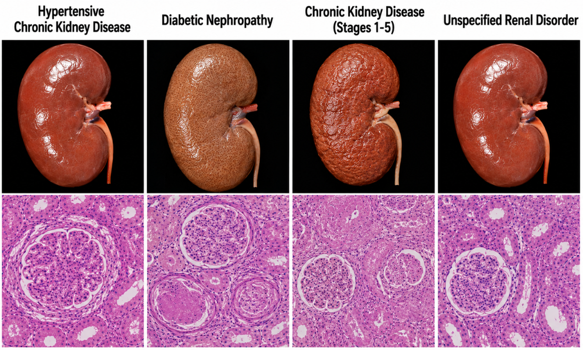
| Hypertensive Chronic Kidney Disease | I12 (Series) | Your kidney filters are under physical strain because your systemic blood pressure is running too high. |
| Diabetic Nephropathy | E11.2 (Series) | The kidney tissue is experiencing chemical and structural stress due to prolonged, unmanaged blood sugar levels. |
| Chronic Kidney Disease (Stages 1-5) | N18 (Series) | A staging code that explicitly tells other doctors exactly how efficiently your kidneys are still filtering your blood. |
| Unspecified Renal Disorder | N28.9 | A temporary placeholder code is used when an ultrasound shows structural changes, but the doctor is waiting for your blood test results to determine the exact cause. |
The Empowering Takeaway is that these codes are not a measure of your personal worth or a reason to panic. They are highly efficient administrative tools.
Categorizing your specific Types of parenchymal disease accurately, your healthcare team ensures that your treatment plan is universally understood by specialists, diagnostic labs, and your insurance provider, leaving zero room for clinical error.
Why Choose Koshikaa? Why Early Screening Matters at Koshikaa
When dealing with structural tissue changes in your vital organs, the most important biological fact to remember is that early stages are almost completely silent. By the time physical symptoms appear, the tissue has often progressed past the point of simple inflammation and into permanent scarring.
This is why transitioning from reactive healthcare (waiting until you feel sick) to proactive healthcare (testing while you feel healthy) is the ultimate protective measure.
As a premier diagnostic facility, Koshikaa is uniquely designed to provide you with a complete, 360-degree view of your internal health before any crisis occurs. Patients choose our center because we offer:
- Complete Diagnostic Clarity: Identifying early tissue stress requires both visual and chemical confirmation. Koshikaa houses state-of-the-art imaging technology for a highly precise Ultrasound scan in Bangalore, seamlessly paired with fully equipped in-house laboratories. This allows your doctors to cross-reference your scan with a comprehensive metabolic Blood test in Bangalore to pinpoint the exact root cause of the tissue change.
- Personalized Screening Paths: We are the first facility in the country to introduce a comprehensive personalized screening questionnaire. We map your exact lifestyle, diet, and family history to recommend only the tests you actually need to monitor your liver and kidney function, preventing unnecessary medical expenses.
- Rapid 24-Hour Reporting: Waiting for medical results is stressful. Our streamlined workflows guarantee that your highly accurate health reports are delivered within 24 hours, giving you and your doctor the immediate data required to make lifestyle adjustments.
True preventive care must be accessible. We offer heavily discounted standard packages and flexible payment options so that monitoring your vital organs never becomes a financial burden.
Conclusion
Seeing medical terminology on a laboratory report does not have to be an intimidating experience. Understanding the various Types of parenchymal disease simply means you now have the biological data necessary to understand how your body is handling its daily metabolic workload. Whether it is early-stage filtration stress in your kidneys or mild fatty congestion in your liver, your body has an incredible capacity to heal and regenerate when given the right support.
Do not wait for a physical symptom to tell you that your functional tissue is struggling. Take control of your long-term vitality today. Book your personalized preventive health check-up with Koshikaa and secure your healthiest tomorrow.