
Receiving a diagnostic report filled with unfamiliar medical terminology can instantly trigger a wave of anxiety. If you have recently opened a medical report and found a reference to structural changes in your organs, your immediate question is likely: What is parenchymal disease? It sounds alarming, but understanding this term is the first step toward taking proactive control of your health.
Often detected completely by surprise during a routine wellness check or a targeted Ultrasound scan in Bangalore, a parenchymal abnormality simply means there is a notable change in the functional tissue of a specific organ.
To demystify this term, it helps to understand how medical professionals look at your body’s anatomy. Every vital organ is broadly divided into two parts: the structural framework (the stroma) and the actual working, functional cells that perform the organ’s daily tasks (the parenchyma).
For instance, the parenchyma of your lungs consists of the tiny air sacs (alveoli) that exchange oxygen, while the parenchyma of your kidneys consists of the microscopic filters (nephrons) that clean your blood.
When a report notes “parenchymal disease,” it simply means that these working cells are experiencing stress, inflammation, or structural alteration. It is not a single, terrifying illness, but rather a descriptive umbrella term. Because these cellular changes often develop silently without causing any initial pain or outward symptoms, catching them early through preventive screening changes the entire trajectory of your wellness journey.
This guide will break down exactly how these changes manifest in your body, specifically within the kidneys and lungs and how modern, proactive healthcare can help you manage and treat them effectively before they impact your quality of life.
Medical Disclaimer
The information provided in this article regarding parenchymal tissue changes, renal pathology, and pulmonary chronic diseases is intended strictly for educational and informational purposes. It does not constitute formal medical advice, a definitive clinical diagnosis, or a personalized treatment protocol. Structural changes in internal organs vary drastically based on individual medical histories, genetics, and co-existing health conditions. Always consult with a qualified healthcare professional, radiologist, or specialist to interpret your diagnostic scans and laboratory reports. Never delay seeking professional medical evaluation, adjusting your medications, or altering your lifestyle based on the informational contents of this guide.
The Kidneys: Renal Parenchymal Disease

The kidneys are master filtration organs, working around the clock to clear toxins, balance electrolytes, and regulate blood pressure.
When a diagnostic report mentions renal parenchymal disease on ultrasound, it indicates that the specialized, working tissue responsible for this daily filtration, the renal cortex and medulla, is experiencing structural stress.
Instead of a localized issue like a kidney stone, parenchymal changes point to cellular-level wear and tear across the organ’s functional zones.
How Cellular Stress Alters Your Ultrasound Scan
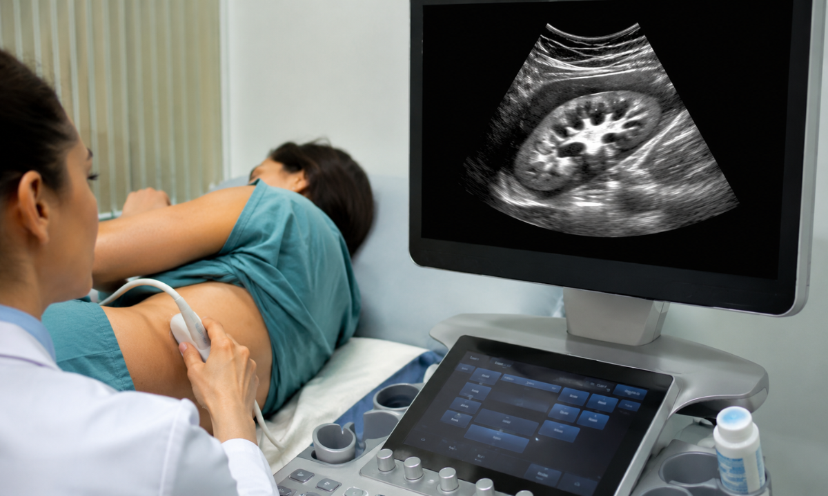
During a standard abdominal scan, a radiologist passes an ultrasound probe over the lower back or abdomen, sending high-frequency sound waves bouncing off your internal organs. Healthy kidney tissue has a specific density that appears relatively dark (hypoechoic) on the screen, contrasting beautifully with the surrounding protective fat.
However, when the delicate filtering units (nephrons) become inflamed, scarred, or congested due to an underlying condition, they change texture. The tissue becomes denser and reflects more sound waves, causing the kidney to appear unusually bright or white on the monitor. Medical professionals refer to this phenomenon as increased echogenicity.
Decoding Renal Parenchymal Disease Grading

To help your physician understand how advanced these structural changes are, radiologists use a standardized framework known as renal parenchymal disease grading. This system evaluates how bright the kidney tissue appears compared to nearby organs like the liver or spleen, and whether the internal architecture remains distinct.
| Disease Grade | Clinical Presentation on Scan | What It Means for Your Health |
|---|---|---|
| Grade 1 | The kidney tissue appears slightly brighter than normal, but the boundary separating the outer cortex from the inner medulla remains perfectly sharp. | Early Warning: Minimal cellular stress. Highly reversible if the underlying trigger is addressed immediately. |
| Grade 2 | The kidney’s brightness matches or slightly exceeds that of the liver. The internal architectural boundaries begin to look blurred or faint. | Moderate Stress: Active tissue strain. Demands targeted medical management to prevent long-term functional decline. |
| Grade 3 | The kidney appears intensely bright (hyperechoic), and the internal structural landmarks are completely obscured. The organ may also appear smaller. | Advanced Changes: Significant scarring or tissue alteration. Requires close, specialized nephrology care to protect remaining function. |
Catching the Silent Shift Early

The most crucial detail about early-stage kidney tissue changes is that they are entirely silent. You cannot feel your nephrons losing density, and your body can comfortably function even when a portion of the tissue is under strain. By the time symptoms like changes in urination, ankle swelling, or chronic fatigue actually appear, the tissue stress has often progressed past Grade 1.
This is precisely why preventive screenings are so transformative. Catching a Grade 1 change on a routine scan provides a golden biological window allowing you to tweak your lifestyle, manage your metabolism, and completely protect your long-term vitality before your body ever flashes a warning light.
The Lungs: Pulmonary Parenchymal Disease

Just as your kidneys rely on specialized tissue to filter your blood, your respiratory system depends on a highly specific biological infrastructure to perform its most vital task: keeping your body oxygenated.
When a medical professional mentions pulmonary parenchymal disease, they are shifting focus away from the main airways (the bronchial tubes) and directing it toward the actual working expansion units of the lungs.
Understanding how this tissue functions and how it can change is key to managing your long-term respiratory vitality with confidence.
The Microscopic Architecture of Breathing
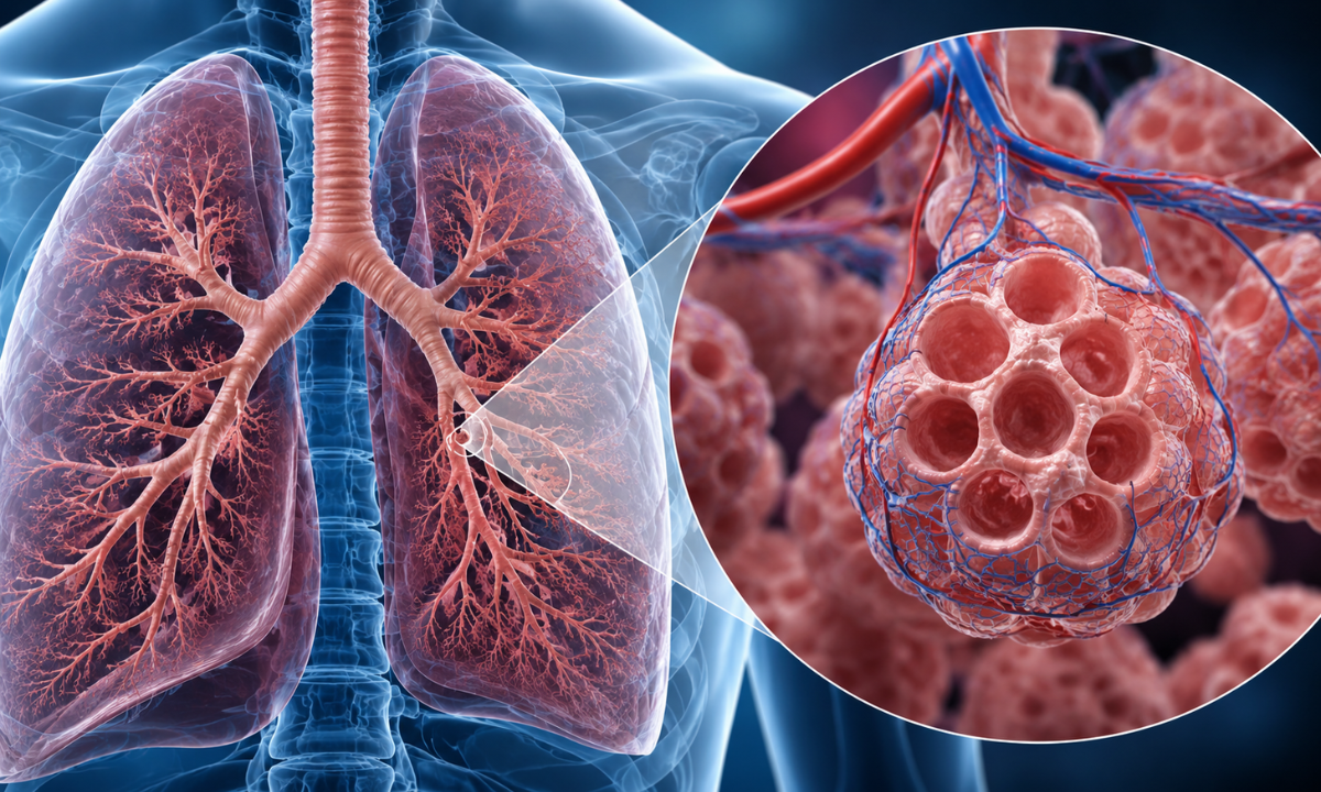
To visualize the lung parenchyma, imagine a massive, sprawling tree. The trunk and thick branches represent your trachea and bronchi, which act as hollow pipes transporting air. The leaves at the very tips of those branches represent the parenchyma.
This functional zone consists of millions of microscopic, elastic air sacs called alveoli, wrapped in an incredibly fine network of tiny blood vessels (capillaries).
Every single time you inhale, these delicate air sacs inflate like miniature balloons, allowing oxygen to cross a tissue barrier into your bloodstream while carbon dioxide passes out to be exhaled.
When a patient develops a lung parenchymal disease, these elastic air sacs and the ultra-thin walls between them become inflamed, congested, or progressively stiffened.
From Elasticity to Stiffness: Cellular Structural Changes
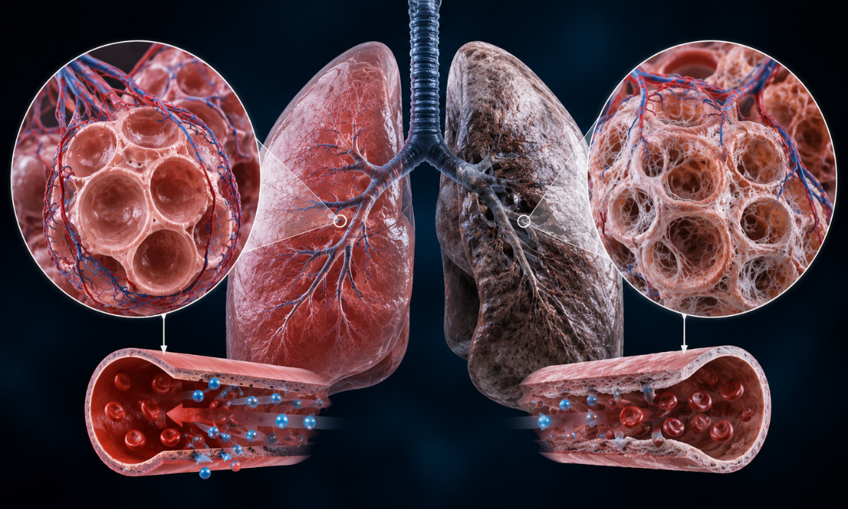
When the functional tissue of the lungs is exposed to sustained stress, whether from environmental toxins, long-term irritation, or an overactive immune response, the body attempts to heal itself by laying down dense collagen fibers.
This process, known as fibrosis, permanently alters the mechanical behavior of the lungs:
- Loss of Compliance: Healthy lung tissue is remarkably compliant and stretchy. As parenchymal changes progress, the tissue becomes rigid, making it feel as though the lungs are restricted by a tight band. It takes significantly more physical effort from your chest muscles just to draw a deep breath.
- Impaired Gas Exchange: As the walls of the air sacs thicken with fibrous tissue, the physical distance oxygen must travel to reach your blood vessels increases. This mismatch means that even if you take a full breath, less oxygen successfully makes it into your systemic circulation.
Managing a Chronic Respiratory Trajectory

Because these structural modifications alter the lung tissue permanently over time, they fall under the broad care umbrella of a chronic lung disease. As the initial phases of tissue stiffness can feel like a subtle lack of stamina or getting unusually winded while climbing stairs, people frequently misattribute early changes to “just getting older” or being slightly out of shape.
However, catching respiratory parenchymal shifts in their earliest stages completely transforms how they are managed.
While advanced tissue changes cannot be entirely reversed, early clinical identification allows medical teams to implement targeted therapies, lifestyle modifications, and antioxidant support that effectively halt the progression of tissue stiffness. By prioritizing early-stage clarity, you protect your breathing capacity and ensure your body retains the stamina to enjoy an active, independent life.
What Causes It and How Does It Present?
When patients see structural changes noted on their diagnostic reports, their first question is naturally about the origin.
Understanding the primary causes of parenchymal disease helps demystify the condition, shifting it from an abstract medical fear into a manageable health metric. Because the parenchyma is the active, working tissue of your organs, it is highly sensitive to sustained systemic stress, prolonged inflammation, and external toxins.
While the exact triggers depend entirely on which specific organ is affected, the root causes generally fall into three major biological categories:
- Metabolic and Cardiovascular Stress: For the kidneys, chronic high blood pressure (hypertension) and unmanaged blood sugar (diabetes) are the leading culprits. These conditions force the delicate microscopic filters to operate under immense vascular pressure, eventually causing them to wear out and scar.
- Environmental and Inhaled Toxins: For the lungs, long-term exposure to cigarette smoke, harsh occupational hazards (such as silica dust, coal, or asbestos), or severe respiratory infections can trigger a biological healing response that goes into overdrive, resulting in thick, fibrotic tissue.
- Autoimmune Activity: In some cases, the body’s own immune system becomes confused and mistakenly attacks healthy functional cells, leading to chronic inflammation in either the respiratory or renal systems.
Decoding Bilateral vs. Unilateral on Your Report
As you read through the radiologist’s notes on your scan, you will likely encounter specific anatomical terms describing the location and symmetry of the tissue changes.
If your diagnostic report explicitly notes parenchymal disease bilateral, there is no need to panic. The term bilateral simply means that the structural changes are present in both the left and right organs simultaneously (e.g., both kidneys or both lungs).
This is incredibly common and entirely expected when the underlying cause is a systemic issue like high blood pressure or a metabolic disorder, because these conditions affect the entire bloodstream equally.
Conversely, if the report is unilateral, it means the changes are isolated to just one side, which usually points to a localized event, such as a past physical injury, a localized blockage, or a severe localized infection.
Ultimately, any form of parenchymal disease is simply your body’s biological warning system. It is a physical indicator that your functional tissue has been pushed beyond its comfortable limits and is actively adapting to chronic stress.
Identifying the root cause early, you can collaborate with your healthcare team to remove the stressor, stabilize the tissue, and protect your remaining organ function.
Early Detection & Clinical Management: Navigating Your Next Steps
When a diagnostic report identifies parenchymal changes, the conversation naturally shifts from understanding the condition to exploring treatment pathways. It is essential to recognize that a parenchymal abnormality is not a fixed, unchanging destination.
As the parenchyma is living, highly active cellular tissue, its trajectory depends entirely on how quickly the underlying cause is identified and managed. Treatment is never a one-size-fits-all prescription; it is a highly tailored strategy aimed at removing tissue stress and halting progression.
The Pathways to Clinical Management
Depending on the specific organ involved and the root cause of the cellular stress, healthcare professionals deploy a combination of medical and lifestyle interventions to protect your organ function:
1. Targeted Pharmaceutical Support: If the tissue changes are driven by metabolic stress, managing the root driver is paramount.
Doctors routinely use precise medications to optimize blood sugar levels or tightly regulate systemic blood pressure, relieving the physical workload on the kidneys’ delicate filters. For respiratory tissue changes driven by inflammation, specialized anti-inflammatory treatments or medications that inhibit tissue stiffening (antifibrotics) may be prescribed to preserve lung compliance.
2. Nutritional and Metabolic Tweak: Your diet directly influences the workload of your vital organs.
For instance, early-stage renal parenchymal changes are often managed by adjusting dietary protein, sodium, and fluid intake to reduce the filtration burden.
3. Elimination of Environmental Stressors: For the lungs, immediately removing toxic triggers such as tobacco smoke, airborne chemical irritants, or occupational dust is the single most effective step to stop the accelerated laying down of rigid collagen fibers.
The Role of Comprehensive Diagnostic Tracking
Because early-stage parenchymal stress does not produce obvious physical symptoms, tracking the health of your tissue requires a combination of advanced medical imaging and precise laboratory evaluation.
For example, if an ultrasound detects a minor structural change in the kidneys, your physician will immediately want to evaluate how well those tissues are actually performing chemically.
This is where advanced laboratory diagnostics become indispensable. A routine, highly precise Blood test in Bangalore allows clinicians to measure critical biological markers, such as Serum Creatinine and Blood Urea Nitrogen (BUN), and calculate your Estimated Glomerular Filtration Rate (eGFR).
Pairing the visual data from a scan with the metabolic data from your blood work, doctors receive a complete, 360-degree map of your health. This comprehensive approach ensures that any subtle shift in tissue vitality is detected instantly, giving you the precise data you need to adjust your lifestyle, protect your organs, and maintain your long-term wellness with absolute confidence.
Why Choose Koshikaa for Your Preventive Health Screenings?
When navigating a potential parenchymal abnormality or simply seeking to establish a baseline for your vital organs, where you get screened matters just as much as the test itself, detecting subtle, cellular-level tissue changes requires advanced diagnostic precision, expert radiological interpretation, and a patient-centric approach that minimizes anxiety.
As a leading Health Screening Centre in Bangalore, Koshikaa is uniquely equipped to partner with you on your preventive healthcare journey. Patients trust our facility for comprehensive diagnostics because we provide:
- India’s First Personalized Screening Questionnaire: We do not believe in cookie-cutter healthcare. Our innovative, personalized questionnaire maps out your exact lifestyle, medical history, and genetic risk factors to tailor a screening package that addresses your body’s specific needs, ensuring you never pay for unnecessary tests.
- Advanced Multi-Modality Imaging: Evaluating parenchymal tissue demands exceptional visual clarity. Koshikaa houses state-of-the-art diagnostic infrastructure under one roof, ranging from low-dose CT scans and high-resolution MRI to precise Ultrasound scan in Bangalore, allowing for meticulous organ evaluations.
- Rapid 24-Hour Report Delivery: We understand that waiting for medical answers can be stressful. Our streamlined laboratory workflows ensure that your highly accurate, comprehensive health reports are finalized and delivered within 24 hours of your screening.
True preventive care must be accessible to everyone. Koshikaa offers transparent, highly competitive pricing alongside flexible payment options and heavily discounted standard packages, making regular wellness checks financially manageable.
Conclusion
A reference to “parenchymal disease” on a medical report can initially feel overwhelming, but it is ultimately a valuable window of opportunity. Whether it indicates early-stage stress in your kidneys or initial structural changes in your lungs, catching these cellular shifts before they manifest as outward symptoms gives you the ultimate advantage: time.
Utilizing routine diagnostic screenings, you shift from a reactive state of fighting illness to a proactive state of preserving wellness. If you are ready to take control of your long-term vitality, establish your baseline, and gain absolute clarity, contact Koshikaa today to select your tailored health screening package.