When scheduling an annual checkup, it is very common to focus exclusively on cardiovascular metrics, prioritizing a routine blood test to evaluate your cholesterol and triglyceride levels.
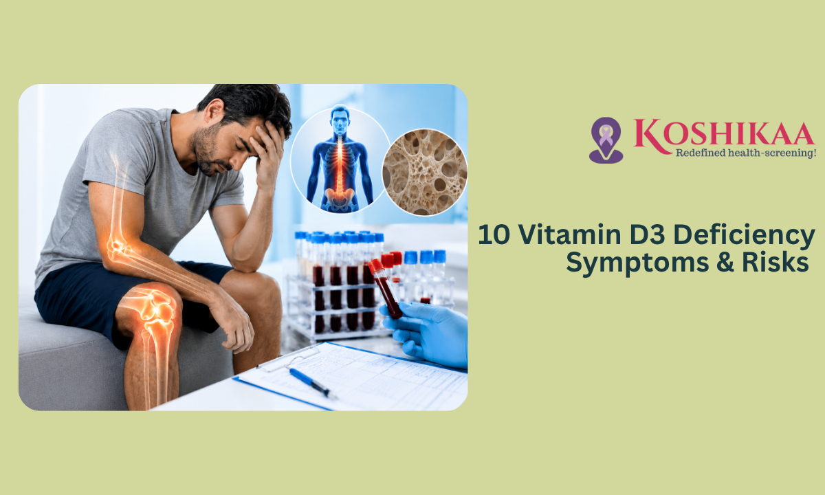
While tracking your heart health is absolutely crucial, many patients completely overlook another essential marker that dictates their daily energy, immunity, and structural strength: Vitamin D3. Securing a comprehensive Blood test in Bangalore is the only definitive way to measure this vital pro-hormone. Unlike standard nutritional vitamins, Vitamin D3 actually functions as a master regulatory switch for your entire body, directly influencing everything from how your body absorbs calcium to how your brain regulates mood.
Let us explore why this essential “sunshine vitamin” is so critical, the silent warning signs your body gives when your levels drop dangerously low, and the severe long-term medical disorders that can develop if this deficiency is left completely unaddressed.
Medical Disclaimer
The information provided in this article is strictly for educational purposes and does not replace professional medical advice, formal diagnosis, or specialized clinical treatment. Standard Vitamin D ranges and required supplementation doses vary significantly based on individual health profiles, age, and existing medical conditions. Always consult directly with a qualified physician regarding your specific laboratory results and before starting any high-dose vitamin supplements. Never ignore medical warnings or delay seeking professional care based on the contents of this guide.
Why Are We So Deficient?

Despite living in a geographically blessed country with abundant sunshine year-round, a massive segment of the urban population is quietly operating empty when it comes to vital micronutrients.
Vitamin D3 is uniquely synthesized by our skin when exposed directly to ultraviolet B (UVB) solar rays. However, modern lifestyle shifts have inadvertently severed this natural biological process, turning what should be a routine metabolic function into a widespread, invisible health crisis.
The Dynamics of Modern Exposure
The prevalence of Vitamin D deficiency in adults has skyrocketed over the last few decades, largely driven by the structural realities of modern daily routines. The shift from open-air occupations to indoor, desk-bound office roles means most working professionals step outside only after the sun has already dipped below the horizon.
Furthermore, our proactive skincare habits, while highly beneficial for preventing surface sun damage, act as a total shield against nutrient synthesis. Applying a standard sunscreen with an SPF as low as 15 blocks over 99% of UVB radiation, completely halting your skin’s capacity to manufacture Vitamin D3.
When you combine minimal sun exposure with urban air pollution, which scatters and filters out vital solar wavelengths, it becomes clear why relying solely on casual outdoor walks is no longer sufficient.
Why Women Face an Elevated Structural Risk?
While this epidemic spans across all age groups, the structural toll of Vitamin D deficiency in women is exceptionally pronounced and carries distinct long-term biological consequences.
Women encounter specific hormonal transitions throughout life that amplify the body’s reliance on this pro-hormone, making any drop in baseline levels far more destructive to their long-term health.
| Life Stage / Biological Driver | Why the Impact is Heightened | Long-Term Structural Consequence |
|---|---|---|
| Pregnancy and Lactation | The developing maternal system must rapidly transfer massive reserves of calcium to support fetal skeletal growth. | Severe depletion of maternal bone density if baseline Vitamin D3 is insufficient to drive intestinal calcium absorption. |
| The Menopausal Transition | Sudden, sharp declines in protective estrogen levels naturally accelerate the rate of bone remodeling and depletion. | Without adequate Vitamin D3 to lock calcium into the skeletal frame, the risk of developing brittle bones climbs exponentially. |
| Modern Metabolic & Lifestyle Overlap | High daily stress levels increase circulating cortisol, a stress hormone that actively competes with and binds to Vitamin D receptors. | Chronic fatigue, weakened cellular defenses, and an increased vulnerability to metabolic and structural imbalances. |
Understanding that our modern environments are structurally designed to limit natural vitamin synthesis shifts the perspective from casual neglect to proactive awareness.
You cannot fix an invisible, body-wide deficiency if you do not know it exists. Now that we understand the environmental and demographic forces driving this widespread shortage, let us examine the ten specific, daily physical symptoms your body utilizes to signal a critically low supply.
The 10 Critical Symptoms You Shouldn’t Ignore

Because Vitamin D3 operates as a body-wide regulatory hormone, a drop in your baseline levels rarely manifests as a single, isolated issue. Instead, the warning signs cascade across multiple physical systems, often mimicking other common illnesses. If you are experiencing several of the following Severe vitamin D deficiency symptoms, your body is actively signaling a critical internal shortage.
- Unexplained Chronic Fatigue: Unlike normal tiredness after a long day, this is a profound, heavy exhaustion that does not improve with sleep. Vitamin D is essential for cellular energy production; without it, your mitochondria (the powerhouses of your cells) simply cannot generate the sustained energy required to get you through the day.
- Persistent Bone and Lower Back Pain: Vitamin D is the exclusive biological key that unlocks your intestines’ ability to absorb calcium from your diet. When levels drop, your body is forced to pull calcium directly from your skeleton to keep your heart beating, resulting in deep, throbbing aches, particularly noticeable in your lower spine, ribs, and legs.
- Proximal Muscle Weakness and Cramps: Your skeletal muscles are packed with Vitamin D receptors. A severe deficiency directly impairs muscle contraction and protein synthesis, leading to unexpected weakness (like struggling to stand up from a low chair), chronic muscle stiffness, and painful nighttime leg cramps.
- Frequent Illnesses and Infections: Vitamin D directly interacts with the cells responsible for fighting off pathogens. A suppressed baseline level drastically lowers your immune response, leaving you highly susceptible to catching every seasonal cold, flu, or respiratory tract infection that circulates in your office or home.
- Depression and Mood Swings: Often nicknamed the “sunshine vitamin,” this pro-hormone plays a pivotal role in the brain’s synthesis of serotonin, the neurotransmitter responsible for mood regulation. A chronic deficiency is heavily linked to sudden mood swings, heightened anxiety, and persistent depressive states.
- Slow and Impaired Wound Healing: If simple cuts, scrapes, or surgical incisions seem to take an unusually long time to close and heal, it points toward a localized cellular deficit. Vitamin D is vital for producing the compounds required to create new skin and suppress harmful inflammation at the wound site.
- Severe Hair Loss: While mild daily shedding is normal, heavy, patchy hair loss (often linked to conditions like alopecia areata) is a severe structural warning. Vitamin D actively regulates the health and life cycle of your hair follicles; without it, new hair growth significantly slows or stops entirely.
- Cognitive Fog and Memory Lapses: Because of its protective role in neurological health, low levels can lead to noticeable cognitive decline. You may experience frequent “brain fog,” an inability to concentrate on complex tasks, or sudden struggles with short-term memory recall.
- Recurrent Dental Problems: Because a deficiency severely hinders calcium absorption, your teeth become just as vulnerable as your bones. Unexplained gum inflammation, weakened enamel, and a sudden increase in cavities are common secondary signs of a system-wide lack of Vitamin D3.
- Unexplained Weight Gain: Vitamin D is a fat-soluble hormone, meaning it is stored in your body’s fat tissues. When levels drop, it can negatively impact your metabolism and insulin sensitivity, making it incredibly difficult to lose weight and frequently leading to unexplained weight accumulation around the midsection.
Ignoring these daily symptoms forces your body to operate in a constant state of structural compromise. In the next section, we will explore exactly what happens when these daily inconveniences evolve into permanent clinical diagnoses.
Health Problems Linked to Low Vitamin D
While daily fatigue, sudden mood drops, and minor muscle cramps are undoubtedly frustrating, they are merely the tip of the iceberg.
These immediate physical complaints are your body’s early warning system. When these signals are consistently ignored or masked with temporary fixes like over-the-counter painkillers or extra caffeine, the sustained lack of this vital pro-hormone forces your internal systems to permanently adapt to a state of depletion.
“A symptom is your body actively asking for help. A chronic disorder is the permanent structural consequence of that request being ignored for too long.”
When discussing the severe health problems linked to low vitamin D, we must look past daily discomforts and examine the long-term clinical diagnoses that arise when cellular regulation completely breaks down.
A chronic deficiency acts as a silent catalyst for several major systemic diseases.
The Major Systemic Clinical Risks
1. Severe Skeletal Degeneration (Osteoporosis and Osteomalacia)
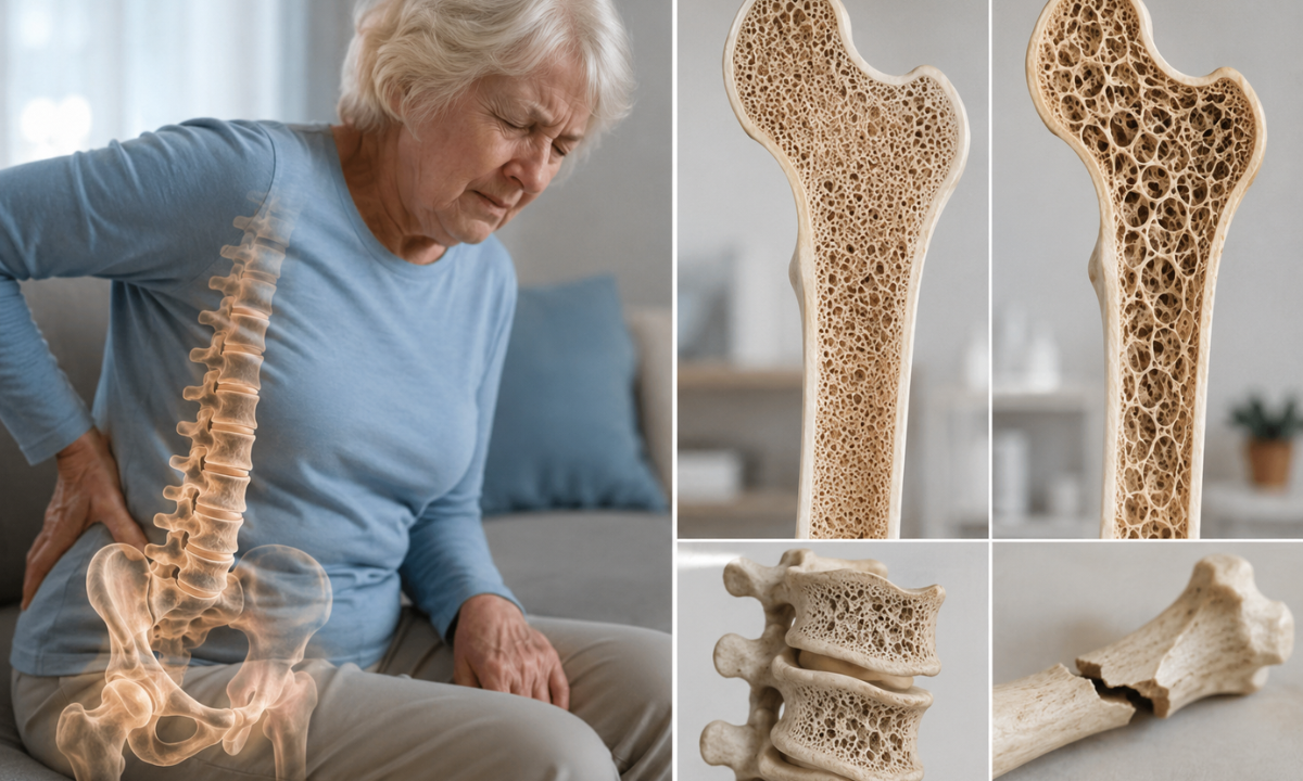
Your bones are not static pillars; they are living tissues that constantly break down and rebuild themselves. Without adequate Vitamin D3, your digestive tract absorbs less than 15% of the calcium you consume.
To maintain vital blood calcium levels, your body aggressively mines your own skeleton. In adults, this prolonged structural theft leads to osteomalacia (softening of the bones) and osteoporosis (porous, brittle bones), making devastating fractures of the hip, spine, and wrist a high-probability event as you age.
2. Autoimmune System Malfunction

Vitamin D is a potent immunomodulator, meaning it acts as the intelligent braking system for your immune cells. It teaches your immune system the difference between a dangerous foreign virus and your own healthy tissue. When levels are critically low, this braking system fails.
The immune system becomes hyperactive and confused, leading to a drastic increase in the risk of developing severe autoimmune disorders such as Multiple Sclerosis (MS), Rheumatoid Arthritis, Hashimoto’s Thyroiditis, and Lupus.
3. Cardiovascular and Endothelial Dysfunction

Your heart and blood vessels are lined with Vitamin D receptors. A prolonged deficiency directly damages the endothelium, the delicate inner lining of your blood vessels. This damage causes vascular stiffness, promotes chronic systemic inflammation, and accelerates the calcification of your arteries.
Over time, this drastically increases your clinical risk for developing uncontrollable hypertension (high blood pressure), congestive heart failure, and sudden ischemic strokes.
4. Metabolic Syndrome and Type 2 Diabetes
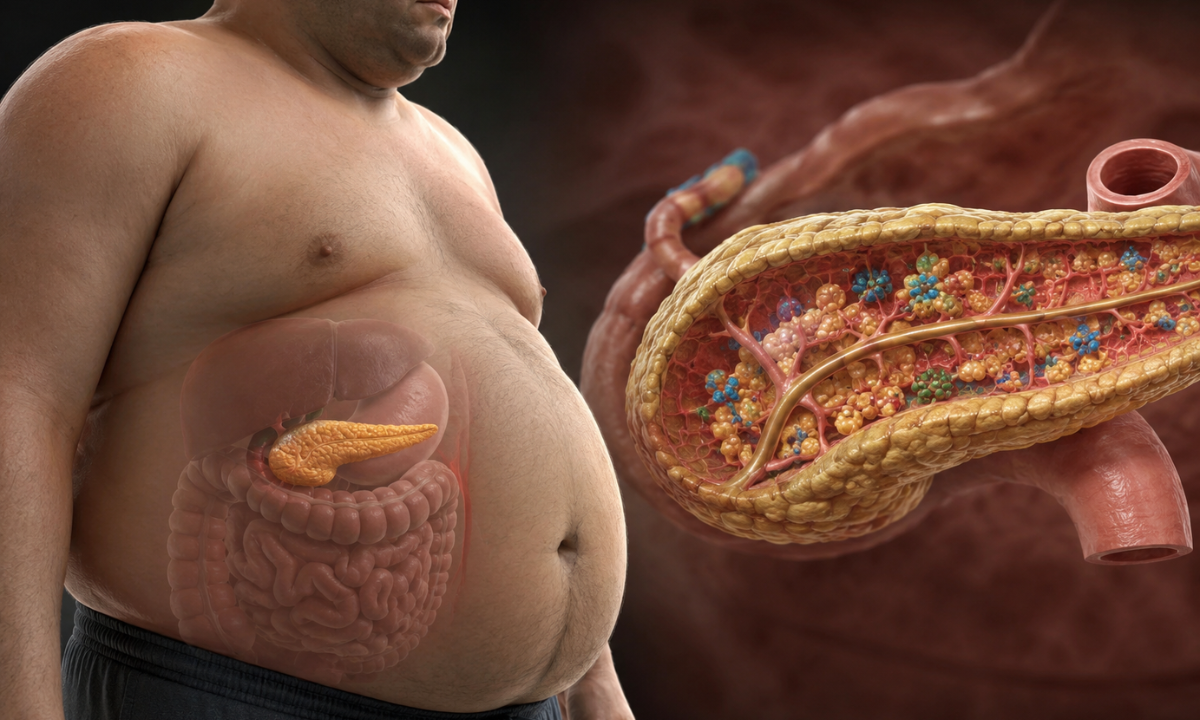
Vitamin D3 plays a direct, hands-on role in pancreatic health. It specifically helps regulate the beta cells in your pancreas that manufacture and secrete insulin. A deep deficiency heavily blunts your body’s insulin sensitivity, forcing your pancreas into overdrive to process basic dietary sugars.
This constant metabolic strain paves a direct path to insulin resistance, severe weight gain, and the eventual onset of Type 2 Diabetes.
The Evolution of Deficit: From Symptom to Disorder
To fully grasp the danger of delay, it is helpful to see exactly how a minor, ignored symptom evolves into a major clinical emergency over time.
| The Ignored Early Symptom | The Affected Biological System | The Long-Term Associated Disorder |
|---|---|---|
| Dull, aching lower back pain | Skeletal & Structural | Osteoporosis and spinal compression fractures |
| Catching frequent seasonal colds | Immune Defense | Autoimmune diseases (Rheumatoid Arthritis, Lupus) |
| Persistent brain fog and fatigue | Neurological & Metabolic | Type 2 Diabetes and accelerated cognitive decline |
| Type 2 Diabetes and accelerated cognitive decline | Cardiovascular | Endothelial dysfunction and hypertension |
Transitioning from a manageable symptom to a permanent, life-altering medical disorder happens silently over years of neglect. The absolute best way to intercept this destructive biological timeline is to stop guessing about your symptoms and secure a definitive clinical answer.
How to Confirm Your Levels: The Diagnostic Process
As the warning signs of low Vitamin D3 perfectly mimic so many other conditions, such as thyroid imbalances, iron deficiency, or simple occupational burnout, you cannot rely on physical symptoms alone to determine your actual health status.
A definitive Vitamin D deficiency diagnosis requires absolute clinical precision. The only medically accurate way to measure your body’s true internal reserves is through a specific blood evaluation known as the 25-hydroxyvitamin D, or 25(OH)D, test.
Attempting to self-diagnose and blindly taking high-dose supplements can actually be dangerous. Because Vitamin D is fat-soluble, your body stores the excess in your tissues rather than flushing it out, which can eventually lead to a rare but severe condition called Vitamin D toxicity.
Instead of guessing, visiting a premier Health Screening centre in Bangalore like Koshikaa allows you to establish a precise baseline safely and quickly.
Decoding Your Lab Results
When you receive your digital report, your Vitamin D levels will typically be measured in nanograms per milliliter (ng/mL). Understanding these ranges helps you instantly grasp where your health currently stands and what level of intervention is required.
Here is the standard clinical reference guide for interpreting your 25(OH)D blood test:
| Clinical Status | Blood Level (ng/mL) | What It Means for Your Body | Required Action |
|---|---|---|---|
| Deficient | Less than 20 ng/mL | Your body is actively pulling calcium from your bones to survive. You are at high risk for the severe symptoms and structural disorders discussed earlier. | Requires immediate clinical intervention and high-dose prescription supplementation under a doctor’s care. |
| Insufficient | 20 – 29 ng/mL | Your levels are borderline. You may not feel severe pain yet, but your immune system and metabolic functions are operating at a reduced capacity. | Requires lifestyle adjustments, increased safe sun exposure, and standard daily supplementation. |
| Optimal (Sufficient) | 30 – 50 ng/mL | Your skeletal structure, immune system, and cardiovascular networks have the exact amount of pro-hormone required to function perfectly. | Maintain your current healthy lifestyle, diet, and outdoor habits. |
| Potential Toxicity | Over 100 ng/mL | Usually caused by extreme over-supplementation. This can cause dangerous calcium buildup in your blood, leading to kidney stones and heart rhythm abnormalities. | Immediately stop all Vitamin D supplements and consult your physician. |
Securing this single, straightforward metric, you completely remove the guesswork from your healthcare routine.
Once you know your exact number, your doctor can prescribe a highly customized recovery plan, whether that involves a slight dietary tweak or a structured prescription to safely guide your levels back into the optimal green zone.
The Koshikaa Advantage
When you are dealing with profound fatigue or unexplained bone pain, the last thing you want is a complicated, stressful healthcare experience.
Identifying a Vitamin D3 deficiency requires pinpoint accuracy, and that is exactly what Koshikaa is built to deliver. As a premier Health Screening centre in Bangalore, we have designed our diagnostic ecosystem to be entirely patient-focused, removing the friction from preventive care.
Our advanced Vitamin D3 blood tests provide precise and reliable measurements of your vitamin levels, helping detect deficiencies early and enabling timely intervention to support stronger bones, improved immunity, and overall wellness.
Here is why patients choose Koshikaa to uncover their hidden health metrics:
- Unmatched Diagnostic Accuracy: Our state-of-the-art laboratory utilizes advanced automated analyzers to measure your 25(OH)D levels with flawless precision, ensuring your doctor receives the exact data needed to prescribe the right dosage.
- Age-Wise Screening Packages: We understand that a 35-year-old woman requires a different health baseline than a 65-year-old man. Our comprehensive checkups are uniquely tailored to your specific age group and biological life stage.
- Personalized Health Care: We are the first screening center in the country to implement a comprehensive personalized questionnaire, ensuring your diagnostic journey is customized to your unique lifestyle, diet, and family history.
You do not have to wait in suspense. Your secure, highly detailed digital health report is delivered directly to your device within 24 hours of your quick blood draw.
Conclusion
You do not have to accept chronic fatigue, persistent bone aches, or a weakened immune system as a normal part of aging or a busy lifestyle. Vitamin D3 deficiency is a widespread epidemic, but it is also one of the easiest clinical conditions to identify and completely reverse. By transitioning from guessing about your symptoms to securing a definitive, precise blood test, you take immediate control of your structural and metabolic future.
Do not wait for a minor, daily inconvenience to evolve into a permanent, chronic disorder. Prioritize your health today, establish your baseline, and give your body exactly what it needs to thrive.