Since the X-ray discovery in 1895, X-rays have remained the most widely used form of medical imaging.
Despite the advent of advanced scans like MRI and CT, the humble X-ray is often the first diagnostic step for everything from a persistent cough to a traumatic injury.
The term Radiography refers to the technique of using X-radiation to view the internal form of an object.
In medicine, this allows doctors to visualize structures inside the human body without making a single incision.
The principle is based on density.
Different tissues absorb radiation at different rates.
Bones, which are dense and calcium-rich, absorb the most radiation and appear white on the film.
Soft tissues like muscles and organs allow more radiation to pass through and appear in shades of gray, while air-filled spaces like the lungs appear black.
Understanding the specific applications of this technology is important for patients.
A dental X-ray uses a completely different frequency and technique than a spine X-ray.
This guide categorizes the various forms of X-ray technology, explains their specific diagnostic roles, and helps you understand what to expect when your doctor prescribes a chest X-ray or other radiographic procedures.
Medical Disclaimer
The information provided in this blog regarding different types of X-rays and diagnostic imaging is for general educational purposes only.
It does not constitute professional medical advice, diagnosis, or treatment.
Radiation exposure, while minimal in modern diagnostics, carries specific contraindications.
Always consult a qualified radiologist or physician before undergoing any imaging procedure.
Pregnancy Warning: If you are pregnant or suspect you might be, you must inform your doctor and technician immediately, as X-rays can be harmful to the developing fetus.
Visit a certified Health screening centre in Bangalore to ensure safety protocols are followed.
Classification by Technology: How Images are Captured
While patients often group all scans under the general term “X-ray,” medical imaging is strictly categorized by how the radiation interacts with the body and how the resulting image is processed.
The three primary technological methods used in a modern X-ray centre in Bangalore are Standard Radiography, Fluoroscopy, and Computed Tomography.
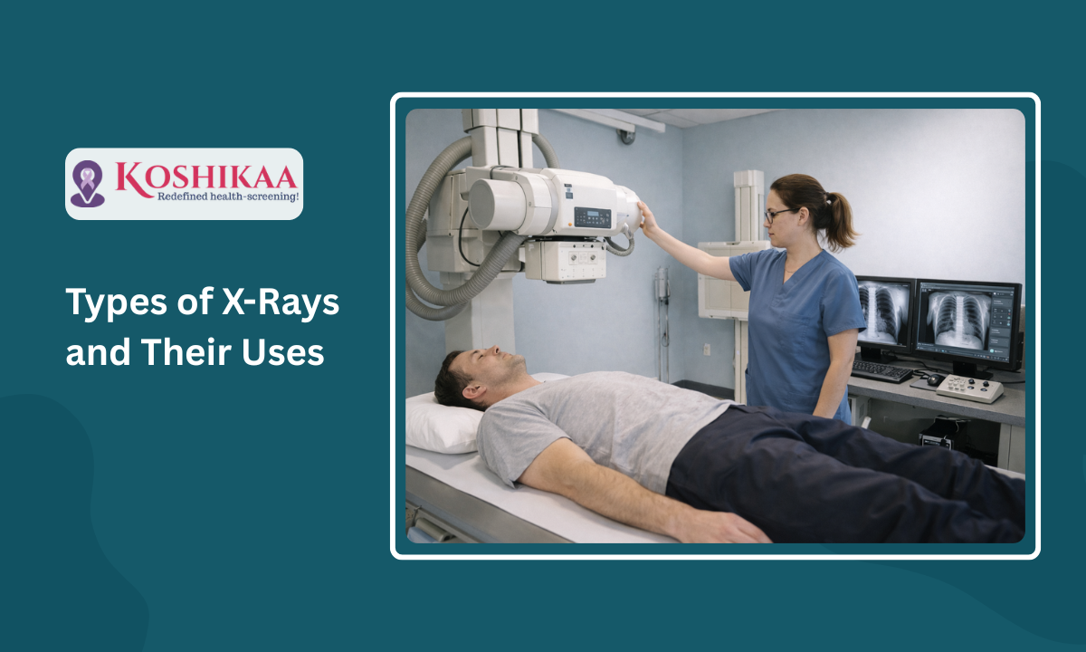
A. Standard Radiography (Static Imaging)
This is the most common form of X-ray. A single burst of radiation passes through the body to capture a static, two-dimensional image on a digital detector or film.
- Mechanism: It functions similarly to a camera shutter.
The machine exposes the sensor for a fraction of a second to capture a specific anatomical structure.
- Applications: It is primarily used for diagnosing bone fractures, joint dislocations, chest infections, and dental issues. A chest X-ray or a limb X-ray falls into this category.
Standard radiography remains the first-line diagnostic tool because it is quick, painless, and involves the lowest radiation dose among all X-ray-based modalities.
It provides a clear snapshot of dense structures like bone but offers limited detail for soft tissues.
B. Fluoroscopy (Real-Time Imaging)
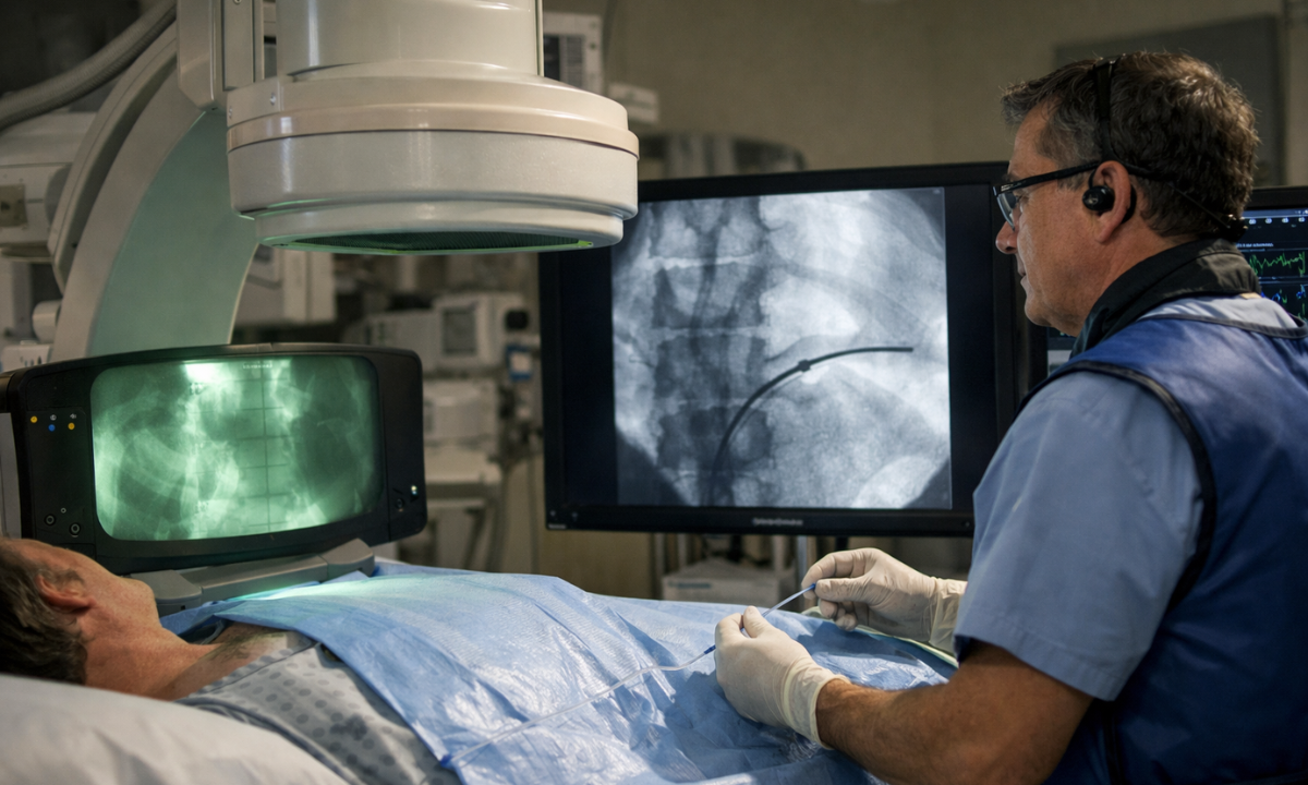
Unlike standard radiography, which takes a static snapshot, fluoroscopy uses a continuous X-ray beam to create a moving video sequence of the interior of the body.
- Mechanism: The X-ray beam is transmitted continuously through the patient to a fluorescent screen, allowing the radiologist to observe internal movement in real-time.
- Applications: It is essential for procedures that require visual guidance, such as positioning a catheter during Angiography, observing the digestive tract during a Barium Swallow, or guiding needle injections into specific joints.
Because fluoroscopy involves continuous exposure to capture motion, the radiation dose is generally higher than that of a standard X-ray.
However, it provides critical functional data that a static image cannot, such as verifying if the esophagus is swallowing correctly or visualizing blood flow through a vessel.
C. Computed Tomography (CT)
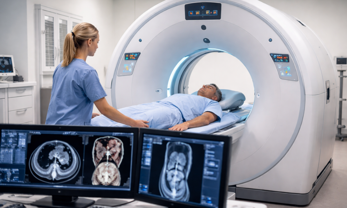
While often discussed as a separate category, a CT scan is technically a sophisticated application of X-ray technology.
- Mechanism: The X-ray source rotates 360 degrees around the patient, taking hundreds of cross-sectional images (slices) from different angles. A computer then processes these slices to reconstruct a detailed 3D model of the internal organs.
- Applications: CT is used for assessing complex internal trauma, detecting tumors, and visualizing blood vessels in the brain or abdomen.
CT scans provide significantly more detail than standard radiography, allowing doctors to distinguish between different types of soft tissues and blood vessels that would otherwise appear invisible or blurred on a regular X-ray film.
Common Clinical Applications
While the underlying technology remains the same, the technique and dosage vary significantly depending on the part of the body being examined.
Radiologists at a Health screening centre in Bangalore adjust these parameters to ensure the clearest image with the lowest possible radiation exposure.
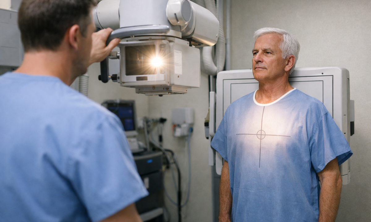
Thoracic Radiography (Chest X-Ray)
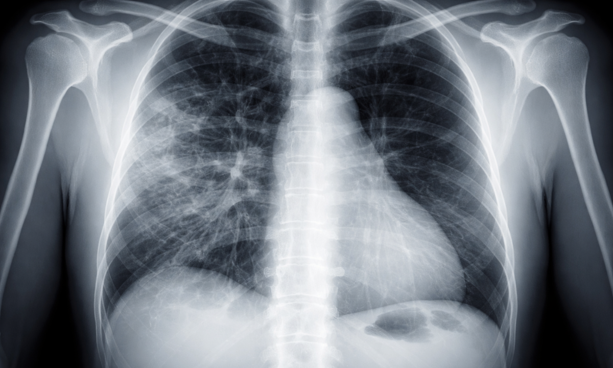
The chest X-ray is the most frequently performed radiographic procedure globally.
It acts as the primary screening tool for heart and lung conditions.
- Lungs: It detects infections such as pneumonia, tuberculosis, or fluid accumulation (pleural effusion).
- Heart: It reveals if the heart is enlarged (cardiomegaly), which can be a sign of heart failure.
- Bones: It identifies fractures in the ribs or clavicle.
An accurate X-ray chest diagnosis requires the patient to take a deep breath and hold it.
This expands the lungs fully, allowing the radiologist to see the fine details of the lung tissue and separate them from the shadows of the ribs.
Skeletal and Spine Radiography
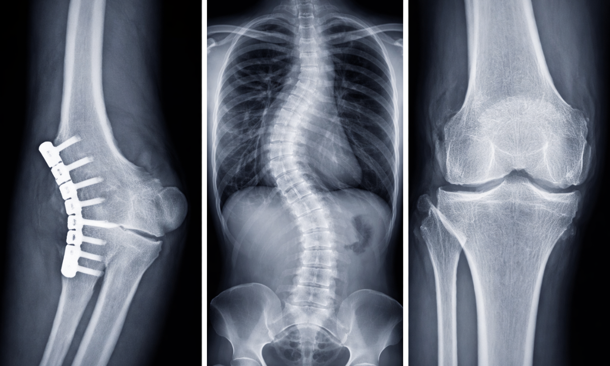
X-rays are the gold standard for assessing the skeletal system.
- Fractures: A simple X-ray can confirm the type and severity of a bone break.
- Spine: A spine X-ray is crucial for diagnosing scoliosis (curvature of the spine), herniated discs, or spinal stenosis. It helps doctors visualize the alignment of the vertebrae.
- Joints: It detects signs of arthritis, dislocations, or bone spurs.
For a spine X-ray, multiple views (front, side, and bending) are often taken to assess the stability of the spinal column under different movements.
This helps orthopedic surgeons plan treatments effectively.
Dental Radiography
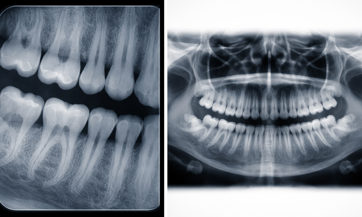
A dental X-ray operates on a much smaller scale but provides high-resolution images of the teeth and jawbone.
- Intraoral X-rays: The film is placed inside the mouth to detect cavities between teeth, check the health of the tooth root, and monitor the surrounding bone level.
- Extraoral X-rays: The film is outside the mouth. This is used to track jaw growth, identify impacted wisdom teeth, or visualize the temporomandibular joint (TMJ).
Dental X-rays use significantly lower radiation doses than medical X-rays.
They are an essential part of routine dental checkups to identify issues like cysts or abscesses that are invisible during a visual exam.
Abdominal Radiography
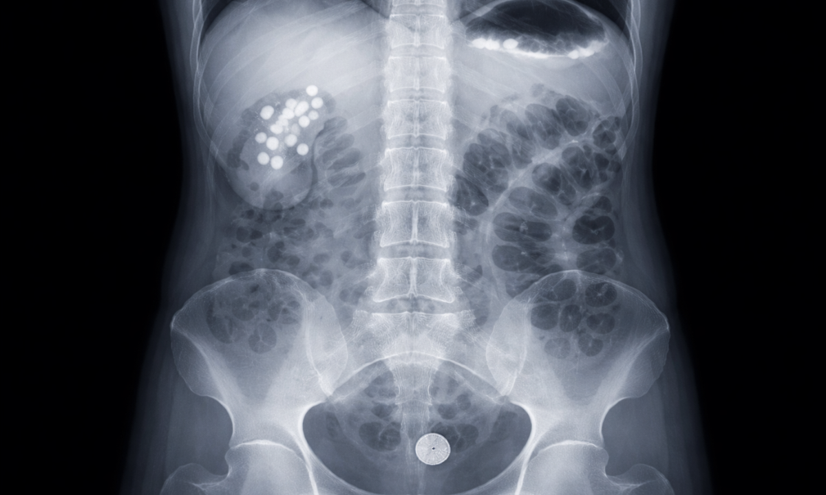
An X-ray of the abdomen, often called a KUB (Kidney, Ureter, Bladder) scan, is frequently used to diagnose digestive and urinary tract issues.
- Kidney Stones: Many stones are calcium-based and show up clearly as white spots.
- Blockages: It can reveal intestinal obstructions or the presence of swallowed foreign objects.
- Perforation: It detects free air in the abdominal cavity, which is a medical emergency indicating a hole in the stomach or intestine.
These specific applications demonstrate the versatility of X-ray technology.
Whether it is a chest X-ray for a cough or a spine X-ray for back pain, the procedure provides immediate, actionable data for physicians.
Specialized Contrast Studies: Fluoroscopy and Angiography
Standard X-rays have a significant limitation.
They are excellent at distinguishing dense bone from air, but they struggle to differentiate between soft tissues of similar density, such as blood vessels, muscles, and organs.
To solve this, radiologists use Contrast substances like iodine or barium that block X-rays and appear white on the image.
By introducing these dyes into the body, doctors can highlight specific internal structures that would otherwise be invisible.
This technique is the foundation of two critical diagnostic procedures: Fluoroscopy and Angiography.
1. Fluoroscopy (Real-Time Functional Imaging)
While a standard X-ray is like a photograph, Fluoroscopy is like a movie.
It passes a continuous X-ray beam through the body to create a live video feed on a monitor.
This allows the doctor to see the internal organs in motion.
- Barium Studies: The patient swallows a barium solution (Barium Swallow) or receives it via enema (Barium Enema).
As the radiopaque liquid moves through the digestive tract, the radiologist can watch for blockages, ulcers, or structural abnormalities in the esophagus, stomach, and intestines.
- Interventional Guidance: Fluoroscopy acts as a live map for surgeons during procedures.
It is used to guide needles into joints for injections, position catheters in arteries, or place stents to open blocked vessels.
This real-time capability makes Fluoroscopy indispensable for diagnosing functional issues, such as swallowing disorders or intestinal motility problems, which a static image would miss completely.
2. Angiography (Vascular Imaging)
Angiography is a specialized type of fluoroscopy focused exclusively on the blood vessels.
Since blood has the same density as surrounding muscle, it does not show up on a regular X-ray.
To visualize it, a doctor inserts a thin tube (catheter) into an artery and injects an iodine-based contrast dye.
- Coronary Angiography: This is the gold standard for detecting heart blockages.
The dye fills the coronary arteries, revealing any narrowing (stenosis) or complete obstruction caused by plaque.
- Cerebral Angiography: This visualizes the blood vessels in the brain to detect aneurysms (weak spots in the vessel wall) or signs of a stroke.
- Peripheral Angiography: This checks for blood flow issues in the legs or arms, often in patients with diabetes or peripheral artery disease.
The resulting images, called angiograms, provide a precise roadmap of the circulatory system.
If a blockage is found, the doctor can often treat it immediately during the same procedure by inflating a balloon (Angioplasty) or placing a stent.
Table: Standard X-Ray vs. Contrast Studies
| Feature | Standard Radiography | Fluoroscopy / Angiography |
|---|---|---|
| Image Type | Static (Single Snapshot) | Dynamic (Live Video) |
| Contrast Agent | Rarely Used | Required (Barium or Iodine) |
| Primary Use | Bones, Lungs, Teeth | Blood Vessels, Digestive Tract, Joint Injections |
| Radiation Dose | Very Low | Moderate to High (Continuous Exposure) |
| Duration | Seconds | Minutes to Hours |
Understanding the distinction between these methods is vital.
While a standard chest X-ray is a quick screening tool, procedures like Angiography are complex, minimally invasive surgeries that require specialized equipment and preparation.
Safety Protocols and Risks: Understanding Radiation
The primary concern for any patient undergoing a radiographic procedure is exposure to ionizing radiation.
While it is true that X-rays carry a small biological risk, modern medical imaging is governed by strict safety protocols to ensure the benefits far outweigh the potential harm.
The ALARA Principle
Radiologists and technicians at every certified X-ray centre in Bangalore follow the ALARA principle.
As Low As Reasonably Achievable.
This means using the minimum amount of radiation necessary to obtain a diagnostic-quality image.
- Shielding: Patients are often given a lead apron or thyroid collar to protect sensitive organs (like reproductive organs and the thyroid gland) from scattered radiation.
- Collimation: The X-ray beam is focused strictly on the area of interest, preventing unnecessary exposure to the rest of the body.
- Digital Sensors: Modern digital X-ray detectors are far more sensitive than older film, requiring significantly less radiation to produce a clear image.
These measures ensure that a routine checkup remains safe.
For example, the radiation dose from a standard chest X-ray is roughly equivalent to the amount of natural background radiation a person is exposed to in 10 days of normal living.
Radiation Dosage Comparison
To understand the risk, it helps to compare medical exposure to natural sources.
Radiation is measured in millisieverts (mSv).
Table: Estimated Effective Doses
| Procedure | Approximate Dose (mSv) | Equivalent Natural Background Radiation |
|---|---|---|
| Dental X-Ray (Bitewing) | 0.005 mSv | 1 Day |
| Chest X-Ray (PA View) | 0.1 mSv | 10 Days |
| Mammogram | 0.4 mSv | 7 Weeks |
| Spine X-Ray | 1.5 mSv | 6 Months |
| CT Scan (Abdomen) | 8.0 mSv | 3 Years |
This comparison highlights that while a simple X-ray is negligible, complex scans like CT involve higher doses and should only be performed when medically necessary.
Special Precautions: Pregnancy and Children
Certain groups require extra caution.
- Pregnancy: Rapidly dividing cells in a developing fetus are highly sensitive to radiation.
If you are pregnant or suspect you might be, you must inform the technician immediately.
In many cases, the doctor will use an alternative like Ultrasound or MRI, which involves no radiation.
- Children: Because children have a longer life expectancy and rapidly growing tissues, they are more sensitive to radiation risks.
Pediatric X-rays use specific low-dose settings to minimize exposure.
By adhering to these safety standards, diagnostic centres ensure that the powerful tool of Radiography detects illness without causing harm.
Why Choose Koshikaa? Reliable Imaging in Bangalore
Finding a dependable X-ray centre in Bangalore is crucial for accurate diagnosis.
At Koshikaa, we integrate advanced radiographic technology with comprehensive preventive care to ensure that every patient receives the highest standard of diagnostic imaging.
Here is why patients trust Koshikaa for their imaging needs:
- Digital Precision: We utilize state-of-the-art Digital Radiography (DR) systems.
Unlike traditional film, digital sensors capture images with significantly higher resolution.
This ensures that even minute fractures, early-stage infections, or subtle bone density changes are detected clearly.
- Expert Analysis: A scan is only as good as the radiologist interpreting it.
Our team of certified technicians and doctors ensures that every chest X-ray or bone scan is reviewed with clinical precision, reducing the risk of misdiagnosis.
- Safety Protocols: We strictly adhere to the ALARA (As Low As Reasonably Achievable) principle.
Every procedure includes proper lead shielding to protect patients from unnecessary radiation exposure.
By combining these diagnostic services with our broader role as a Health screening centre in Bangalore, Koshikaa offers a holistic approach to your health.
Whether you require a standalone X-ray in Bangalore for an injury or a comprehensive chest scan as part of a master health checkup, our facility provides a seamless and professional experience.
Conclusion
From a simple dental X-ray to a complex angiography, radiography remains the backbone of modern medical diagnosis.
It allows doctors to see beyond the surface symptoms and identify the root cause of pain or illness within the body’s internal structures.
While the technology is powerful, it must be used responsibly.
Understanding the different types of X-rays helps you as a patient to ask the right questions and feel more comfortable during the procedure.
- Do not delay: If you have a persistent cough, bone pain, or a dental issue, a simple scan can provide the answers you need.
- Choose wisely: Ensure you visit a certified centre that prioritizes safety and image quality.
For reliable, low-radiation diagnostic services, visit Koshikaa.
Our advanced imaging technology ensures that you get the clear picture your health deserves.
Sources:
- General Radiography & Chest X-Rays: RadiologyInfo: Chest X-ray (Radiography)
- Dental Radiography (Intraoral & Extraoral): ADA: X-Rays/Radiographs
- Fluoroscopy & Contrast Studies: Johns Hopkins: Fluoroscopy Procedure
- Angiography & Vascular Imaging: Cleveland Clinic: Coronary Angiogram
- Radiation Safety & Dosage (ALARA): FDA: Medical X-ray Imaging
- Client Profile: Koshikaa