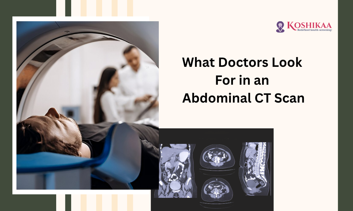
A CT scan of the abdomen is one of the most advanced and reliable imaging procedures for diagnosing internal organ issues. It allows doctors to get a clear view of your stomach, kidneys, liver, and surrounding tissues.
If you are wondering what doctors specifically look for in a CT scan, you have come to the right place. At our best health screening centre in Bangalore, we ensure accurate and timely imaging to help detect potential health concerns. Curious about what your scan might reveal? Keep reading.
What is a CT Scan of the Abdomen?
A CT scan of the abdomen uses X-ray technology combined with computer processing to produce detailed cross-sectional images of your abdominal organs. It is faster and more precise than regular X-rays.
A CT scan of the abdomen and pelvis can capture multiple organs at once, helping doctors identify problems in both the stomach and surrounding structures.
Example: A patient experiencing persistent abdominal pain underwent a CT scan, which revealed early-stage kidney stones that were not detected in an ultrasound.
How Does a CT Scan Work?
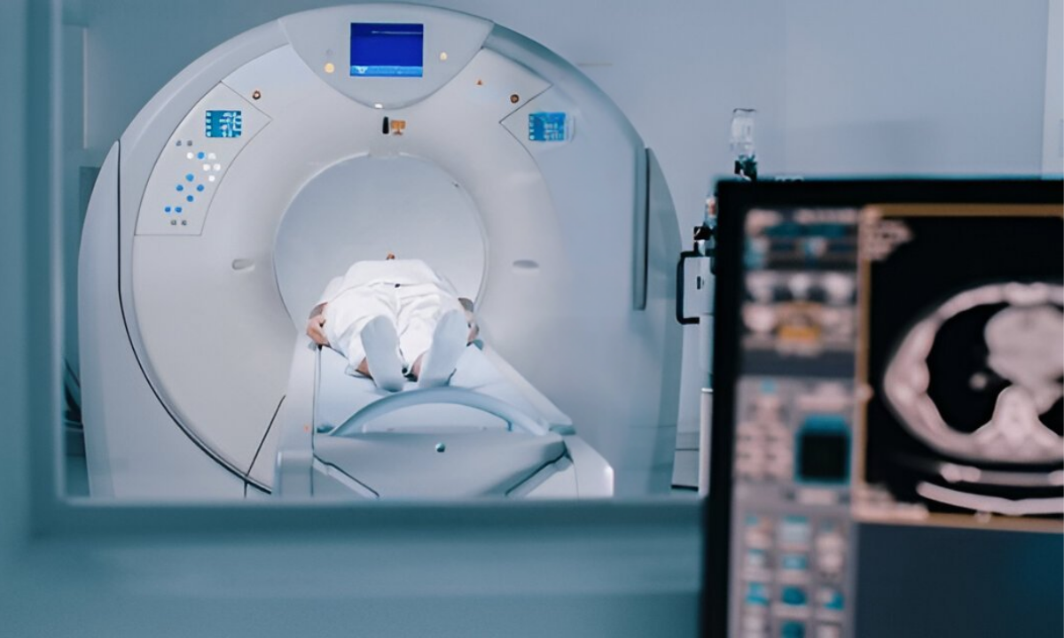
During a CT scan procedure, you lie on a table that slides into a doughnut-shaped machine. The scanner rotates around your body, capturing multiple images. Sometimes, a CT scan contrast is used to enhance the visibility of blood vessels and internal organs.
Tip: Drinking plenty of water before your scan can improve image clarity, especially if contrast dye is used.
What Doctors Look for in a CT Scan of the Abdomen
- Kidney Stones: Small, hard mineral deposits in your kidneys can cause pain and urinary problems. CT scans detect even tiny stones.
- Appendicitis: Inflammation of the appendix is a medical emergency. CT scans help confirm appendicitis and avoid unnecessary surgery.
- Neoplasm: Any abnormal tissue growth or tumor can be detected early with a CT scan of the abdomen, which helps in timely treatment planning.
- Organ Size and Shape: Enlarged liver, spleen, or abnormal bowel structures can indicate underlying conditions like infections or liver disease.
- Internal Bleeding or Injury: Trauma patients often require a CT scan to detect hidden internal injuries that aren’t visible externally.
- Inflammation and Infections: Conditions like pancreatitis or diverticulitis can be accurately diagnosed using abdominal CT imaging.
Tip: Always follow your doctor’s advice on fasting before a CT scan for accurate results.
Scanning Stomach: What It Can and Cannot Detect
While a CT scan for the stomach is highly effective, it has certain limitations:
| Can Detect | Cannot Detect |
|---|---|
| Tumors and masses | Microscopic infections |
| Stomach ulcers (indirectly) | Minor functional disorders like acid reflux |
| Inflammation or swelling | Certain food-related digestive issues |
| Kidney stones or gallstones | Early mucosal lesions |
Example: A patient with chronic stomach pain had a CT scan that revealed gallstones, but acid reflux required a separate endoscopy for diagnosis.
CT Scan Abdomen and Pelvis: Benefits
- Quick and painless procedure
- Detects a wide range of conditions
- Provides clear images of multiple organs simultaneously
- Helps doctors plan surgeries or treatments effectively
Tip: Always bring your previous reports to your scan to help doctors compare changes over time.
How to Understand Your CT Scan Report
A CT scan report includes details about organ size, structure, and any abnormal findings. Doctors interpret the images, noting anything unusual, like kidney stones, inflammation, or tumors.
Tip: Ask your radiologist to explain terms if you find the report confusing; understanding your results helps in timely treatment.
Example: A normal report usually mentions “no abnormal mass or lesion detected”, while abnormal findings might note “small hypodense lesion in liver, recommend follow-up.”
Choosing the Best Scan for Abdominal Pain
For persistent abdominal pain, the best scan for abdominal pain depends on your symptoms:
- CT scan for abdomen: Best for kidney stones, tumors, and appendicitis
- Ultrasound: Initial screening for gallstones or fluid
- MRI: Detailed soft tissue imaging when CT is inconclusive
At our health screening centre in Bangalore, we guide patients on the right scan to ensure accurate diagnosis without unnecessary tests.
When is a CT Scan Recommended?
Doctors may recommend a CT scan of the abdomen if you have:
- Severe abdominal pain
- Unexplained weight loss
- Persistent nausea or vomiting
- Blood in urine or stool
- History of kidney stones or abdominal trauma
Tip: Don’t ignore recurring stomach discomfort. Early scanning can prevent complications.
Safety and Preparation
- Fasting: Usually required 4–6 hours before the scan.
- Contrast Allergy: Inform your doctor if you have a history of allergies
- Pregnancy: Avoid unless essential
CT scan contrast is safe for most patients but might cause mild nausea or warmth during injection. Drinking plenty of water helps flush it out.
Examples of Conditions Detected
| Condition | Detection by CT Scan |
|---|---|
| Kidney Stones | High accuracy for stones as small as 1–2 mm |
| Appendicitis | Detects inflammation and early rupture risk |
| Tumors / Neoplasm | Detects masses and helps in staging cancer |
| Liver or Spleen Enlargement | Measures organ size accurately |
| Internal Bleeding | Detects trauma-related injuries quickly |
Wrapping Up
A CT scan of the abdomen is an essential tool in modern diagnostics, helping doctors detect serious conditions early. Choosing the best CT scan in Bangalore ensures high-quality imaging, accurate interpretation, and expert guidance.
At Koshikaa, we combine advanced technology with patient comfort to make your scanning experience smooth and informative. For your home or hospital visit, our team ensures a stress-free process.
By understanding what doctors look for in a CT scan, you can take proactive steps for your health and avoid complications. For comprehensive abdominal screening and expert care, Koshikaa remains the best health screening centre in Bangalore.
FAQs
1. What can a CT scan detect in the abdomen?
It can detect kidney stones, tumors, appendicitis, organ enlargement, internal bleeding, inflammation, and infections for accurate diagnosis and treatment planning.
2. What can an abdominal CT scan not detect?
It cannot detect minor functional disorders, microscopic infections, early mucosal lesions, or certain digestive problems like acid reflux without additional tests.
3. Can a CT scan detect all stomach problems?
No, CT scans detect structural and organ abnormalities, but functional or microscopic issues, such as ulcers or acid reflux, may require endoscopy or other specialized tests.
4. How to know the CT scan report is normal?
A normal report will state, “No abnormal mass, lesion, or organ enlargement detected,” indicating all organs are within size and structure limits.
5. What is the best scan for abdominal pain?
A CT scan for the abdomen is ideal for stones, tumors, or appendicitis; an ultrasound is good for gallstones; and an MRI helps when soft tissue details are needed.