Top 10 Symptoms Indicating the Need for an X-Ray 

Pet CT scan in Bangalore

Table of Contents

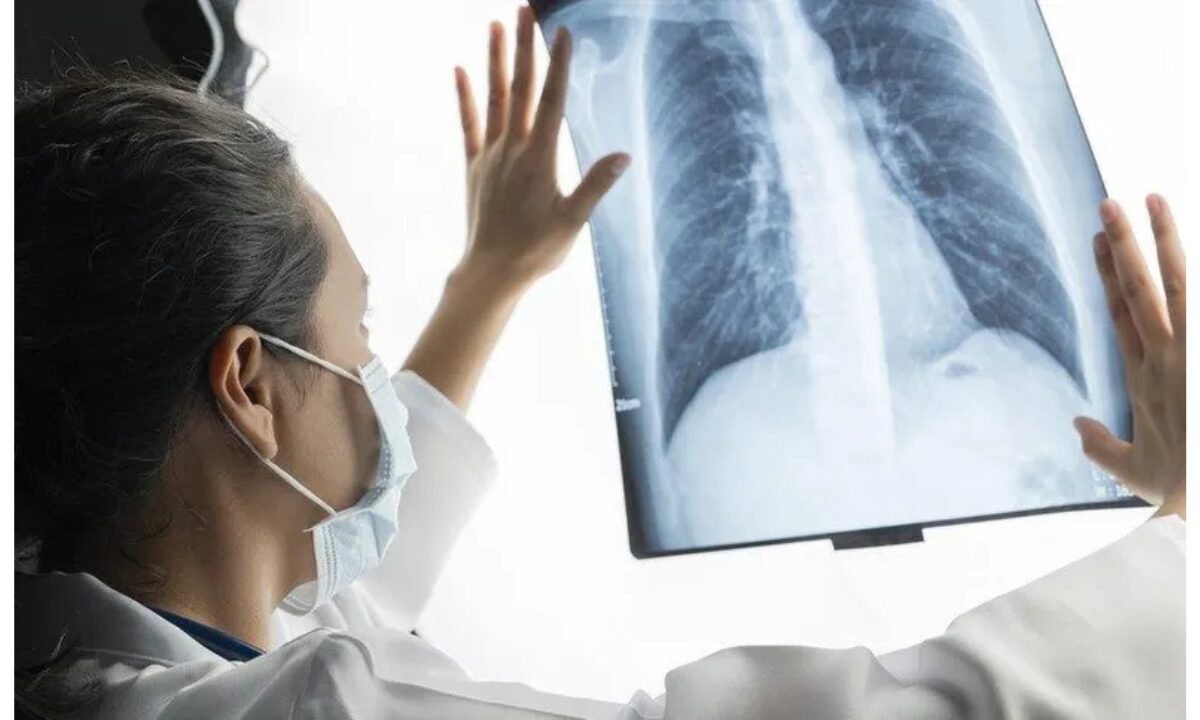
The diagnostic use of X-ray technology has become a typical component in medical examinations of multiple health conditions. Medical images through X-rays give physicians the ability to diagnose problems with bones and internal organs so they can provide a proper medical diagnosis. X-ray examinations serve to identify a wide range of medical issues beyond bone fractures even though they remain associated with broken bone diagnosis. The following symptoms provide indications to help you decide if you should get an X-ray.  

1. Persistent Shortness of Breath

Pet CT scan in Bangalore

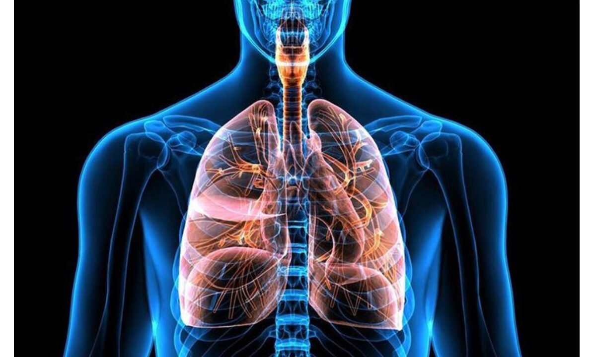
Individuals experiencing ongoing shortness of breath must seek medical attention because this might indicate severe health issues. Both pulmonary conditions and serious lung problems including COPD could be detected when you experience these symptoms. The evaluation of respiratory problems frequently requires a chest x-ray for shortness of breath & for diagnosing potential obstructions or infections and examining lung structures because doctors use this method to determine the causes of shortness of breath.  

The numerous clinics that provide services for X-ray in Bangalore services allow residents to obtain proper testing whenever they need it.  

2. Chronic Cough 

Medical evaluation is needed for a cough that persists for more than weeks and up to months. A continuous cough beyond eight weeks means your body may be dealing with lung damage or any severe condition including tuberculosis or lung cancer. A chest X-ray might become necessary to detect unseen respiratory system diseases because the lungs extensively impact both immune and respiratory functions.  

A visit to a doctor with modern healthcare options allows you to obtain quick and efficient services for X-ray in Bangalore that diagnose your enduring cough.   

3. Severe Chest Pain  

All signs of chest pain require immediate medical attention. The cause of chest pain may begin in the heart but further examination reveals possible abnormalities of the lungs or ribs or diaphragm. Chest X-rays become essential for a person with chest pain to evaluate both pneumonia and rib fractures in addition to pleurisy (inflammation of the lung lining).  

A combination of shortness of breath with chest pain typically requires medical staff to conduct a chest X-ray examination.  

4. Injuries or Fractures  

Accidents and sports-related accidents along with falls are the key reasons individuals require X-ray evaluations for their physical injuries. The specialized equipment of an X-ray machine enables doctors to visualize clearly any area that may demonstrate fractures as well as sprains or bone dislocations.  

The diagnostic X-ray image shows doctors if patients require surgical intervention or need rest with rehabilitation. People in Bangalore should urgently seek X-ray procedures if they have suffered an injury because it promotes accurate medical treatment.  

5. Swelling or Unexplained Pain  

New swelling that seems to appear without reason in any body part should prompt you to consider a problem with bones or soft tissues beneath the skin. Unidentified persistent pain could point towards a dangerous medical condition like infection or tumor in bones.  

Doctors can detect tumors or cysts as well as eliminate severe conditions of the affected area through X-ray imaging. Seek medical help from a healthcare provider as soon as you notice any unexplained swelling together with pain.  

6. Difficulty Breathing or Wheezing  

When wheezing along with breathing troubles appears, physicians need to think about asthma, bronchitis or persistent lung infections. The diagnostic tool of a chest x-ray serves as essential examination for shortness of breath cases to monitor lung capacity and detect lung obstructions along with examining for fluid accumulation.  

People living in Bangalore can discover a straightforward and efficient process to obtain X-ray services in the city because of its streamlined medical infrastructure.   

7. Head or Skull Injury 

A medical check-up is required if you experience dizziness alongside blurred vision and persistent headaches after sustaining a head injury. Medical tests are needed to diagnose a brain injury and skull fracture or internal bleeding. Doctors can use cranial X-rays to check the skull bones for fractures together with detecting any anomalies present. 

X-rays remain essential in determining further investigation because they provide diagnostic information although they do not offer the same level of image detail as CT scans

8. Joint Stiffness or Pain 

Drastically persistent joint stiffness or discomfort requires medical attention because it might be due to arthritis a fracture or degenerative joint disease. The structural analysis of bones together with joints through X-ray imaging allows doctors to detect both abnormalities and inflammation as well as physical damage within these skeletal components.  

Joint pain patients in Bangalore can receive a quick diagnosis through X-ray in Bangalore services to determine their medical condition precisely.  

9. Unexplained Fevers that Won’t Subside 

Failure to treat fever with normal methods could indicate the existence of an infection or disease condition. Inspection image tests are necessary to detect infectious causes within lung and bone areas along with other locations where infections might be hiding. For instance, a chest x-ray for shortness of breath could also reveal if fever-related symptoms stem from pneumonia or tuberculosis.  

Combining blood work and imaging helps doctors rule out potential causes more effectively. For prolonged fever accompanied by other symptoms, consider consulting a healthcare specialist immediately.

10. Symptoms of Tumors or Cancer  

Few of the most critical situations warrant X-ray examinations when you display signs of tumors or cancer development. Doctors should examine medical causes when patients develop unexplained skin growths and unexplained weight change combined with intense fatigue. The initial medical procedure for identifying tissue deformities begins with X-ray diagnosis of both bones and lungs and other body organs. Healthcare facilities in Bangalore deliver premium-quality diagnostic services that enable residents to easily receive early-detection X-ray procedures. 

Why X-rays Are Critical for Diagnosis 

The internal body examination of structures is possible through painless X-ray procedures that operate quickly without requiring invasive approaches to the body. These medical tools allow physicians to detect various illnesses particularly the diseases which affect bones or the respiratory system diseases.  Whether it’s a chest x-ray for shortness of breath, bone injuries, or unusual swelling, the results guide doctors toward the right treatment plan.  

When Should You See a Doctor?

While these symptoms are strong indicators of when to get an X-ray, consult a healthcare provider if you’re unsure. Putting off imaging procedures results in increased difficulty during treatment at a later stage. The accessibility of X-ray facilities in Bangalore ensures patients can quickly obtain an X-ray diagnosis thus professionals recommend they avoid delays in getting medical care.  

Summarizing 

Persistent breathing problems along with shortness of breath merit a chest x-ray for shortness of breath evaluation to eliminate the possibility of pneumonia or lung diseases.  

  • The diagnosis of X-ray imaging should be considered when you present with symptoms such as chronic cough, chest pain or injuries.  
  • X-ray imaging shows the potential to detect serious medical problems when patients experience tumors combined with fractures and infections along with fever-related swelling.  
  • Patients can obtain reliable X-ray services at a quick pace from contemporary healthcare settings in Bangalore.  

Seeking early diagnosis is crucial for treating conditions effectively because you should never delay examining symptoms which might signal more serious health issues. Always prioritize your health!    

Koshikaa Screening centre delivers superior service for x-ray in Bangalore through their advanced imaging equipment that produces accurate outcomes for patients.

MRI of the uterus is primarily used for diagnosing and evaluating conditions such as uterine fibroids, adenomyosis, and malignancies. It is particularly useful in the planning of surgical procedures and assessing the extent of gynecological diseases. This imaging test is valuable for its ability to deliver detailed cross-sectional images of the uterus and surrounding structures.
An MRI of the spleen can help diagnose various disorders affecting the spleen including splenomegaly (enlarged spleen), tumors, and other abnormalities. It provides clear, detailed images that are crucial for assessing the health of the spleen and planning potential surgical interventions.
Prostate MRI is a non-invasive imaging technique that provides detailed pictures of the prostate gland and surrounding tissues. It helps in the detection, staging, and monitoring of prostate cancer. Prostate MRIs are increasingly used to guide biopsy procedures and assist in the planning of treatment strategies for prostate health.
An MRI of the pancreas is used to identify issues such as pancreatitis, pancreatic cancer, and other pancreatic disorders. It offers high-resolution images that help in detecting small lesions and assessing the pancreatic ducts. This imaging technique is valuable for guiding biopsies and planning surgeries involving the pancreas.
MRI scans of the liver, kidney, and bile ducts provide detailed images that help diagnose various conditions, such as tumors, cysts, and blockages in the bile ducts. They are crucial for assessing organ damage, staging cancer, and planning liver or kidney-related surgeries. This type of MRI can also evaluate the health and function of these organs comprehensively.
Breast MRI is highly sensitive for detecting breast cancer, especially in individuals with dense breast tissue or a high risk of cancer. It is often used in conjunction with mammography to provide a more comprehensive evaluation. Breast MRIs can also monitor the response to breast cancer treatments and provide detailed pre-surgical mapping.
Brain and spine MRIs are critical tools for diagnosing and monitoring neurological conditions, including tumors, spinal cord injuries, and multiple sclerosis. These MRIs can reveal abnormalities in brain tissue and spinal cord structure that might not be visible with other imaging methods. They are essential for planning surgeries and assessing the effectiveness of treatments in neurology.
PET-CT scans of transplant organs like the liver, kidney, or pancreas are performed to assess metabolic activity, detect complications, and monitor post-transplant function. These scans play a vital role in the early detection of issues such as organ rejection, recurrence of disease, or infection, guiding timely interventions and optimizing transplant outcomes for patients.
PET-CT examinations of organs such as the liver, kidney, or prostate are crucial for assessing metabolic activity, detecting abnormalities, and monitoring disease progression. These scans help diagnose conditions such as liver metastases, renal cell carcinoma, or prostate cancer, guiding treatment decisions and monitoring response to therapy over time.
PET-CT scans of soft-tissue masses are utilized to assess metabolic activity and detect malignant tumors or other abnormalities. By combining PET imaging with CT imaging, these scans provide detailed information about tumor location, size, and metabolic characteristics, aiding in accurate diagnosis and treatment planning for soft-tissue cancers.
PET-CT scans are valuable for detecting cysts or fluid collections within the body, including cysts in the liver, kidneys, or other organs. These scans help characterize the nature and metabolic activity of cysts, guiding diagnosis and treatment planning for conditions such as cystic liver disease, renal cysts, or pancreatic cysts.
PET-CT scans of muscles are performed to assess metabolic activity and detect abnormalities such as inflammation, infection, or tumors. These scans provide valuable information about muscle function and pathology, aiding in the diagnosis and management of conditions like myositis, muscular dystrophy, or soft-tissue sarcomas.
PET-CT scans of the brain are utilized to evaluate brain metabolism and detect abnormalities such as tumors, epilepsy, or neurodegenerative diseases. By combining functional PET imaging with anatomical CT imaging, these scans offer comprehensive information about brain function and structure, guiding neurosurgical interventions and treatment decisions for neurological disorders
PET-CT scans of bones are commonly used to detect bone metastases in cancer patients, assess bone health in conditions like osteoporosis, and evaluate response to treatment. By combining PET and CT imaging, these scans provide detailed information about metabolic activity and anatomical changes in bone structures, aiding in accurate diagnosis and treatment planning for various bone-related conditions.
CT scans of the spine are conducted to assess the vertebrae, spinal cord, and surrounding soft tissues for conditions like herniated discs, spinal fractures, or spinal stenosis, aiding in the diagnosis and management of spinal disorders with detailed imaging essential for surgical planning and intervention.
CT scans of the pelvis are utilized to visualize the pelvic bones, reproductive organs, bladder, and surrounding structures for conditions such as fractures, tumors, or pelvic inflammatory disease, guiding treatment plans and surgical interventions with precise anatomical information.
CT scans of the neck are performed to evaluate structures such as the throat, thyroid gland, lymph nodes, and blood vessels for abnormalities like tumors, infections, or injuries, assisting in diagnosis and management of head and neck disorders with detailed imaging.
CT scans of the extremities are employed to assess bones, joints, and soft tissues in the arms or legs, aiding in the diagnosis of fractures, arthritis, or other musculoskeletal conditions, guiding orthopedic treatment and rehabilitation plans for optimal recovery.
CT scans of the chest are utilized to examine the lungs, heart, blood vessels, and surrounding structures for conditions such as lung nodules, pneumonia, or pulmonary embolism, facilitating accurate diagnosis and management of respiratory and cardiovascular diseases.
CT scans of the brain are conducted to assess brain structures, identify abnormalities like tumors, hemorrhages, or strokes, and aid in diagnosing neurological conditions, guiding neurosurgical interventions and treatment strategies with precise imaging of the brain’s anatomy.
CT scans of the abdomen are performed to visualize and evaluate the internal organs such as the liver, kidneys, pancreas, and intestines for conditions like tumors, infections, or injuries, providing detailed images essential for diagnosis and treatment planning.
Transplant organ ultrasound scans, including those of the liver, kidney, or pancreas, are performed to assess organ viability, detect complications, and monitor post-transplant function. These scans play a vital role in the early detection of issues such as organ rejection, vascular complications, or fluid collections, guiding timely interventions and optimizing transplant outcomes for patients.
Ultrasound examinations of organs such as the liver, kidney, or prostate are crucial for assessing organ structure, detecting abnormalities, and monitoring disease progression. These scans help diagnose conditions such as liver cirrhosis, kidney stones, or prostate enlargement, guiding treatment decisions and monitoring response to therapy over time.
Soft-tissue masses can be evaluated using ultrasound to determine their size, location, and characteristics. Ultrasound imaging helps differentiate between benign and malignant masses, guiding further diagnostic evaluation and treatment planning. By providing real-time imaging, ultrasound assists in the precise localization of soft-tissue masses for biopsy or surgical removal if needed.
Ultrasound scans are valuable for detecting cysts or fluid collections within the body, including cysts in the liver, kidneys, or other organs. These scans help characterize the nature and size of cysts, aiding in diagnosis and treatment planning for conditions such as cystic liver disease or renal cysts, and guiding interventions such as cyst drainage or sclerotherapy.
Musculoskeletal ultrasound scans are used to assess the health and integrity of muscles, identifying issues such as strains, tears, or muscle inflammation. These scans provide detailed images of muscle structures, allowing healthcare providers to diagnose muscle injuries accurately and develop personalized rehabilitation plans to promote recovery and restore function.
Ultrasound examinations of joints are essential for evaluating joint health, detecting abnormalities such as inflammation, fluid accumulation, or cartilage damage. By visualizing the joint structures in real-time, ultrasound helps diagnose conditions like arthritis, bursitis, or ligament injuries, guiding treatment decisions and monitoring disease progression over time.
Ultrasound scans of tendons are performed to assess for injuries, inflammation, or tears, aiding in the diagnosis and management of conditions such as tendonitis or tendon tears. These scans provide detailed images of tendon structures, helping healthcare providers determine the extent of damage and plan appropriate treatment strategies, including physical therapy or surgical intervention if necessary.
Pelvis X-rays play a crucial role in diagnosing injuries, diseases, and developmental conditions affecting the pelvic region. They provide detailed images of the hip bones, sacrum, and coccyx, aiding in the assessment of fractures, dislocations, or signs of arthritis. Pelvic X-rays are instrumental in guiding surgical interventions, monitoring healing progress, and evaluating conditions such as osteoporosis or tumors. By providing precise anatomical information, these imaging studies enable healthcare professionals to deliver personalized care and improve patient outcomes.
Teeth X-rays, known as dental radiographs, are indispensable tools for dentists to assess oral health. They aid in diagnosing dental decay, gum disease, and abnormalities in tooth structure or alignment. These images provide valuable insights into hidden dental issues, allowing for early intervention and prevention of complications. Additionally, dental X-rays are essential for planning treatments such as orthodontics, root canals, or dental implants, ensuring optimal outcomes and patient satisfaction.
Spine X-rays are pivotal in diagnosing spinal fractures, degenerative conditions, and spinal misalignments, providing critical information for treatment planning and monitoring spinal health. They offer detailed images of the vertebrae and surrounding structures, enabling healthcare professionals to identify issues such as scoliosis, disc herniation, or spinal stenosis
Skull X-rays are valuable for detecting fractures, assessing head trauma, and evaluating the alignment of facial bones, crucial in diagnosing conditions like fractures, tumors, or developmental abnormalities in the skull. They provide detailed views of the cranial structure, aiding clinicians in determining appropriate treatment strategies and ensuring patient safety.
For the heart, X-ray solutions can aid in the detection of various cardiac problems, ranging from mild to severe, including – Heart failure, Cardiac congestion and other heart-related conditions. In addition to cardiac assessments, X-ray solutions are also used for breast examinations to identify tumors or signs of breast cancer.

Bone X-rays are essential for diagnosing fractures and assessing bone alignment, aiding doctors in developing treatment plans and monitoring healing progress. They provide detailed images of the skeletal system, helping identify abnormalities or injuries that may not be visible through other imaging techniques

Abdominal X-rays are performed when patients experience symptoms such as significant nausea, ongoing vomiting, abdominal pain, flank or back pain, or abdominal swelling. These X-rays can help diagnose various conditions.
Search Bar

Frequently Booked Tests

Frequently Booked Checkups