CT Scan Preparation: What You Should Know in 2025   

ct scan preparation

Table of Contents

The CT scan demonstrates how medical technology industries deliver both precise diagnostic results and operation efficiency. Many patients understand CT scans or computed tomography scans as a cause for anxiety. Knowledge about the procedure, detailed preparation information and expected experience reduce worries throughout the diagnostic process. The article provides complete information about CT scans that patients need to know in 2025 during preparation.  

What Is a CT Scan? 

CT scanners enable medical professionals to generate accurate detailed images which show different sections of the human body. Advanced computer processing applies to X-ray images taken from many angles to generate cross-sectional images. Medical staff can examine the body by its layers with CT scans because this helps them identify diseases and check patient treatment progress and surgical intervention requirements. The specific purposes of CT scan operations require description.  

  • CT scanners provide multiple uses for medical diagnosis both during examination and planning of treatments and surgical operations.  
  • CT scans evaluate conditions by identifying both infections and fractures as well as blood clots tumours and internal injuries in patients.  
  • CT imaging serves the purpose of monitoring treatment success by assessing therapies such as cancer management.  
  • CT scan in Bangalore provide doctors with a complete view of patient-internal anatomy during operations through detailed planning procedures.  

Your medical professional recommends CT scans because these imaging tools provide better visibility than X-rays or MRIs fail to provide.  

How to Prepare for a CT Scan?

ct scan in bangalore

The successful execution of both results accuracy and patient-friendly outcomes depends on appropriate preparation steps. These essential steps should be performed before your CT scan booking appointment.  

1.  Understand the CT scan procedure  

CT scans enable concentration on different body parts the chest and abdomen alongside the head and spine along with particular organs. Based on what your doctor suspects or needs to rule out, they will request a particular type of scan. 

For instance, if you’re doing an abdominal CT scan, you might be asked to avoid food or drinks for a few hours ahead of time. The procedure of scanning your head does not need the same limitations regarding diet as other scans.  

2. Clothing Options for the Scan  

Choose items of clothing that are both flowing and easy to wear when selecting an outfit. Medical staff will typically provide hospital gowns to patients because metal materials within clothing can create unwanted interference during imaging procedures.  

Before the test all metal objects need removal from your body since they produce interference during image capturing.  

3. Fasting for Certain Scans 

CT scans with contrast require patients to avoid eating anything during a specific period. The unique substance known as contrast dye provides an improved visual examination of organs and blood vessels or tissues for your medical professional. The healthcare provider will instruct the length of time before eating or drinking when fasting becomes required.  

4. Let Your Provider Know About Allergies and Medical History 

You need to notify your doctor about previous reactions to iodine, shellfish, and contrast dye before undergoing any medical procedure. Your complete medical background must be disclosed to doctors because diabetes kidney problems and persistent illness medication use require specific attention.

What to Expect During the CT Scan?

Most people want to know the step-by-step process of CT scan testing along with the expected patient experience. The CT scan procedure follows a simple process that provides a comfortable experience to most people.  

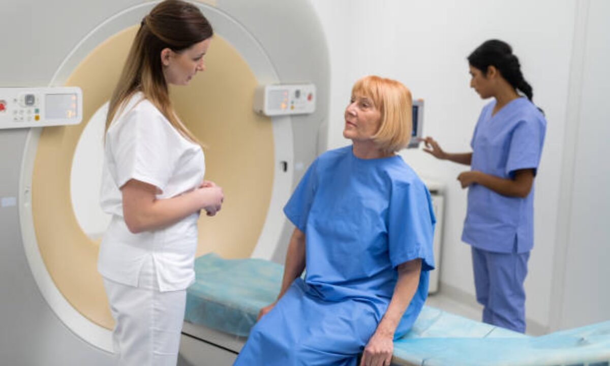
1. Arrival and Briefing 

When you reach the venue a technician will clarify the examination steps and then address every question you have. The type of positioning for the CT scan on the examination table depends on which body region needs to be examined. Patients rest in back, stomach, or side positions.  

2. Use of Contrast Dye  

The medical staff will administer contrast dye by either using an injection or through oral consumption or by using an enema as needed. Receiving the injection results in a brief body warmth together with a metallic aftertaste which disappears quickly and does no harm.  

3. The Scanning Process  

The CT scanner consists of a large doughnut-shaped structure that the scanning table will bring you before starting image acquisition. During the picture-taking process, you must remain motionless inside the machine. The staff might request you to pause breathing for brief intervals as this helps produce clearer images during the scan process.  

The total scanning process duration varies from 10 to 30 minutes according to different CT scan types. The process requires no pain so you can sit back and implement the instructions from the technician.  

How to Prepare After a CT Scan?

A person who underwent the procedure can begin their regular activities unless they receive contrary directions from medical personnel. However, if contrast dye is used, you’ll be asked to stay hydrated to help flush it out of your system. Drinking plenty of fluids is essential in the hours following the scan. 

If you experience any mild side effects like nausea or dizziness, inform the technician or your doctor right away.

CT Scan Cost in Bangalore and Accessibility 

A wide array of world-class medical facilities along with advanced diagnostic services welcome patients who want to perform a CT scan in Bangalore. The diagnostic process is served by trained medical specialists through both private and hospital-based facilities and diagnostic centres.  

The CT scan cost in Bangalore depends on three main elements including the scan type, the presence or absence of contrast dye and the selected facility. Potential patients should ask diagnostic centres for procedure cost details as well as their insurance policies and available discount packages during their appointment scheduling process.  

Tips for a Smooth and Stress-Free Experience

Ask your doctor whatever questions you have regarding the CT scan procedure and its risks alongside alternative diagnostics. More knowledge uncovers the way to decrease your anxiety levels.  

Comfortable dresses should be worn because they simplify the gown change process.  

Daily follow all instructions that include medication restrictions which must be avoided alongside instructions for fasting as well as appropriate hydration protocols. By following these instructions the diagnostic accuracy of the results will be maximized.  

Inform the technician when you witness symptoms of claustrophobia or have equipment-related worries.  

Why CT Scans Remain Essential in 2025  

Medical imaging technology developments have not diminished the clinical value of CT scans because they continue to deliver precise diagnostic results in healthcare treatments. Medicine has achieved unprecedented speed and precision in CT scanning technology which became more widely available in 2025. The healthcare industry in Bangalore introduced safer scanners with lower radiation protection while maintaining results quality.  

The process of undergoing a CT scan at any facility in Bangalore should not cause you stress. Learning about the preparation process and procedure beforehand allows you to enter your appointment more firmly and relaxed.  

Your medical team alongside your doctor will assist with your concerns about CT scan functions alongside its procedure so you can feel confident about your understanding. Services from the medical field become straightforward and empowering when you communicate openly while following basic preparation steps.  

Koshikaa operates offering dependable CT scan in Bangalore services to patients at reasonable prices assisted by latest equipment and specialist talent.

MRI of the uterus is primarily used for diagnosing and evaluating conditions such as uterine fibroids, adenomyosis, and malignancies. It is particularly useful in the planning of surgical procedures and assessing the extent of gynecological diseases. This imaging test is valuable for its ability to deliver detailed cross-sectional images of the uterus and surrounding structures.
An MRI of the spleen can help diagnose various disorders affecting the spleen including splenomegaly (enlarged spleen), tumors, and other abnormalities. It provides clear, detailed images that are crucial for assessing the health of the spleen and planning potential surgical interventions.
Prostate MRI is a non-invasive imaging technique that provides detailed pictures of the prostate gland and surrounding tissues. It helps in the detection, staging, and monitoring of prostate cancer. Prostate MRIs are increasingly used to guide biopsy procedures and assist in the planning of treatment strategies for prostate health.
An MRI of the pancreas is used to identify issues such as pancreatitis, pancreatic cancer, and other pancreatic disorders. It offers high-resolution images that help in detecting small lesions and assessing the pancreatic ducts. This imaging technique is valuable for guiding biopsies and planning surgeries involving the pancreas.
MRI scans of the liver, kidney, and bile ducts provide detailed images that help diagnose various conditions, such as tumors, cysts, and blockages in the bile ducts. They are crucial for assessing organ damage, staging cancer, and planning liver or kidney-related surgeries. This type of MRI can also evaluate the health and function of these organs comprehensively.
Breast MRI is highly sensitive for detecting breast cancer, especially in individuals with dense breast tissue or a high risk of cancer. It is often used in conjunction with mammography to provide a more comprehensive evaluation. Breast MRIs can also monitor the response to breast cancer treatments and provide detailed pre-surgical mapping.
Brain and spine MRIs are critical tools for diagnosing and monitoring neurological conditions, including tumors, spinal cord injuries, and multiple sclerosis. These MRIs can reveal abnormalities in brain tissue and spinal cord structure that might not be visible with other imaging methods. They are essential for planning surgeries and assessing the effectiveness of treatments in neurology.
PET-CT scans of transplant organs like the liver, kidney, or pancreas are performed to assess metabolic activity, detect complications, and monitor post-transplant function. These scans play a vital role in the early detection of issues such as organ rejection, recurrence of disease, or infection, guiding timely interventions and optimizing transplant outcomes for patients.
PET-CT examinations of organs such as the liver, kidney, or prostate are crucial for assessing metabolic activity, detecting abnormalities, and monitoring disease progression. These scans help diagnose conditions such as liver metastases, renal cell carcinoma, or prostate cancer, guiding treatment decisions and monitoring response to therapy over time.
PET-CT scans of soft-tissue masses are utilized to assess metabolic activity and detect malignant tumors or other abnormalities. By combining PET imaging with CT imaging, these scans provide detailed information about tumor location, size, and metabolic characteristics, aiding in accurate diagnosis and treatment planning for soft-tissue cancers.
PET-CT scans are valuable for detecting cysts or fluid collections within the body, including cysts in the liver, kidneys, or other organs. These scans help characterize the nature and metabolic activity of cysts, guiding diagnosis and treatment planning for conditions such as cystic liver disease, renal cysts, or pancreatic cysts.
PET-CT scans of muscles are performed to assess metabolic activity and detect abnormalities such as inflammation, infection, or tumors. These scans provide valuable information about muscle function and pathology, aiding in the diagnosis and management of conditions like myositis, muscular dystrophy, or soft-tissue sarcomas.
PET-CT scans of the brain are utilized to evaluate brain metabolism and detect abnormalities such as tumors, epilepsy, or neurodegenerative diseases. By combining functional PET imaging with anatomical CT imaging, these scans offer comprehensive information about brain function and structure, guiding neurosurgical interventions and treatment decisions for neurological disorders
PET-CT scans of bones are commonly used to detect bone metastases in cancer patients, assess bone health in conditions like osteoporosis, and evaluate response to treatment. By combining PET and CT imaging, these scans provide detailed information about metabolic activity and anatomical changes in bone structures, aiding in accurate diagnosis and treatment planning for various bone-related conditions.
CT scans of the spine are conducted to assess the vertebrae, spinal cord, and surrounding soft tissues for conditions like herniated discs, spinal fractures, or spinal stenosis, aiding in the diagnosis and management of spinal disorders with detailed imaging essential for surgical planning and intervention.
CT scans of the pelvis are utilized to visualize the pelvic bones, reproductive organs, bladder, and surrounding structures for conditions such as fractures, tumors, or pelvic inflammatory disease, guiding treatment plans and surgical interventions with precise anatomical information.
CT scans of the neck are performed to evaluate structures such as the throat, thyroid gland, lymph nodes, and blood vessels for abnormalities like tumors, infections, or injuries, assisting in diagnosis and management of head and neck disorders with detailed imaging.
CT scans of the extremities are employed to assess bones, joints, and soft tissues in the arms or legs, aiding in the diagnosis of fractures, arthritis, or other musculoskeletal conditions, guiding orthopedic treatment and rehabilitation plans for optimal recovery.
CT scans of the chest are utilized to examine the lungs, heart, blood vessels, and surrounding structures for conditions such as lung nodules, pneumonia, or pulmonary embolism, facilitating accurate diagnosis and management of respiratory and cardiovascular diseases.
CT scans of the brain are conducted to assess brain structures, identify abnormalities like tumors, hemorrhages, or strokes, and aid in diagnosing neurological conditions, guiding neurosurgical interventions and treatment strategies with precise imaging of the brain’s anatomy.
CT scans of the abdomen are performed to visualize and evaluate the internal organs such as the liver, kidneys, pancreas, and intestines for conditions like tumors, infections, or injuries, providing detailed images essential for diagnosis and treatment planning.
Transplant organ ultrasound scans, including those of the liver, kidney, or pancreas, are performed to assess organ viability, detect complications, and monitor post-transplant function. These scans play a vital role in the early detection of issues such as organ rejection, vascular complications, or fluid collections, guiding timely interventions and optimizing transplant outcomes for patients.
Ultrasound examinations of organs such as the liver, kidney, or prostate are crucial for assessing organ structure, detecting abnormalities, and monitoring disease progression. These scans help diagnose conditions such as liver cirrhosis, kidney stones, or prostate enlargement, guiding treatment decisions and monitoring response to therapy over time.
Soft-tissue masses can be evaluated using ultrasound to determine their size, location, and characteristics. Ultrasound imaging helps differentiate between benign and malignant masses, guiding further diagnostic evaluation and treatment planning. By providing real-time imaging, ultrasound assists in the precise localization of soft-tissue masses for biopsy or surgical removal if needed.
Ultrasound scans are valuable for detecting cysts or fluid collections within the body, including cysts in the liver, kidneys, or other organs. These scans help characterize the nature and size of cysts, aiding in diagnosis and treatment planning for conditions such as cystic liver disease or renal cysts, and guiding interventions such as cyst drainage or sclerotherapy.
Musculoskeletal ultrasound scans are used to assess the health and integrity of muscles, identifying issues such as strains, tears, or muscle inflammation. These scans provide detailed images of muscle structures, allowing healthcare providers to diagnose muscle injuries accurately and develop personalized rehabilitation plans to promote recovery and restore function.
Ultrasound examinations of joints are essential for evaluating joint health, detecting abnormalities such as inflammation, fluid accumulation, or cartilage damage. By visualizing the joint structures in real-time, ultrasound helps diagnose conditions like arthritis, bursitis, or ligament injuries, guiding treatment decisions and monitoring disease progression over time.
Ultrasound scans of tendons are performed to assess for injuries, inflammation, or tears, aiding in the diagnosis and management of conditions such as tendonitis or tendon tears. These scans provide detailed images of tendon structures, helping healthcare providers determine the extent of damage and plan appropriate treatment strategies, including physical therapy or surgical intervention if necessary.
Pelvis X-rays play a crucial role in diagnosing injuries, diseases, and developmental conditions affecting the pelvic region. They provide detailed images of the hip bones, sacrum, and coccyx, aiding in the assessment of fractures, dislocations, or signs of arthritis. Pelvic X-rays are instrumental in guiding surgical interventions, monitoring healing progress, and evaluating conditions such as osteoporosis or tumors. By providing precise anatomical information, these imaging studies enable healthcare professionals to deliver personalized care and improve patient outcomes.
Teeth X-rays, known as dental radiographs, are indispensable tools for dentists to assess oral health. They aid in diagnosing dental decay, gum disease, and abnormalities in tooth structure or alignment. These images provide valuable insights into hidden dental issues, allowing for early intervention and prevention of complications. Additionally, dental X-rays are essential for planning treatments such as orthodontics, root canals, or dental implants, ensuring optimal outcomes and patient satisfaction.
Spine X-rays are pivotal in diagnosing spinal fractures, degenerative conditions, and spinal misalignments, providing critical information for treatment planning and monitoring spinal health. They offer detailed images of the vertebrae and surrounding structures, enabling healthcare professionals to identify issues such as scoliosis, disc herniation, or spinal stenosis
Skull X-rays are valuable for detecting fractures, assessing head trauma, and evaluating the alignment of facial bones, crucial in diagnosing conditions like fractures, tumors, or developmental abnormalities in the skull. They provide detailed views of the cranial structure, aiding clinicians in determining appropriate treatment strategies and ensuring patient safety.
For the heart, X-ray solutions can aid in the detection of various cardiac problems, ranging from mild to severe, including – Heart failure, Cardiac congestion and other heart-related conditions. In addition to cardiac assessments, X-ray solutions are also used for breast examinations to identify tumors or signs of breast cancer.

Bone X-rays are essential for diagnosing fractures and assessing bone alignment, aiding doctors in developing treatment plans and monitoring healing progress. They provide detailed images of the skeletal system, helping identify abnormalities or injuries that may not be visible through other imaging techniques

Abdominal X-rays are performed when patients experience symptoms such as significant nausea, ongoing vomiting, abdominal pain, flank or back pain, or abdominal swelling. These X-rays can help diagnose various conditions.
Search Bar

Frequently Booked Tests

Frequently Booked Checkups