Understanding the Difference Between Tumor and Cancer

cancer detection

Table of Contents

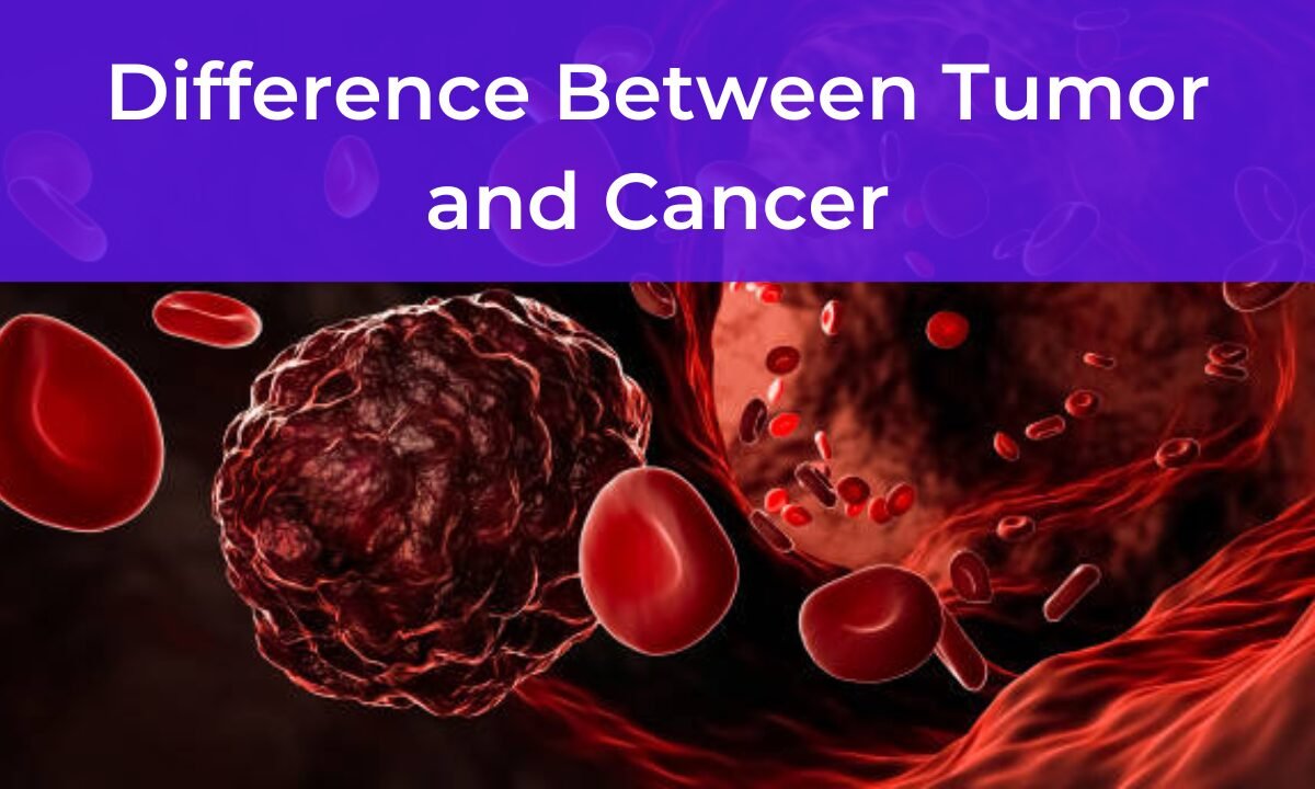
The words ‘tumor,’ and ‘cancer’, sound almost the same to many people, so they automatically assume that the two have the same meaning. As the two terms seem interchangeable, they are not. The difference between cancer and tumor needs to be understood for clarity, prevention, and above all, early detection and treatment. This article simply explains concepts, including what they mean and how they are related, as well as their differences.

What is a Tumor?

CT scan in bangalore

A tumor is a mass of tissue that results from the excessive and abnormal multiplication and division of cells. Normally, cell growth is a precisely controlled process that goes wrong when cells start dividing uncontrollably and form a tumor. 

Yet not all tumors are bad. Tumors can be classified into two main categories:

1. Benign Tumors (Non-cancerous):

Most benign tumors are not life-threatening. They don’t spread elsewhere in the body and often stay put. For example, a lipoma, which is a benign tumor composed mostly of fat cells, doesn’t spread into neighboring tissue. Benign tumors aren’t cancer, but because they can grow too large or press on vital organs, blood vessels, or nerves, they can cause problems.

2. Malignant Tumors (Cancerous):

This is where the confusion between cancer and tumor is so often made. A malignant tumor is a cancer. Malignant means the tumors will grow uncontrollably and can spread to nearby tissues damaging healthy cells. And they can spread to other parts of the body, a process called metastasis.

What is Cancer?

Cancer is a group of diseases in which cells grow uncontrollably and can spread to other parts of the body. Cancer starts when the DNA of cells becomes damaged and tumors can develop in almost any part of your body. That damage might be due to genetic mutations, exposure to UV radiation during skin sun damage, or certain lifestyle choices such as smoking.

Cancer is also able to spread, meaning it’s different from a benign tumor. If the cancerous cells behave differently than normal cells your body may be changed and put under serious threat due to this.

What’s important to understand is not all tumors are cancerous, but all cancer begins as a tumor. With that in mind, we can begin to understand more clearly the difference between cancer and tumor as we proceed.

The Big Difference—Between Cancer and Tumor

To gain a clear understanding of the difference between cancer and tumor, let us dive deeper into their unique characteristics:

1. Definition:

Tumor: A tumor is a mass due to abnormal cell growth. It can be either malignant or benign.

Cancer: Cancer is a disease in which cells overgrow and spread uncontrollably, beginning as a tumor.

2. Nature:

Tumor: Not all tumors are harmful. Other benign tumors are noninvasive and are considered harmless unless they grow extremely large or press upon vital organs.

Cancer: Cancer is always harmful. Tumors are classified as malignant (cancerous) tumors that destroy tissues, can spread, and are life-threatening.

3. Spread:

Tumor: A benign tumor doesn’t spread to other parts of the body.

Cancer: Malignancies are cancers that metastasize or spread to different organs and tissues.

4. Aggressiveness:

Tumor: Tumors that grow slowly and are less aggressive are called benign tumors.

Cancer: Cancer cells do not always die, they continue to grow quickly and are aggressive, growing through surrounding tissue and causing severe damage.

5. Treatment:

Tumor: Tumors may not be treated if they are Benign. They may be surgically removed when they cause discomfort or represent health risks.

Cancer: Treatments for cancer detection, like surgery, chemotherapy, radiation therapy, immunotherapy, or some combination of all these approaches, are more intense.

Tumor and Cancer Similarities.

There are a few overlaps, but it’s crucial to clarify the two terms. Let’s address those:

1. Both Involve Abnormal Cell Growth: Unregulated cell growth in the body is the cause of both tumors and cancers.

2. Detection Methods Are Similar: Often imaging tests, biopsies, and scans are used to diagnose benign and malignant tumors as well as cancer.

3. Both May Require Medical Attention: A tumor can be benign or malignant, so based on the size, location, and impact on the body it may or may not require intervention.

How do Tumors and Cancer form?

Now we can dig deeper to find out what causes cancer to grow and what causes a tumor to grow.

Causes of Tumors:

  • Genetic predisposition
  • Infections
  • Hormonal imbalances
  • Chronic inflammation
  • Examples include environmental or lifestyle factors (such as smoking, alcohol, etc.)

Causes of Cancer:

  • Genetic mutations
  • Or carcinogens such as tobacco smoke or asbestos
  • Excessive alcohol consumption
  • This means long-term exposure to UV radiation.
  • Human Papilloma Virus (HPV)
  • Lack of exercise and poor diet

How are Tumors or Cancer diagnosed by doctors?

Doctors find out if a tumor is benign or malignant, and if a person has cancer, by doing a combination of tests and procedures. These diagnostic tools may include:

1. Physical Examination: Looking for lumps or abnormalities.

2. Imaging Tests: The characteristics of a tumor can be revealed by X-rays, MRIs, CT scans, or ultrasounds.

3. Biopsy: An analysis of a small tissue sample is made under a microscope to find out if it is cancer or not.

4. Blood Tests: Certain cancers release specific marker products into the bloodstream for diagnosis.  

Should a Benign Tumour Be Treated?

Not all benign tumors require treatment. Benign tumors may remain stable and cause no symptoms and yet many live their whole lives with them. But a benign tumor that begins to grow enough to press on vital structures such as blood vessels or nerves or creates problems might have to be removed surgically.

Whereas malignant tumors are always medical intervention required. The earlier a cancer is found and treated, the better.

Is a Benign Tumor Oddly Able to Turn into Cancer?

Sometimes a benign tumor can turn cancerous. The term for this, malignant transformation, is rare and depends on the type of tumor and its location. To give an example, some polyps in the colon, even though they may begin as benign tumors, can turn into colorectal cancer. That’s why doctors often have people remove these growths despite them not being cancerous.

Importance of Early Detection

Tumor and cancer are when early detection is critical in managing them. If you find a strange lump or if you experience any other unusual symptoms, you must see the doctor as soon as possible. Mammograms, colonoscopies, and Pap smears are regular screenings that can be done to find out whether a problem is present.

Understanding the difference between cancer and tumors means you can spot early warning signs and get the right care without going into unnecessary panic.

Conclusion

In conclusion, the knowledge of the difference between cancer and tumors will make a big difference in health knowledge. Tumor, simply, is an abnormal growth of cells that can be either benign or malignant. In contrast, cancer is the name for malignant tumors occurring in any tissues that invade surrounding tissues and spread into the rest of the body.

Though both terms may sound daunting, knowledge and rapid action are essential to effective treatment. Knowing the differences can help you to work with your healthcare provider if you ever have a diagnosis of a tumor or cancer.

 A reliable and comprehensive cancer screening test Bangalore is what Koshikaa offers, aimed at early detection and prompt intervention to help increase your chances of better health.

MRI of the uterus is primarily used for diagnosing and evaluating conditions such as uterine fibroids, adenomyosis, and malignancies. It is particularly useful in the planning of surgical procedures and assessing the extent of gynecological diseases. This imaging test is valuable for its ability to deliver detailed cross-sectional images of the uterus and surrounding structures.
An MRI of the spleen can help diagnose various disorders affecting the spleen including splenomegaly (enlarged spleen), tumors, and other abnormalities. It provides clear, detailed images that are crucial for assessing the health of the spleen and planning potential surgical interventions.
Prostate MRI is a non-invasive imaging technique that provides detailed pictures of the prostate gland and surrounding tissues. It helps in the detection, staging, and monitoring of prostate cancer. Prostate MRIs are increasingly used to guide biopsy procedures and assist in the planning of treatment strategies for prostate health.
An MRI of the pancreas is used to identify issues such as pancreatitis, pancreatic cancer, and other pancreatic disorders. It offers high-resolution images that help in detecting small lesions and assessing the pancreatic ducts. This imaging technique is valuable for guiding biopsies and planning surgeries involving the pancreas.
MRI scans of the liver, kidney, and bile ducts provide detailed images that help diagnose various conditions, such as tumors, cysts, and blockages in the bile ducts. They are crucial for assessing organ damage, staging cancer, and planning liver or kidney-related surgeries. This type of MRI can also evaluate the health and function of these organs comprehensively.
Breast MRI is highly sensitive for detecting breast cancer, especially in individuals with dense breast tissue or a high risk of cancer. It is often used in conjunction with mammography to provide a more comprehensive evaluation. Breast MRIs can also monitor the response to breast cancer treatments and provide detailed pre-surgical mapping.
Brain and spine MRIs are critical tools for diagnosing and monitoring neurological conditions, including tumors, spinal cord injuries, and multiple sclerosis. These MRIs can reveal abnormalities in brain tissue and spinal cord structure that might not be visible with other imaging methods. They are essential for planning surgeries and assessing the effectiveness of treatments in neurology.
PET-CT scans of transplant organs like the liver, kidney, or pancreas are performed to assess metabolic activity, detect complications, and monitor post-transplant function. These scans play a vital role in the early detection of issues such as organ rejection, recurrence of disease, or infection, guiding timely interventions and optimizing transplant outcomes for patients.
PET-CT examinations of organs such as the liver, kidney, or prostate are crucial for assessing metabolic activity, detecting abnormalities, and monitoring disease progression. These scans help diagnose conditions such as liver metastases, renal cell carcinoma, or prostate cancer, guiding treatment decisions and monitoring response to therapy over time.
PET-CT scans of soft-tissue masses are utilized to assess metabolic activity and detect malignant tumors or other abnormalities. By combining PET imaging with CT imaging, these scans provide detailed information about tumor location, size, and metabolic characteristics, aiding in accurate diagnosis and treatment planning for soft-tissue cancers.
PET-CT scans are valuable for detecting cysts or fluid collections within the body, including cysts in the liver, kidneys, or other organs. These scans help characterize the nature and metabolic activity of cysts, guiding diagnosis and treatment planning for conditions such as cystic liver disease, renal cysts, or pancreatic cysts.
PET-CT scans of muscles are performed to assess metabolic activity and detect abnormalities such as inflammation, infection, or tumors. These scans provide valuable information about muscle function and pathology, aiding in the diagnosis and management of conditions like myositis, muscular dystrophy, or soft-tissue sarcomas.
PET-CT scans of the brain are utilized to evaluate brain metabolism and detect abnormalities such as tumors, epilepsy, or neurodegenerative diseases. By combining functional PET imaging with anatomical CT imaging, these scans offer comprehensive information about brain function and structure, guiding neurosurgical interventions and treatment decisions for neurological disorders
PET-CT scans of bones are commonly used to detect bone metastases in cancer patients, assess bone health in conditions like osteoporosis, and evaluate response to treatment. By combining PET and CT imaging, these scans provide detailed information about metabolic activity and anatomical changes in bone structures, aiding in accurate diagnosis and treatment planning for various bone-related conditions.
CT scans of the spine are conducted to assess the vertebrae, spinal cord, and surrounding soft tissues for conditions like herniated discs, spinal fractures, or spinal stenosis, aiding in the diagnosis and management of spinal disorders with detailed imaging essential for surgical planning and intervention.
CT scans of the pelvis are utilized to visualize the pelvic bones, reproductive organs, bladder, and surrounding structures for conditions such as fractures, tumors, or pelvic inflammatory disease, guiding treatment plans and surgical interventions with precise anatomical information.
CT scans of the neck are performed to evaluate structures such as the throat, thyroid gland, lymph nodes, and blood vessels for abnormalities like tumors, infections, or injuries, assisting in diagnosis and management of head and neck disorders with detailed imaging.
CT scans of the extremities are employed to assess bones, joints, and soft tissues in the arms or legs, aiding in the diagnosis of fractures, arthritis, or other musculoskeletal conditions, guiding orthopedic treatment and rehabilitation plans for optimal recovery.
CT scans of the chest are utilized to examine the lungs, heart, blood vessels, and surrounding structures for conditions such as lung nodules, pneumonia, or pulmonary embolism, facilitating accurate diagnosis and management of respiratory and cardiovascular diseases.
CT scans of the brain are conducted to assess brain structures, identify abnormalities like tumors, hemorrhages, or strokes, and aid in diagnosing neurological conditions, guiding neurosurgical interventions and treatment strategies with precise imaging of the brain’s anatomy.
CT scans of the abdomen are performed to visualize and evaluate the internal organs such as the liver, kidneys, pancreas, and intestines for conditions like tumors, infections, or injuries, providing detailed images essential for diagnosis and treatment planning.
Transplant organ ultrasound scans, including those of the liver, kidney, or pancreas, are performed to assess organ viability, detect complications, and monitor post-transplant function. These scans play a vital role in the early detection of issues such as organ rejection, vascular complications, or fluid collections, guiding timely interventions and optimizing transplant outcomes for patients.
Ultrasound examinations of organs such as the liver, kidney, or prostate are crucial for assessing organ structure, detecting abnormalities, and monitoring disease progression. These scans help diagnose conditions such as liver cirrhosis, kidney stones, or prostate enlargement, guiding treatment decisions and monitoring response to therapy over time.
Soft-tissue masses can be evaluated using ultrasound to determine their size, location, and characteristics. Ultrasound imaging helps differentiate between benign and malignant masses, guiding further diagnostic evaluation and treatment planning. By providing real-time imaging, ultrasound assists in the precise localization of soft-tissue masses for biopsy or surgical removal if needed.
Ultrasound scans are valuable for detecting cysts or fluid collections within the body, including cysts in the liver, kidneys, or other organs. These scans help characterize the nature and size of cysts, aiding in diagnosis and treatment planning for conditions such as cystic liver disease or renal cysts, and guiding interventions such as cyst drainage or sclerotherapy.
Musculoskeletal ultrasound scans are used to assess the health and integrity of muscles, identifying issues such as strains, tears, or muscle inflammation. These scans provide detailed images of muscle structures, allowing healthcare providers to diagnose muscle injuries accurately and develop personalized rehabilitation plans to promote recovery and restore function.
Ultrasound examinations of joints are essential for evaluating joint health, detecting abnormalities such as inflammation, fluid accumulation, or cartilage damage. By visualizing the joint structures in real-time, ultrasound helps diagnose conditions like arthritis, bursitis, or ligament injuries, guiding treatment decisions and monitoring disease progression over time.
Ultrasound scans of tendons are performed to assess for injuries, inflammation, or tears, aiding in the diagnosis and management of conditions such as tendonitis or tendon tears. These scans provide detailed images of tendon structures, helping healthcare providers determine the extent of damage and plan appropriate treatment strategies, including physical therapy or surgical intervention if necessary.
Pelvis X-rays play a crucial role in diagnosing injuries, diseases, and developmental conditions affecting the pelvic region. They provide detailed images of the hip bones, sacrum, and coccyx, aiding in the assessment of fractures, dislocations, or signs of arthritis. Pelvic X-rays are instrumental in guiding surgical interventions, monitoring healing progress, and evaluating conditions such as osteoporosis or tumors. By providing precise anatomical information, these imaging studies enable healthcare professionals to deliver personalized care and improve patient outcomes.
Teeth X-rays, known as dental radiographs, are indispensable tools for dentists to assess oral health. They aid in diagnosing dental decay, gum disease, and abnormalities in tooth structure or alignment. These images provide valuable insights into hidden dental issues, allowing for early intervention and prevention of complications. Additionally, dental X-rays are essential for planning treatments such as orthodontics, root canals, or dental implants, ensuring optimal outcomes and patient satisfaction.
Spine X-rays are pivotal in diagnosing spinal fractures, degenerative conditions, and spinal misalignments, providing critical information for treatment planning and monitoring spinal health. They offer detailed images of the vertebrae and surrounding structures, enabling healthcare professionals to identify issues such as scoliosis, disc herniation, or spinal stenosis
Skull X-rays are valuable for detecting fractures, assessing head trauma, and evaluating the alignment of facial bones, crucial in diagnosing conditions like fractures, tumors, or developmental abnormalities in the skull. They provide detailed views of the cranial structure, aiding clinicians in determining appropriate treatment strategies and ensuring patient safety.
For the heart, X-ray solutions can aid in the detection of various cardiac problems, ranging from mild to severe, including – Heart failure, Cardiac congestion and other heart-related conditions. In addition to cardiac assessments, X-ray solutions are also used for breast examinations to identify tumors or signs of breast cancer.

Bone X-rays are essential for diagnosing fractures and assessing bone alignment, aiding doctors in developing treatment plans and monitoring healing progress. They provide detailed images of the skeletal system, helping identify abnormalities or injuries that may not be visible through other imaging techniques

Abdominal X-rays are performed when patients experience symptoms such as significant nausea, ongoing vomiting, abdominal pain, flank or back pain, or abdominal swelling. These X-rays can help diagnose various conditions.
Search Bar

Frequently Booked Tests

Frequently Booked Checkups