Role of Regular Mammography Tests in Breast Cancer Treatment

mammography test

Table of Contents

Women around the world are at risk of developing breast cancer. It is one of the most frequently diagnosed cancers and the leading cause of death from cancer in women. A mammography test is one of the most effective methods of detection in the early stage. This simple examination has prevented many breast cancers around the world in the early stages, before they become life-threatening, by finding breast cancer.

In this article, we will find out the role of regular mammography tests in treating breast cancer and why they are one of the best tools for the fight against this terrible disease.

Breast Cancer and its Challenges.

mammography in bangalore

The term breast cancer can refer to a tumor that develops when cells in the breast tissue grow uncontrollably. The more cancerous cells it has, the more potential it has to spread to other areas of the body; the cancer is more difficult to treat. Successful outcomes depend greatly on early detection because the cancer is found in a stage where it is still manageable.

What is a Mammography Test?

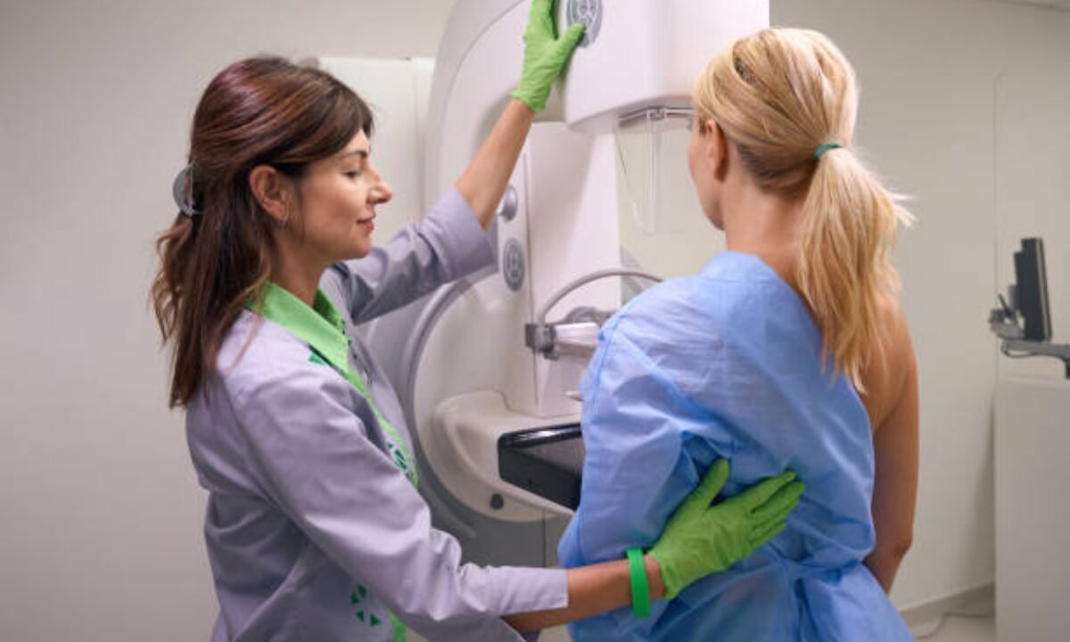
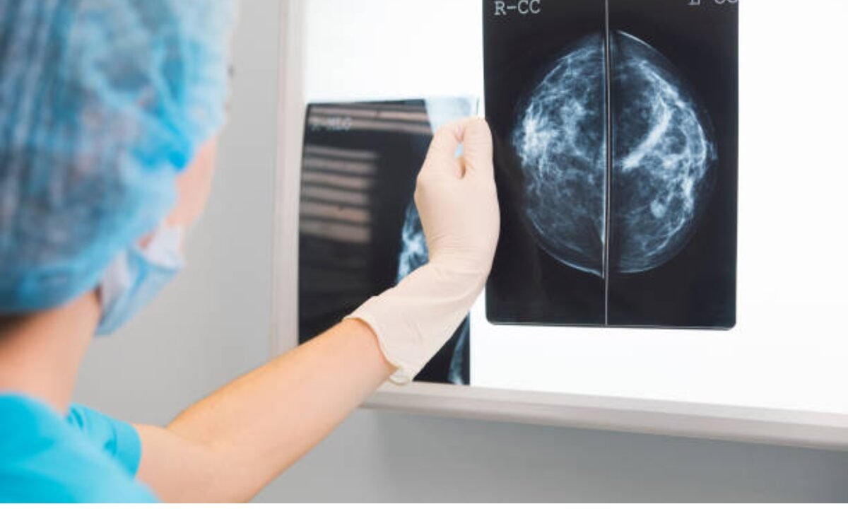
A mammography is an X-ray performed to examine breast tissue. It is a quick, noninvasive test well suited to detect abnormalities like calcifications, cysts, or tumors of the breast. In particular, it is beneficial since the test can detect small tumors or changes in breast tissue that are not felt during a physical exam.

If you are having a mammography test, the breast is flattened into two plates to get clear X-ray images. While many women may find the procedure uncomfortable, the temporary discomfort is well worth the possible lifesaving advantages of finding cancer earlier. 

Role of Mammography in Early Detection

Early detection is crucial in the treatment of breast cancer. Numerous studies and medical experts have emphasized that identifying cancer before it spreads not only improves survival rates but also enables less invasive treatment options. A mammography test can detect tumors as small as a grain of rice, well before they can be felt by hand. 

In most cases, patients who catch breast cancer at its earliest stage, either stage 0 or stage 1, have a much better prognosis. Early detection by a mammography test can also reduce the necessity for aggressive treatments such as chemotherapy or mastectomy. 

When Should You Have Regular Mammography Tests?

It all depends on several factors, including age, personal history of any health problems, and risk factors like family history of breast cancer. These criteria determine when and to what age the recommendations suggest you should have regular mammography tests.

1. Women above the age of 40

Doctors say women older than 40 would be wise to get screened for breast cancer every one to two years, and recommend they speak with their family doctor about what could be the best way to do so. Women in this age group are at increased risk for developing breast cancer, and regular screening can substantially decrease the incidence of late-stage disease.

2. High-Risk Individuals

Eventually, some women may start screening for early stages of breast cancer in their 30s or when their doctor orders them to do so if they have a family history or are carriers of the genetic marker BRCA1 or BRCA2 mutation.

3. Post-Menopausal Women

Routine mammography tests for women are especially important especially after menopause as post-menopause breast cancer risk increases. One can detect and treat cancer on time with vigilant screening during this phase of life.

How are the Routine Mammography Tests Advantageous to you?

Indeed, the benefits of routine mammography tests include the following advantages:

1. Early Detection Saves Lives

The tests for regular mammography have been proven to reduce breast cancer death risk by finding the disease early. Early detection often means patients don’t have to suffer more painful treatments, and therefore improve their quality of life.

2. Improved Treatment Options

Unless you’re in the later stages of breast cancer, most of them can only be treated surgically or through localized radiation. The mammography test, if the cancer is caught early enough, may even save more of the breast tissue.

3. Reassurance for Women

Routine mammography exams give many women peace of mind. Once women know that the health of their breasts has been looked after and that potential problems will be spotted early, their anxiety about cancer is much diminished.

What are Mammograms and Why are they Important?

Unfortunately, however, even with the benefits, some women may be afraid or misinformed about mammography tests. The most common concerns are that they are exposed to radiation, the procedure is painful, or they do not need the test. These concerns should be addressed to encourage more women to prioritize this screening method.

1. Radiation Risks Are Minimal

The quantity of radiation in a mammography test is very low, like that which you could have from natural sources in the environment, and within a few months, it does not cause much discomfort. 

2. Discomfort is Momentary.

Not everyone experiences a little bit of discomfort during the compression process. But the procedure is very short, only a few minutes. This is nothing compared to the potential benefits.

3. Not all abnormalities are cancer.

Also, know that regardless of an abnormal mammography result, it doesn’t always mean cancer. Unfortunately, the result in actuality, is often benign, or it is not cancerous in most instances.

Mammography with Other Screening Tools

A mammography test is a good, but far from perfect, way to detect breast cancer early. In some cases, specifically in women with dense breast tissue some further imaging, such as ultrasound or MRI, may be needed to confirm findings. And self-examination and regular clinical breast exams are also part of a woman’s total breast health program.

Instead of working against a one-size-fits-all healthcare approach, the goal is to proactively combat the risk of breast cancer by working with healthcare providers to create a personalized screening plan that may include regular mammography tests.

What is the Future of Mammography Tests?

Technology continues to advance mammography tests becoming more effective. Our innovations such as 3D mammography — also known as tomosynthesis — provide more detailed images of the breast so that smaller tumors can be detected, particularly in women with dense breast tissue. The future offers greater accuracy and fewer false positives.

Conclusion

Regular mammography tests are an invaluable assistance in the fight against breast cancer. When they detect the disease early, they give women better treatment options, better chances of survival, and a greater chance of being cured. Mammograms are a tool to empower women to take control over their breast health, and by doing so encourage women to return yearly and become proactive when they need to be. 

There is no one-size-fits-all all, but healthcare providers suggest screening in a personalized fashion for each woman based on unique risk factors. Performing mammography can save lives, and the simple act of one does so much to catch breast cancer early. Mammograms are proof that preventive care is powerful.

At Koshikaa we offer state-of-the-art facilities for mammography in Bangalore to diagnose breast abnormalities accurately and at an early stage, facilitating the treatment.

MRI of the uterus is primarily used for diagnosing and evaluating conditions such as uterine fibroids, adenomyosis, and malignancies. It is particularly useful in the planning of surgical procedures and assessing the extent of gynecological diseases. This imaging test is valuable for its ability to deliver detailed cross-sectional images of the uterus and surrounding structures.
An MRI of the spleen can help diagnose various disorders affecting the spleen including splenomegaly (enlarged spleen), tumors, and other abnormalities. It provides clear, detailed images that are crucial for assessing the health of the spleen and planning potential surgical interventions.
Prostate MRI is a non-invasive imaging technique that provides detailed pictures of the prostate gland and surrounding tissues. It helps in the detection, staging, and monitoring of prostate cancer. Prostate MRIs are increasingly used to guide biopsy procedures and assist in the planning of treatment strategies for prostate health.
An MRI of the pancreas is used to identify issues such as pancreatitis, pancreatic cancer, and other pancreatic disorders. It offers high-resolution images that help in detecting small lesions and assessing the pancreatic ducts. This imaging technique is valuable for guiding biopsies and planning surgeries involving the pancreas.
MRI scans of the liver, kidney, and bile ducts provide detailed images that help diagnose various conditions, such as tumors, cysts, and blockages in the bile ducts. They are crucial for assessing organ damage, staging cancer, and planning liver or kidney-related surgeries. This type of MRI can also evaluate the health and function of these organs comprehensively.
Breast MRI is highly sensitive for detecting breast cancer, especially in individuals with dense breast tissue or a high risk of cancer. It is often used in conjunction with mammography to provide a more comprehensive evaluation. Breast MRIs can also monitor the response to breast cancer treatments and provide detailed pre-surgical mapping.
Brain and spine MRIs are critical tools for diagnosing and monitoring neurological conditions, including tumors, spinal cord injuries, and multiple sclerosis. These MRIs can reveal abnormalities in brain tissue and spinal cord structure that might not be visible with other imaging methods. They are essential for planning surgeries and assessing the effectiveness of treatments in neurology.
PET-CT scans of transplant organs like the liver, kidney, or pancreas are performed to assess metabolic activity, detect complications, and monitor post-transplant function. These scans play a vital role in the early detection of issues such as organ rejection, recurrence of disease, or infection, guiding timely interventions and optimizing transplant outcomes for patients.
PET-CT examinations of organs such as the liver, kidney, or prostate are crucial for assessing metabolic activity, detecting abnormalities, and monitoring disease progression. These scans help diagnose conditions such as liver metastases, renal cell carcinoma, or prostate cancer, guiding treatment decisions and monitoring response to therapy over time.
PET-CT scans of soft-tissue masses are utilized to assess metabolic activity and detect malignant tumors or other abnormalities. By combining PET imaging with CT imaging, these scans provide detailed information about tumor location, size, and metabolic characteristics, aiding in accurate diagnosis and treatment planning for soft-tissue cancers.
PET-CT scans are valuable for detecting cysts or fluid collections within the body, including cysts in the liver, kidneys, or other organs. These scans help characterize the nature and metabolic activity of cysts, guiding diagnosis and treatment planning for conditions such as cystic liver disease, renal cysts, or pancreatic cysts.
PET-CT scans of muscles are performed to assess metabolic activity and detect abnormalities such as inflammation, infection, or tumors. These scans provide valuable information about muscle function and pathology, aiding in the diagnosis and management of conditions like myositis, muscular dystrophy, or soft-tissue sarcomas.
PET-CT scans of the brain are utilized to evaluate brain metabolism and detect abnormalities such as tumors, epilepsy, or neurodegenerative diseases. By combining functional PET imaging with anatomical CT imaging, these scans offer comprehensive information about brain function and structure, guiding neurosurgical interventions and treatment decisions for neurological disorders
PET-CT scans of bones are commonly used to detect bone metastases in cancer patients, assess bone health in conditions like osteoporosis, and evaluate response to treatment. By combining PET and CT imaging, these scans provide detailed information about metabolic activity and anatomical changes in bone structures, aiding in accurate diagnosis and treatment planning for various bone-related conditions.
CT scans of the spine are conducted to assess the vertebrae, spinal cord, and surrounding soft tissues for conditions like herniated discs, spinal fractures, or spinal stenosis, aiding in the diagnosis and management of spinal disorders with detailed imaging essential for surgical planning and intervention.
CT scans of the pelvis are utilized to visualize the pelvic bones, reproductive organs, bladder, and surrounding structures for conditions such as fractures, tumors, or pelvic inflammatory disease, guiding treatment plans and surgical interventions with precise anatomical information.
CT scans of the neck are performed to evaluate structures such as the throat, thyroid gland, lymph nodes, and blood vessels for abnormalities like tumors, infections, or injuries, assisting in diagnosis and management of head and neck disorders with detailed imaging.
CT scans of the extremities are employed to assess bones, joints, and soft tissues in the arms or legs, aiding in the diagnosis of fractures, arthritis, or other musculoskeletal conditions, guiding orthopedic treatment and rehabilitation plans for optimal recovery.
CT scans of the chest are utilized to examine the lungs, heart, blood vessels, and surrounding structures for conditions such as lung nodules, pneumonia, or pulmonary embolism, facilitating accurate diagnosis and management of respiratory and cardiovascular diseases.
CT scans of the brain are conducted to assess brain structures, identify abnormalities like tumors, hemorrhages, or strokes, and aid in diagnosing neurological conditions, guiding neurosurgical interventions and treatment strategies with precise imaging of the brain’s anatomy.
CT scans of the abdomen are performed to visualize and evaluate the internal organs such as the liver, kidneys, pancreas, and intestines for conditions like tumors, infections, or injuries, providing detailed images essential for diagnosis and treatment planning.
Transplant organ ultrasound scans, including those of the liver, kidney, or pancreas, are performed to assess organ viability, detect complications, and monitor post-transplant function. These scans play a vital role in the early detection of issues such as organ rejection, vascular complications, or fluid collections, guiding timely interventions and optimizing transplant outcomes for patients.
Ultrasound examinations of organs such as the liver, kidney, or prostate are crucial for assessing organ structure, detecting abnormalities, and monitoring disease progression. These scans help diagnose conditions such as liver cirrhosis, kidney stones, or prostate enlargement, guiding treatment decisions and monitoring response to therapy over time.
Soft-tissue masses can be evaluated using ultrasound to determine their size, location, and characteristics. Ultrasound imaging helps differentiate between benign and malignant masses, guiding further diagnostic evaluation and treatment planning. By providing real-time imaging, ultrasound assists in the precise localization of soft-tissue masses for biopsy or surgical removal if needed.
Ultrasound scans are valuable for detecting cysts or fluid collections within the body, including cysts in the liver, kidneys, or other organs. These scans help characterize the nature and size of cysts, aiding in diagnosis and treatment planning for conditions such as cystic liver disease or renal cysts, and guiding interventions such as cyst drainage or sclerotherapy.
Musculoskeletal ultrasound scans are used to assess the health and integrity of muscles, identifying issues such as strains, tears, or muscle inflammation. These scans provide detailed images of muscle structures, allowing healthcare providers to diagnose muscle injuries accurately and develop personalized rehabilitation plans to promote recovery and restore function.
Ultrasound examinations of joints are essential for evaluating joint health, detecting abnormalities such as inflammation, fluid accumulation, or cartilage damage. By visualizing the joint structures in real-time, ultrasound helps diagnose conditions like arthritis, bursitis, or ligament injuries, guiding treatment decisions and monitoring disease progression over time.
Ultrasound scans of tendons are performed to assess for injuries, inflammation, or tears, aiding in the diagnosis and management of conditions such as tendonitis or tendon tears. These scans provide detailed images of tendon structures, helping healthcare providers determine the extent of damage and plan appropriate treatment strategies, including physical therapy or surgical intervention if necessary.
Pelvis X-rays play a crucial role in diagnosing injuries, diseases, and developmental conditions affecting the pelvic region. They provide detailed images of the hip bones, sacrum, and coccyx, aiding in the assessment of fractures, dislocations, or signs of arthritis. Pelvic X-rays are instrumental in guiding surgical interventions, monitoring healing progress, and evaluating conditions such as osteoporosis or tumors. By providing precise anatomical information, these imaging studies enable healthcare professionals to deliver personalized care and improve patient outcomes.
Teeth X-rays, known as dental radiographs, are indispensable tools for dentists to assess oral health. They aid in diagnosing dental decay, gum disease, and abnormalities in tooth structure or alignment. These images provide valuable insights into hidden dental issues, allowing for early intervention and prevention of complications. Additionally, dental X-rays are essential for planning treatments such as orthodontics, root canals, or dental implants, ensuring optimal outcomes and patient satisfaction.
Spine X-rays are pivotal in diagnosing spinal fractures, degenerative conditions, and spinal misalignments, providing critical information for treatment planning and monitoring spinal health. They offer detailed images of the vertebrae and surrounding structures, enabling healthcare professionals to identify issues such as scoliosis, disc herniation, or spinal stenosis
Skull X-rays are valuable for detecting fractures, assessing head trauma, and evaluating the alignment of facial bones, crucial in diagnosing conditions like fractures, tumors, or developmental abnormalities in the skull. They provide detailed views of the cranial structure, aiding clinicians in determining appropriate treatment strategies and ensuring patient safety.
For the heart, X-ray solutions can aid in the detection of various cardiac problems, ranging from mild to severe, including – Heart failure, Cardiac congestion and other heart-related conditions. In addition to cardiac assessments, X-ray solutions are also used for breast examinations to identify tumors or signs of breast cancer.

Bone X-rays are essential for diagnosing fractures and assessing bone alignment, aiding doctors in developing treatment plans and monitoring healing progress. They provide detailed images of the skeletal system, helping identify abnormalities or injuries that may not be visible through other imaging techniques

Abdominal X-rays are performed when patients experience symptoms such as significant nausea, ongoing vomiting, abdominal pain, flank or back pain, or abdominal swelling. These X-rays can help diagnose various conditions.
Search Bar

Frequently Booked Tests

Frequently Booked Checkups