5 Reasons Why PET CT Scans Are Recommended by Doctors  

PET CT scan in bangalore

Table of Contents

Today, technology is as essential to the modern medical world as it is to virtually any industry. PET CT scan is one such advanced diagnostic tool. If you or someone you know has been told you should have this scan done, you may wonder why your doctor has suggested it. 

Read on as this article will give you a simple yet detailed comprehension of when medical professionals rely on PET CT scans and why they are irreplaceable in the healthcare industry. Also, if you’re looking for expensive health care in Bangalore, or if you want to undergo a PET CT scan, you will find all the information related to the PET CT scan cost Bangalore.  

What is a PET CT Scan?

cost of pet ct scan in bangalore

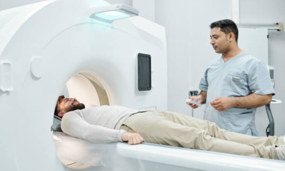
A PET CT scan combines the benefits of both technologies—one that shows what is happening inside your body—to allow doctors to capture a very precise image. Because of this precision, PET CT scans are often the gold standard to diagnose difficult conditions.  

Accurate Diagnosis for Cancer  

A PET CT scan is one of the most common reasons for having the scan. The disease of cancer occurs when cells in our body behave abnormally.  A PET CT scan is critical for effective treatment, and so it is crucial in detecting cancer in those early stages.  

Tumors can be detected in their very earliest stages of growth using this type of test. Unlike other imaging tests, a PET CT scan shows not just where the tumor is — but also how active it is, and whether it’s malignant or benign.  

PET CT scans are also important to doctors for determining if cancer has spread to other areas of the body. Deciding the best treatment plan is also helpful. A PET CT scan is vital to improving survival rates for such diseases as breast cancer and lung cancer by providing earlier diagnosis. While technological advances always have a price, the PET CT scan cost in Bangalore is extremely cheap in comparison to other cities around the world and thus available to most.  

Monitoring Treatment Progress  

A PET CT scan may be needed if patients already have cancer or other serious conditions and are being treated for that. For example, chemotherapy or radiation therapy can chemically weaken a tumor, shrink it, or reduce its activity, and this progress must be checked periodically.  

A PET CT scan provides a clear image of what’s going on with the area affected and also gives doctors a good indication of whether they should stick with the current treatment plan or modify it.  

Small but important markers of improvement might not be so easy to see on standard imaging tests such as X-rays and ultrasounds, but a PET scan might offer a clearer picture. 

Hidden Medical Condition Detection

Not all illnesses have clear symptoms. Standard diagnostic tools do not expose some problems, such as inflammation, infections, or particular abnormality of organs.  

If a diagnosis can’t be found with other imaging tests, doctors may recommend a PET CT scan if they think there is a hidden condition that’s causing symptoms for no reason. That scan can even identify infections, neurological disorders, and sometimes heart abnormalities that standard CT or MRI cannot. 

The PET CT scan cost Bangalore is very much lower than the cost of any other metropolitan city and thus, more patients can avail of this technology sadly.  

Pre-Surgical Planning

PET CT scans are often performed before surgery — especially for critical diseases like cancer — because that’s exactly why.  

Doctors might recommend a PET CT scan before surgery to learn how far the disease has spread and how to proceed with the surgery in as precise a manner as possible. Knowing the exact location of abnormal tissue or the size of a tumor and how far it’s spread increases the chances of a successful outcome when choosing to have surgery.  

With this level of detailed preparation, patients will recover much quicker and can minimize the damage to healthy tissues. Because of that, health screening centers everywhere in the country, especially Bangalore, reap the opportunity to provide PET CT scans at an affordable rate, making this scan in Bangalore a good choice to spend on your health.   

Safe and Non-Invasive Procedure

As it is a non-invasive procedure, PET CT scans are another vital reason doctors recommend them. It is unlike surgical biopsies, which involve removing a portion of tissue to be tested, in that an injection of a small, harmless radioactive tracer is administered into the body. This tracer lights up areas of abnormal activity during the scan, and the whole thing should take less than a couple of hours.  

Additionally, a PET CT scan is a very low-dose radiation device and is safe. It’s no comparison, the risk is small compared to the advantage of gaining critical medical insight. This is why many older adults, people with weakened immunity, and those who avoid invasive procedures prefer PET CT scans.  

On the one hand, patients and families can find yet another combination of a low PET CT scan cost Bangalore rates combined with the highest class of standards in the healthcare system of Bangalore if safety and affordability are your concerns.  

A Final Word on PET CT Scans 

Today’s medical world uses a revolutionary tool called the PET CT scan that doctors can trust to diagnose and treat all kinds of conditions. This test gives you an unparalleled perspective into the body without the pain or discomfort — of catching cancer early; knowing how treatment is going; finding hidden illnesses; and planning for surgery.

Cost is a concern for many, and so it makes sense that Bangalore is becoming a base for affordable healthcare technology. Many patients can afford the PET CT scan cost Bangalore and thus have the opportunity to benefit from the use of this life-saving diagnostic tool.  

If your doctor advises a PET CT scan, remember it is not just a test but one of the most accurate ways to assess, diagnose, and tackle health conditions effectively. By investing in this technology, you’re investing in health, clarity, and well-being for years to come.   

Koshikaa is a trusted healthcare provider offering comprehensive services for full-body checkup in Bangalore, ensuring accurate diagnostics and personalized care for patients.

MRI of the uterus is primarily used for diagnosing and evaluating conditions such as uterine fibroids, adenomyosis, and malignancies. It is particularly useful in the planning of surgical procedures and assessing the extent of gynecological diseases. This imaging test is valuable for its ability to deliver detailed cross-sectional images of the uterus and surrounding structures.
An MRI of the spleen can help diagnose various disorders affecting the spleen including splenomegaly (enlarged spleen), tumors, and other abnormalities. It provides clear, detailed images that are crucial for assessing the health of the spleen and planning potential surgical interventions.
Prostate MRI is a non-invasive imaging technique that provides detailed pictures of the prostate gland and surrounding tissues. It helps in the detection, staging, and monitoring of prostate cancer. Prostate MRIs are increasingly used to guide biopsy procedures and assist in the planning of treatment strategies for prostate health.
An MRI of the pancreas is used to identify issues such as pancreatitis, pancreatic cancer, and other pancreatic disorders. It offers high-resolution images that help in detecting small lesions and assessing the pancreatic ducts. This imaging technique is valuable for guiding biopsies and planning surgeries involving the pancreas.
MRI scans of the liver, kidney, and bile ducts provide detailed images that help diagnose various conditions, such as tumors, cysts, and blockages in the bile ducts. They are crucial for assessing organ damage, staging cancer, and planning liver or kidney-related surgeries. This type of MRI can also evaluate the health and function of these organs comprehensively.
Breast MRI is highly sensitive for detecting breast cancer, especially in individuals with dense breast tissue or a high risk of cancer. It is often used in conjunction with mammography to provide a more comprehensive evaluation. Breast MRIs can also monitor the response to breast cancer treatments and provide detailed pre-surgical mapping.
Brain and spine MRIs are critical tools for diagnosing and monitoring neurological conditions, including tumors, spinal cord injuries, and multiple sclerosis. These MRIs can reveal abnormalities in brain tissue and spinal cord structure that might not be visible with other imaging methods. They are essential for planning surgeries and assessing the effectiveness of treatments in neurology.
PET-CT scans of transplant organs like the liver, kidney, or pancreas are performed to assess metabolic activity, detect complications, and monitor post-transplant function. These scans play a vital role in the early detection of issues such as organ rejection, recurrence of disease, or infection, guiding timely interventions and optimizing transplant outcomes for patients.
PET-CT examinations of organs such as the liver, kidney, or prostate are crucial for assessing metabolic activity, detecting abnormalities, and monitoring disease progression. These scans help diagnose conditions such as liver metastases, renal cell carcinoma, or prostate cancer, guiding treatment decisions and monitoring response to therapy over time.
PET-CT scans of soft-tissue masses are utilized to assess metabolic activity and detect malignant tumors or other abnormalities. By combining PET imaging with CT imaging, these scans provide detailed information about tumor location, size, and metabolic characteristics, aiding in accurate diagnosis and treatment planning for soft-tissue cancers.
PET-CT scans are valuable for detecting cysts or fluid collections within the body, including cysts in the liver, kidneys, or other organs. These scans help characterize the nature and metabolic activity of cysts, guiding diagnosis and treatment planning for conditions such as cystic liver disease, renal cysts, or pancreatic cysts.
PET-CT scans of muscles are performed to assess metabolic activity and detect abnormalities such as inflammation, infection, or tumors. These scans provide valuable information about muscle function and pathology, aiding in the diagnosis and management of conditions like myositis, muscular dystrophy, or soft-tissue sarcomas.
PET-CT scans of the brain are utilized to evaluate brain metabolism and detect abnormalities such as tumors, epilepsy, or neurodegenerative diseases. By combining functional PET imaging with anatomical CT imaging, these scans offer comprehensive information about brain function and structure, guiding neurosurgical interventions and treatment decisions for neurological disorders
PET-CT scans of bones are commonly used to detect bone metastases in cancer patients, assess bone health in conditions like osteoporosis, and evaluate response to treatment. By combining PET and CT imaging, these scans provide detailed information about metabolic activity and anatomical changes in bone structures, aiding in accurate diagnosis and treatment planning for various bone-related conditions.
CT scans of the spine are conducted to assess the vertebrae, spinal cord, and surrounding soft tissues for conditions like herniated discs, spinal fractures, or spinal stenosis, aiding in the diagnosis and management of spinal disorders with detailed imaging essential for surgical planning and intervention.
CT scans of the pelvis are utilized to visualize the pelvic bones, reproductive organs, bladder, and surrounding structures for conditions such as fractures, tumors, or pelvic inflammatory disease, guiding treatment plans and surgical interventions with precise anatomical information.
CT scans of the neck are performed to evaluate structures such as the throat, thyroid gland, lymph nodes, and blood vessels for abnormalities like tumors, infections, or injuries, assisting in diagnosis and management of head and neck disorders with detailed imaging.
CT scans of the extremities are employed to assess bones, joints, and soft tissues in the arms or legs, aiding in the diagnosis of fractures, arthritis, or other musculoskeletal conditions, guiding orthopedic treatment and rehabilitation plans for optimal recovery.
CT scans of the chest are utilized to examine the lungs, heart, blood vessels, and surrounding structures for conditions such as lung nodules, pneumonia, or pulmonary embolism, facilitating accurate diagnosis and management of respiratory and cardiovascular diseases.
CT scans of the brain are conducted to assess brain structures, identify abnormalities like tumors, hemorrhages, or strokes, and aid in diagnosing neurological conditions, guiding neurosurgical interventions and treatment strategies with precise imaging of the brain’s anatomy.
CT scans of the abdomen are performed to visualize and evaluate the internal organs such as the liver, kidneys, pancreas, and intestines for conditions like tumors, infections, or injuries, providing detailed images essential for diagnosis and treatment planning.
Transplant organ ultrasound scans, including those of the liver, kidney, or pancreas, are performed to assess organ viability, detect complications, and monitor post-transplant function. These scans play a vital role in the early detection of issues such as organ rejection, vascular complications, or fluid collections, guiding timely interventions and optimizing transplant outcomes for patients.
Ultrasound examinations of organs such as the liver, kidney, or prostate are crucial for assessing organ structure, detecting abnormalities, and monitoring disease progression. These scans help diagnose conditions such as liver cirrhosis, kidney stones, or prostate enlargement, guiding treatment decisions and monitoring response to therapy over time.
Soft-tissue masses can be evaluated using ultrasound to determine their size, location, and characteristics. Ultrasound imaging helps differentiate between benign and malignant masses, guiding further diagnostic evaluation and treatment planning. By providing real-time imaging, ultrasound assists in the precise localization of soft-tissue masses for biopsy or surgical removal if needed.
Ultrasound scans are valuable for detecting cysts or fluid collections within the body, including cysts in the liver, kidneys, or other organs. These scans help characterize the nature and size of cysts, aiding in diagnosis and treatment planning for conditions such as cystic liver disease or renal cysts, and guiding interventions such as cyst drainage or sclerotherapy.
Musculoskeletal ultrasound scans are used to assess the health and integrity of muscles, identifying issues such as strains, tears, or muscle inflammation. These scans provide detailed images of muscle structures, allowing healthcare providers to diagnose muscle injuries accurately and develop personalized rehabilitation plans to promote recovery and restore function.
Ultrasound examinations of joints are essential for evaluating joint health, detecting abnormalities such as inflammation, fluid accumulation, or cartilage damage. By visualizing the joint structures in real-time, ultrasound helps diagnose conditions like arthritis, bursitis, or ligament injuries, guiding treatment decisions and monitoring disease progression over time.
Ultrasound scans of tendons are performed to assess for injuries, inflammation, or tears, aiding in the diagnosis and management of conditions such as tendonitis or tendon tears. These scans provide detailed images of tendon structures, helping healthcare providers determine the extent of damage and plan appropriate treatment strategies, including physical therapy or surgical intervention if necessary.
Pelvis X-rays play a crucial role in diagnosing injuries, diseases, and developmental conditions affecting the pelvic region. They provide detailed images of the hip bones, sacrum, and coccyx, aiding in the assessment of fractures, dislocations, or signs of arthritis. Pelvic X-rays are instrumental in guiding surgical interventions, monitoring healing progress, and evaluating conditions such as osteoporosis or tumors. By providing precise anatomical information, these imaging studies enable healthcare professionals to deliver personalized care and improve patient outcomes.
Teeth X-rays, known as dental radiographs, are indispensable tools for dentists to assess oral health. They aid in diagnosing dental decay, gum disease, and abnormalities in tooth structure or alignment. These images provide valuable insights into hidden dental issues, allowing for early intervention and prevention of complications. Additionally, dental X-rays are essential for planning treatments such as orthodontics, root canals, or dental implants, ensuring optimal outcomes and patient satisfaction.
Spine X-rays are pivotal in diagnosing spinal fractures, degenerative conditions, and spinal misalignments, providing critical information for treatment planning and monitoring spinal health. They offer detailed images of the vertebrae and surrounding structures, enabling healthcare professionals to identify issues such as scoliosis, disc herniation, or spinal stenosis
Skull X-rays are valuable for detecting fractures, assessing head trauma, and evaluating the alignment of facial bones, crucial in diagnosing conditions like fractures, tumors, or developmental abnormalities in the skull. They provide detailed views of the cranial structure, aiding clinicians in determining appropriate treatment strategies and ensuring patient safety.
For the heart, X-ray solutions can aid in the detection of various cardiac problems, ranging from mild to severe, including – Heart failure, Cardiac congestion and other heart-related conditions. In addition to cardiac assessments, X-ray solutions are also used for breast examinations to identify tumors or signs of breast cancer.

Bone X-rays are essential for diagnosing fractures and assessing bone alignment, aiding doctors in developing treatment plans and monitoring healing progress. They provide detailed images of the skeletal system, helping identify abnormalities or injuries that may not be visible through other imaging techniques

Abdominal X-rays are performed when patients experience symptoms such as significant nausea, ongoing vomiting, abdominal pain, flank or back pain, or abdominal swelling. These X-rays can help diagnose various conditions.
Search Bar

Frequently Booked Tests

Frequently Booked Checkups