Understanding Sinus CT Scan: Process, Benefits, and More

sinus ct scan

Table of Contents

A Sinus CT Scan remains among the standard medical procedures for obtaining comprehensive sinus images throughout the facial structures. The medical technique enables proper diagnosis of infection-related issues alongside inflammation conditions and blockages along with structural abnormalities in the sinuses. This article will explain key aspects of Sinus CT Scans by explaining their functioning process and preparation guidelines as well as their significance in modern medical diagnostics.

What Is a Sinus CT Scan?

A Sinus CT Scan functions through X-ray technology to generate multi-dimensional pictures of facial sinus structures together with adjacent anatomical regions. A CT scan differs from standard X-rays because it generates three-dimensional internal images while traditional X-rays provide only two-dimensional visibility. Through an improved imaging perspective, doctors perform more precise diagnoses of sinus conditions.

What is it used for? 

Medical professionals perform sinus CT scans for several diagnostic purposes. A sinus CT scan serves physicians in their evaluation of sinus infections together with chronic sinusitis and nasal polyps along with tumours and any facial discomfort with no explanation. The procedure enables both surgeons and doctors to assess structural abnormalities including septal deviations and examine sinus health before operating on patients.

Normal Sinus CT Scan vs Abnormal: What’s the Difference?

ct scan in bangalore

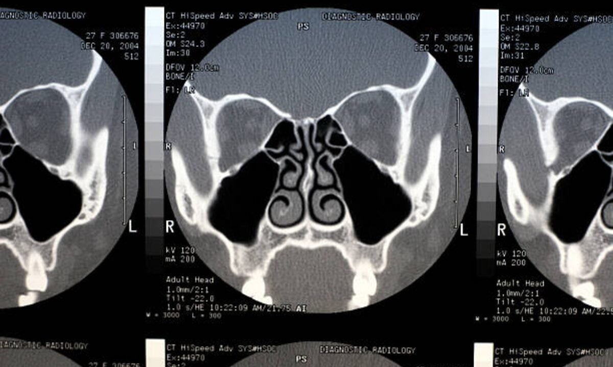
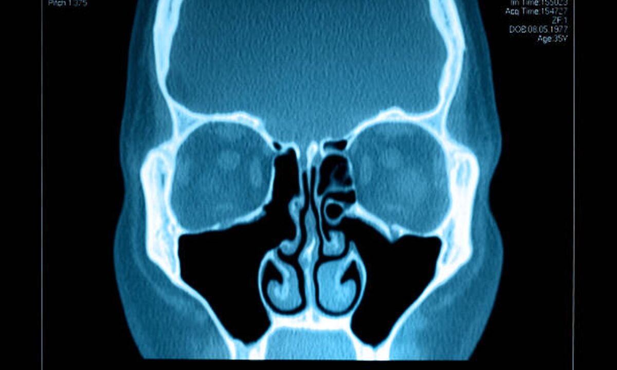
Medical professionals need to evaluate CT scan results by deciding if the viewed sinuses show normal or abnormal characteristics. The sinuses appear normal in standard CT imaging because they show both air pockets and lack any indication of swelling as well as growth abnormalities or blockages. There are no infections, and all sinus passages appear in their natural state.

On the other hand, an abnormal sinus CT scan indicates some kind of problem. This could be a sinus infection, fluid accumulation, nasal polyps, or structural deformities. 

The appearance of sinus clouding along with blocked tissue usually means the patient has sinusitis or inflammation. A sinus CT scan enables doctors to make precise diagnoses through normal versus abnormal distinction which leads to correct treatment decisions.

How Do I Prepare for a Sinus CT Scan?

A sinus CT scan requires simple steps of preparation, although following these procedures leads to successful results.

1. Discuss all your existing medical situations and allergy history with your doctor.  

Let your doctor be aware of active medical conditions including allergies together with diabetes and pregnancy before the scheduled scan. It becomes crucial to inform the doctor about potential adverse reactions to contrast dye because the scan process involves its use.

2. Wear Comfortable, Metal-Free Clothing  

The imaging process will face disruptions because of metal items such as jewelry including glasses or hairpins. A simple non-metal clothing selection is recommended since screening technicians might need you to remove items.

3. Specific directions about using contrast dye must be followed by patients

Your doctor will require you to fast food and beverages before obtaining a contrast-enhanced sinus CT scan. Post-procedure drinking ample amounts of water aids in clearing the contrast dye from your system.

4. Your scanning process will be optimized by staying motionless while keeping calm throughout the test duration. 

The examination process consumes around 10-15 minutes of time. Clear results will be attained by staying motionless throughout the imaging process. The technician will assist you throughout the process therefore remaining calm and relaxed.

The simple instructions presented here will enable you to enter your sinus CT scan experience without anxiety.

Benefits of a Sinus CT Scan

Using a sinus CT scan provides patients with multiple distinct benefits, which establishes it as a top-diagnostic method for managing sinus complaints.

Accurate Diagnosis  

CT scanning technology reveals far superior image details than regular procedures which helps doctors identify tiny nasal abnormalities.

Non-Invasive and Painless 

Your sinus CT scan takes place without any invasive procedures because the complete process is mechanically performed and completely painless. It serves as the top diagnostic method for population groups of any age.

Time-Efficient  

CT scans that assess the sinuses usually finish within 15 minutes of the procedure time. Treatment decisions become speedier because results become available shortly after the completion of the examination.

Pre-Surgical Mapping

The scan creates essential guidance that surgeons need before performing sinus surgery on patients experiencing recurring sinus conditions. The procedure pinpoints important areas that surgeons should handle throughout the surgical process.

CT Scan in Bangalore: Why Location Matters

Any person recommended to undergo a sinus CT scan in Bangalore can find multiple trusted healthcare facilities with modern imaging technology across the city. Patients who select health screening centre that employ professional radiologists access both exact diagnosis and dependable care. The established status of Bangalore as a medical center provides you with multiple healthcare facilities that deliver international standard diagnostic services.

When Is a Full Body Checkup in Bangalore Beneficial?

Your attention to sinus care needs to be paired with a full body checkup in Bangalore particularly when you have existing medical issues. Health screening facilities throughout the city supply complete diagnostic bundles to evaluate different body systems. Through complete body testing you gain complete health insight and simultaneously detect additional health problems affecting your respiratory system which influences your sinus function.

Is a Sinus CT Scan Safe?

People express security concerns about medical imaging testing because radiation exposure frequently comes into play. The low radiation levels within sinus CT scans make them secure for general patients. CT scanners today achieve maximum radiation safety standards alongside their goals for producing clear imaging data. Women who are pregnant as well as patients with medical issues should speak with their doctor before getting this procedure because it involves radiation exposure.

The important diagnostic advantages of sinus CT scans substantially surpass the minimal safety risks that exist during medical procedures for the general patient population. 

Where to Get a Sinus CT Scan in Bangalore? 

A reliable site for sinus CT scan purposes in Bangalore requires patients to select a centre with established professional excellence. Avoid facilities that lack experienced radiologists or modern medical equipment because these conditions provide enhanced care to patients. You should always look at patient reviews and also ask your doctor to suggest the most reliable facilities in Bangalore.

Conclusion

Sinus CT scanning stands as an essential examination method to deliver critical information about your sinus health. This diagnostic imaging technology enables doctors to understand both chronic sinus infections and unidentified facial discomfort to decide appropriate medical treatment. Understanding the difference between a normal sinus CT scan vs abnormal findings allows patients to better comprehend their diagnosis and treatment plan.

Finally, if you’re located in Bangalore, rejoice in the fact that there are excellent facilities for CT scan as well as the option of a full body checkup in Bangalore, offered by many reputable health screening centers.

Your health control begins when you prepare with expert guidance which enables you to improve your life quality. 

Koshikaa provides patients with extensive packages for full body checkups in Bangalore and personalized health evaluations through their diagnostic processes to promote patient wellness.

MRI of the uterus is primarily used for diagnosing and evaluating conditions such as uterine fibroids, adenomyosis, and malignancies. It is particularly useful in the planning of surgical procedures and assessing the extent of gynecological diseases. This imaging test is valuable for its ability to deliver detailed cross-sectional images of the uterus and surrounding structures.
An MRI of the spleen can help diagnose various disorders affecting the spleen including splenomegaly (enlarged spleen), tumors, and other abnormalities. It provides clear, detailed images that are crucial for assessing the health of the spleen and planning potential surgical interventions.
Prostate MRI is a non-invasive imaging technique that provides detailed pictures of the prostate gland and surrounding tissues. It helps in the detection, staging, and monitoring of prostate cancer. Prostate MRIs are increasingly used to guide biopsy procedures and assist in the planning of treatment strategies for prostate health.
An MRI of the pancreas is used to identify issues such as pancreatitis, pancreatic cancer, and other pancreatic disorders. It offers high-resolution images that help in detecting small lesions and assessing the pancreatic ducts. This imaging technique is valuable for guiding biopsies and planning surgeries involving the pancreas.
MRI scans of the liver, kidney, and bile ducts provide detailed images that help diagnose various conditions, such as tumors, cysts, and blockages in the bile ducts. They are crucial for assessing organ damage, staging cancer, and planning liver or kidney-related surgeries. This type of MRI can also evaluate the health and function of these organs comprehensively.
Breast MRI is highly sensitive for detecting breast cancer, especially in individuals with dense breast tissue or a high risk of cancer. It is often used in conjunction with mammography to provide a more comprehensive evaluation. Breast MRIs can also monitor the response to breast cancer treatments and provide detailed pre-surgical mapping.
Brain and spine MRIs are critical tools for diagnosing and monitoring neurological conditions, including tumors, spinal cord injuries, and multiple sclerosis. These MRIs can reveal abnormalities in brain tissue and spinal cord structure that might not be visible with other imaging methods. They are essential for planning surgeries and assessing the effectiveness of treatments in neurology.
PET-CT scans of transplant organs like the liver, kidney, or pancreas are performed to assess metabolic activity, detect complications, and monitor post-transplant function. These scans play a vital role in the early detection of issues such as organ rejection, recurrence of disease, or infection, guiding timely interventions and optimizing transplant outcomes for patients.
PET-CT examinations of organs such as the liver, kidney, or prostate are crucial for assessing metabolic activity, detecting abnormalities, and monitoring disease progression. These scans help diagnose conditions such as liver metastases, renal cell carcinoma, or prostate cancer, guiding treatment decisions and monitoring response to therapy over time.
PET-CT scans of soft-tissue masses are utilized to assess metabolic activity and detect malignant tumors or other abnormalities. By combining PET imaging with CT imaging, these scans provide detailed information about tumor location, size, and metabolic characteristics, aiding in accurate diagnosis and treatment planning for soft-tissue cancers.
PET-CT scans are valuable for detecting cysts or fluid collections within the body, including cysts in the liver, kidneys, or other organs. These scans help characterize the nature and metabolic activity of cysts, guiding diagnosis and treatment planning for conditions such as cystic liver disease, renal cysts, or pancreatic cysts.
PET-CT scans of muscles are performed to assess metabolic activity and detect abnormalities such as inflammation, infection, or tumors. These scans provide valuable information about muscle function and pathology, aiding in the diagnosis and management of conditions like myositis, muscular dystrophy, or soft-tissue sarcomas.
PET-CT scans of the brain are utilized to evaluate brain metabolism and detect abnormalities such as tumors, epilepsy, or neurodegenerative diseases. By combining functional PET imaging with anatomical CT imaging, these scans offer comprehensive information about brain function and structure, guiding neurosurgical interventions and treatment decisions for neurological disorders
PET-CT scans of bones are commonly used to detect bone metastases in cancer patients, assess bone health in conditions like osteoporosis, and evaluate response to treatment. By combining PET and CT imaging, these scans provide detailed information about metabolic activity and anatomical changes in bone structures, aiding in accurate diagnosis and treatment planning for various bone-related conditions.
CT scans of the spine are conducted to assess the vertebrae, spinal cord, and surrounding soft tissues for conditions like herniated discs, spinal fractures, or spinal stenosis, aiding in the diagnosis and management of spinal disorders with detailed imaging essential for surgical planning and intervention.
CT scans of the pelvis are utilized to visualize the pelvic bones, reproductive organs, bladder, and surrounding structures for conditions such as fractures, tumors, or pelvic inflammatory disease, guiding treatment plans and surgical interventions with precise anatomical information.
CT scans of the neck are performed to evaluate structures such as the throat, thyroid gland, lymph nodes, and blood vessels for abnormalities like tumors, infections, or injuries, assisting in diagnosis and management of head and neck disorders with detailed imaging.
CT scans of the extremities are employed to assess bones, joints, and soft tissues in the arms or legs, aiding in the diagnosis of fractures, arthritis, or other musculoskeletal conditions, guiding orthopedic treatment and rehabilitation plans for optimal recovery.
CT scans of the chest are utilized to examine the lungs, heart, blood vessels, and surrounding structures for conditions such as lung nodules, pneumonia, or pulmonary embolism, facilitating accurate diagnosis and management of respiratory and cardiovascular diseases.
CT scans of the brain are conducted to assess brain structures, identify abnormalities like tumors, hemorrhages, or strokes, and aid in diagnosing neurological conditions, guiding neurosurgical interventions and treatment strategies with precise imaging of the brain’s anatomy.
CT scans of the abdomen are performed to visualize and evaluate the internal organs such as the liver, kidneys, pancreas, and intestines for conditions like tumors, infections, or injuries, providing detailed images essential for diagnosis and treatment planning.
Transplant organ ultrasound scans, including those of the liver, kidney, or pancreas, are performed to assess organ viability, detect complications, and monitor post-transplant function. These scans play a vital role in the early detection of issues such as organ rejection, vascular complications, or fluid collections, guiding timely interventions and optimizing transplant outcomes for patients.
Ultrasound examinations of organs such as the liver, kidney, or prostate are crucial for assessing organ structure, detecting abnormalities, and monitoring disease progression. These scans help diagnose conditions such as liver cirrhosis, kidney stones, or prostate enlargement, guiding treatment decisions and monitoring response to therapy over time.
Soft-tissue masses can be evaluated using ultrasound to determine their size, location, and characteristics. Ultrasound imaging helps differentiate between benign and malignant masses, guiding further diagnostic evaluation and treatment planning. By providing real-time imaging, ultrasound assists in the precise localization of soft-tissue masses for biopsy or surgical removal if needed.
Ultrasound scans are valuable for detecting cysts or fluid collections within the body, including cysts in the liver, kidneys, or other organs. These scans help characterize the nature and size of cysts, aiding in diagnosis and treatment planning for conditions such as cystic liver disease or renal cysts, and guiding interventions such as cyst drainage or sclerotherapy.
Musculoskeletal ultrasound scans are used to assess the health and integrity of muscles, identifying issues such as strains, tears, or muscle inflammation. These scans provide detailed images of muscle structures, allowing healthcare providers to diagnose muscle injuries accurately and develop personalized rehabilitation plans to promote recovery and restore function.
Ultrasound examinations of joints are essential for evaluating joint health, detecting abnormalities such as inflammation, fluid accumulation, or cartilage damage. By visualizing the joint structures in real-time, ultrasound helps diagnose conditions like arthritis, bursitis, or ligament injuries, guiding treatment decisions and monitoring disease progression over time.
Ultrasound scans of tendons are performed to assess for injuries, inflammation, or tears, aiding in the diagnosis and management of conditions such as tendonitis or tendon tears. These scans provide detailed images of tendon structures, helping healthcare providers determine the extent of damage and plan appropriate treatment strategies, including physical therapy or surgical intervention if necessary.
Pelvis X-rays play a crucial role in diagnosing injuries, diseases, and developmental conditions affecting the pelvic region. They provide detailed images of the hip bones, sacrum, and coccyx, aiding in the assessment of fractures, dislocations, or signs of arthritis. Pelvic X-rays are instrumental in guiding surgical interventions, monitoring healing progress, and evaluating conditions such as osteoporosis or tumors. By providing precise anatomical information, these imaging studies enable healthcare professionals to deliver personalized care and improve patient outcomes.
Teeth X-rays, known as dental radiographs, are indispensable tools for dentists to assess oral health. They aid in diagnosing dental decay, gum disease, and abnormalities in tooth structure or alignment. These images provide valuable insights into hidden dental issues, allowing for early intervention and prevention of complications. Additionally, dental X-rays are essential for planning treatments such as orthodontics, root canals, or dental implants, ensuring optimal outcomes and patient satisfaction.
Spine X-rays are pivotal in diagnosing spinal fractures, degenerative conditions, and spinal misalignments, providing critical information for treatment planning and monitoring spinal health. They offer detailed images of the vertebrae and surrounding structures, enabling healthcare professionals to identify issues such as scoliosis, disc herniation, or spinal stenosis
Skull X-rays are valuable for detecting fractures, assessing head trauma, and evaluating the alignment of facial bones, crucial in diagnosing conditions like fractures, tumors, or developmental abnormalities in the skull. They provide detailed views of the cranial structure, aiding clinicians in determining appropriate treatment strategies and ensuring patient safety.
For the heart, X-ray solutions can aid in the detection of various cardiac problems, ranging from mild to severe, including – Heart failure, Cardiac congestion and other heart-related conditions. In addition to cardiac assessments, X-ray solutions are also used for breast examinations to identify tumors or signs of breast cancer.

Bone X-rays are essential for diagnosing fractures and assessing bone alignment, aiding doctors in developing treatment plans and monitoring healing progress. They provide detailed images of the skeletal system, helping identify abnormalities or injuries that may not be visible through other imaging techniques

Abdominal X-rays are performed when patients experience symptoms such as significant nausea, ongoing vomiting, abdominal pain, flank or back pain, or abdominal swelling. These X-rays can help diagnose various conditions.
Search Bar

Frequently Booked Tests

Frequently Booked Checkups