How to Prepare for a Full Body Checkup Test for Accurate Results

Table of Contents

Understanding how to properly prepare for a full-body checkup test can significantly impact the accuracy of your health screening results.

Whether you are scheduling your annual health assessment or addressing specific concerns, proper preparation ensures that your test results reflect your true health status.

Many people underestimate the importance of pre-test preparation, which can lead to inaccurate readings and unnecessary follow-up tests.

If you are looking for a full body checkup in Bangalore, knowing these preparation guidelines will help you get the most reliable results from your health screening.

But what exactly should you do before your appointment to ensure everything goes smoothly?

What is a Full Body Checkup

A full body checkup is a comprehensive health screening that evaluates your overall physical condition through various diagnostic tests.

It typically includes blood tests, urine analysis, imaging studies, and physical examinations to detect potential health issues early.

These checkups help identify risk factors for diseases like diabetes, heart conditions, thyroid disorders, and other metabolic problems.

Regular screenings are essential for preventive healthcare and maintaining long-term wellness.

Understanding the Full Body Check-Up Tests List

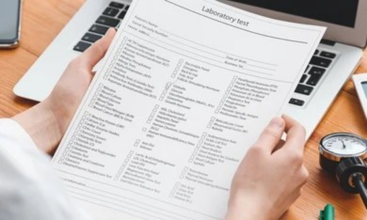
A standard full-body check-up test list includes several essential diagnostic procedures.

The most common is a blood test for a full body checkup, which measures cholesterol levels, blood sugar, liver function, kidney function, and complete blood count.

Other tests typically include urine analysis, thyroid profile, vitamin D levels, and lipid profile.

Some packages also offer ECG, chest X-ray, ultrasound abdomen, and stress tests depending on age and health requirements.

Advanced screenings may include tumor markers, as many people wonder can full body checkup can detect cancer – yes, certain markers and imaging can identify early warning signs.

When reviewing the full body checkup test list with price, you’ll notice variations based on the comprehensiveness of the package.

Basic packages start from affordable rates, while comprehensive ones with advanced imaging and specialized tests cost more.

How to Prepare Before Your Health Screening

1. Fasting Requirements

  • Most blood tests require fasting for 8-12 hours before sample collection.
  • This means avoiding all food and beverages except water during this period.
  • Fasting ensures accurate measurement of blood sugar, cholesterol, and triglyceride levels.
  • Schedule your appointment early in the morning so fasting doesn’t disrupt your entire day.

2. Hydration Guidelines

  • While fasting from food is necessary, staying hydrated is crucial.
  • Drinking adequate water before your test helps in easy blood sample collection and doesn’t affect most test results.
  • Avoid alcohol for at least 24-48 hours before your checkup, as it can alter liver function tests.
  • Also, limit caffeine intake, as it may affect heart rate and blood pressure readings.

3. Medication and Supplements

  • Inform the health screening centre in Bangalore about all medications and supplements you’re currently taking.
  • Some medications may need to be temporarily paused, while others should be continued as prescribed.
  • Never stop medications without consulting your doctor, especially for chronic conditions like diabetes or hypertension.
  • Carry a list of your current medications to share with the healthcare team.

4. Lifestyle Considerations

  • Avoid strenuous exercise 24 hours before your checkup, as it can temporarily elevate certain enzyme levels.
  • Get adequate sleep the night before, as fatigue can affect blood pressure and other vital signs.
  • Wear comfortable, loose-fitting clothes that allow easy access for blood collection.
  • Arrive well-rested and relaxed to ensure accurate readings of vital parameters.

5. Special Preparations for Women

  • Women should ideally schedule their full-body checkup in Bangalore when they are not menstruating, if possible.
  • Menstruation can affect certain blood test results and urine analysis.
  • If you’re pregnant or suspect pregnancy, inform the healthcare provider before any X-rays or imaging tests.

6. What to Bring

  • Carry any previous medical reports or full body test report documents for comparison.
  • Bring your identification, insurance documents, and prescription list.
  • Having past reports helps doctors identify trends and changes in your health parameters over time.
  • This historical data provides valuable context for interpreting your current results.

Tips for Accurate Test Results

  • Stick to your regular diet for 2-3 days before the test – sudden dietary changes can affect results.
  • Avoid smoking for at least 2 hours before your appointment, as it impacts lung function tests.
  • Don’t apply lotions or oils on your skin before the checkup, especially if an ultrasound is included.
  • Inform about recent illnesses, infections, or vaccinations, as these can temporarily alter test values.

Understanding Your Results

After your tests are completed, the laboratory processes your samples and generates a comprehensive report.

Your full body test report typically becomes available within 24-48 hours.

The report includes reference ranges alongside your values, making it easier to identify abnormalities.

Schedule a follow-up consultation with your doctor to understand the results thoroughly.

Don’t panic if some values are slightly outside the normal range – context matters, and your doctor will interpret results based on your overall health.

Final Thoughts

Proper preparation is the foundation of obtaining accurate and meaningful results from your health screening.

By following these guidelines, you ensure that your investment in preventive healthcare yields reliable insights into your well-being.

Whether you are scheduling a routine checkup or addressing specific health concerns, remember that preparation begins days before your actual appointment.

If you are searching for a reliable full-body checkup in Bangalore, Koshikaa offers comprehensive health screening packages with advanced diagnostic facilities.

Taking charge of your health through regular checkups is one of the best decisions you can make for your long-term wellness.

Start preparing today, and take the first step toward a healthier tomorrow.

Frequently Asked Questions

Q1: What should we do before a full body checkup?

Fast for 8-12 hours, stay hydrated with water, avoid alcohol for 24-48 hours, get adequate sleep, wear comfortable clothing, and carry previous medical reports. Inform your doctor about current medications and any recent illnesses for accurate interpretation of results.

Q2: How to prepare for a blood test to get the best results?

Fast for the recommended duration, drink plenty of water, avoid strenuous exercise 24 hours prior, don’t smoke before the test, and stay relaxed. Ensure you’re well-rested and inform the technician about any medications you’re taking for proper sample collection.

Q3: Should we go empty stomach for a full body checkup?

Yes, most full-body checkups require 8-12 hours of fasting to ensure accurate blood sugar, cholesterol, and triglyceride measurements. However, you can drink water during the fasting period. Schedule your appointment for early morning to minimize fasting discomfort and inconvenience.

Q4: Can I drink water before a full body check-up?

Yes, drinking water is allowed and encouraged before your full-body checkup. Adequate hydration helps with easier blood sample collection and doesn’t interfere with most test results. However, avoid other beverages like coffee, tea, juice, or milk during the fasting period.

Q5: What are the 5 main tests for a full body checkup?

The five main tests include Complete Blood Count (CBC), Lipid Profile for cholesterol levels, Blood Sugar tests (fasting and post-meal), Liver Function Test (LFT), and Kidney Function Test (KFT). These cover major health parameters and help detect common health conditions effectively.

MRI of the uterus is primarily used for diagnosing and evaluating conditions such as uterine fibroids, adenomyosis, and malignancies. It is particularly useful in the planning of surgical procedures and assessing the extent of gynecological diseases. This imaging test is valuable for its ability to deliver detailed cross-sectional images of the uterus and surrounding structures.
An MRI of the spleen can help diagnose various disorders affecting the spleen including splenomegaly (enlarged spleen), tumors, and other abnormalities. It provides clear, detailed images that are crucial for assessing the health of the spleen and planning potential surgical interventions.
Prostate MRI is a non-invasive imaging technique that provides detailed pictures of the prostate gland and surrounding tissues. It helps in the detection, staging, and monitoring of prostate cancer. Prostate MRIs are increasingly used to guide biopsy procedures and assist in the planning of treatment strategies for prostate health.
An MRI of the pancreas is used to identify issues such as pancreatitis, pancreatic cancer, and other pancreatic disorders. It offers high-resolution images that help in detecting small lesions and assessing the pancreatic ducts. This imaging technique is valuable for guiding biopsies and planning surgeries involving the pancreas.
MRI scans of the liver, kidney, and bile ducts provide detailed images that help diagnose various conditions, such as tumors, cysts, and blockages in the bile ducts. They are crucial for assessing organ damage, staging cancer, and planning liver or kidney-related surgeries. This type of MRI can also evaluate the health and function of these organs comprehensively.
Breast MRI is highly sensitive for detecting breast cancer, especially in individuals with dense breast tissue or a high risk of cancer. It is often used in conjunction with mammography to provide a more comprehensive evaluation. Breast MRIs can also monitor the response to breast cancer treatments and provide detailed pre-surgical mapping.
Brain and spine MRIs are critical tools for diagnosing and monitoring neurological conditions, including tumors, spinal cord injuries, and multiple sclerosis. These MRIs can reveal abnormalities in brain tissue and spinal cord structure that might not be visible with other imaging methods. They are essential for planning surgeries and assessing the effectiveness of treatments in neurology.
PET-CT scans of transplant organs like the liver, kidney, or pancreas are performed to assess metabolic activity, detect complications, and monitor post-transplant function. These scans play a vital role in the early detection of issues such as organ rejection, recurrence of disease, or infection, guiding timely interventions and optimizing transplant outcomes for patients.
PET-CT examinations of organs such as the liver, kidney, or prostate are crucial for assessing metabolic activity, detecting abnormalities, and monitoring disease progression. These scans help diagnose conditions such as liver metastases, renal cell carcinoma, or prostate cancer, guiding treatment decisions and monitoring response to therapy over time.
PET-CT scans of soft-tissue masses are utilized to assess metabolic activity and detect malignant tumors or other abnormalities. By combining PET imaging with CT imaging, these scans provide detailed information about tumor location, size, and metabolic characteristics, aiding in accurate diagnosis and treatment planning for soft-tissue cancers.
PET-CT scans are valuable for detecting cysts or fluid collections within the body, including cysts in the liver, kidneys, or other organs. These scans help characterize the nature and metabolic activity of cysts, guiding diagnosis and treatment planning for conditions such as cystic liver disease, renal cysts, or pancreatic cysts.
PET-CT scans of muscles are performed to assess metabolic activity and detect abnormalities such as inflammation, infection, or tumors. These scans provide valuable information about muscle function and pathology, aiding in the diagnosis and management of conditions like myositis, muscular dystrophy, or soft-tissue sarcomas.
PET-CT scans of the brain are utilized to evaluate brain metabolism and detect abnormalities such as tumors, epilepsy, or neurodegenerative diseases. By combining functional PET imaging with anatomical CT imaging, these scans offer comprehensive information about brain function and structure, guiding neurosurgical interventions and treatment decisions for neurological disorders
PET-CT scans of bones are commonly used to detect bone metastases in cancer patients, assess bone health in conditions like osteoporosis, and evaluate response to treatment. By combining PET and CT imaging, these scans provide detailed information about metabolic activity and anatomical changes in bone structures, aiding in accurate diagnosis and treatment planning for various bone-related conditions.
CT scans of the spine are conducted to assess the vertebrae, spinal cord, and surrounding soft tissues for conditions like herniated discs, spinal fractures, or spinal stenosis, aiding in the diagnosis and management of spinal disorders with detailed imaging essential for surgical planning and intervention.
CT scans of the pelvis are utilized to visualize the pelvic bones, reproductive organs, bladder, and surrounding structures for conditions such as fractures, tumors, or pelvic inflammatory disease, guiding treatment plans and surgical interventions with precise anatomical information.
CT scans of the neck are performed to evaluate structures such as the throat, thyroid gland, lymph nodes, and blood vessels for abnormalities like tumors, infections, or injuries, assisting in diagnosis and management of head and neck disorders with detailed imaging.
CT scans of the extremities are employed to assess bones, joints, and soft tissues in the arms or legs, aiding in the diagnosis of fractures, arthritis, or other musculoskeletal conditions, guiding orthopedic treatment and rehabilitation plans for optimal recovery.
CT scans of the chest are utilized to examine the lungs, heart, blood vessels, and surrounding structures for conditions such as lung nodules, pneumonia, or pulmonary embolism, facilitating accurate diagnosis and management of respiratory and cardiovascular diseases.
CT scans of the brain are conducted to assess brain structures, identify abnormalities like tumors, hemorrhages, or strokes, and aid in diagnosing neurological conditions, guiding neurosurgical interventions and treatment strategies with precise imaging of the brain’s anatomy.
CT scans of the abdomen are performed to visualize and evaluate the internal organs such as the liver, kidneys, pancreas, and intestines for conditions like tumors, infections, or injuries, providing detailed images essential for diagnosis and treatment planning.
Transplant organ ultrasound scans, including those of the liver, kidney, or pancreas, are performed to assess organ viability, detect complications, and monitor post-transplant function. These scans play a vital role in the early detection of issues such as organ rejection, vascular complications, or fluid collections, guiding timely interventions and optimizing transplant outcomes for patients.
Ultrasound examinations of organs such as the liver, kidney, or prostate are crucial for assessing organ structure, detecting abnormalities, and monitoring disease progression. These scans help diagnose conditions such as liver cirrhosis, kidney stones, or prostate enlargement, guiding treatment decisions and monitoring response to therapy over time.
Soft-tissue masses can be evaluated using ultrasound to determine their size, location, and characteristics. Ultrasound imaging helps differentiate between benign and malignant masses, guiding further diagnostic evaluation and treatment planning. By providing real-time imaging, ultrasound assists in the precise localization of soft-tissue masses for biopsy or surgical removal if needed.
Ultrasound scans are valuable for detecting cysts or fluid collections within the body, including cysts in the liver, kidneys, or other organs. These scans help characterize the nature and size of cysts, aiding in diagnosis and treatment planning for conditions such as cystic liver disease or renal cysts, and guiding interventions such as cyst drainage or sclerotherapy.
Musculoskeletal ultrasound scans are used to assess the health and integrity of muscles, identifying issues such as strains, tears, or muscle inflammation. These scans provide detailed images of muscle structures, allowing healthcare providers to diagnose muscle injuries accurately and develop personalized rehabilitation plans to promote recovery and restore function.
Ultrasound examinations of joints are essential for evaluating joint health, detecting abnormalities such as inflammation, fluid accumulation, or cartilage damage. By visualizing the joint structures in real-time, ultrasound helps diagnose conditions like arthritis, bursitis, or ligament injuries, guiding treatment decisions and monitoring disease progression over time.
Ultrasound scans of tendons are performed to assess for injuries, inflammation, or tears, aiding in the diagnosis and management of conditions such as tendonitis or tendon tears. These scans provide detailed images of tendon structures, helping healthcare providers determine the extent of damage and plan appropriate treatment strategies, including physical therapy or surgical intervention if necessary.
Pelvis X-rays play a crucial role in diagnosing injuries, diseases, and developmental conditions affecting the pelvic region. They provide detailed images of the hip bones, sacrum, and coccyx, aiding in the assessment of fractures, dislocations, or signs of arthritis. Pelvic X-rays are instrumental in guiding surgical interventions, monitoring healing progress, and evaluating conditions such as osteoporosis or tumors. By providing precise anatomical information, these imaging studies enable healthcare professionals to deliver personalized care and improve patient outcomes.
Teeth X-rays, known as dental radiographs, are indispensable tools for dentists to assess oral health. They aid in diagnosing dental decay, gum disease, and abnormalities in tooth structure or alignment. These images provide valuable insights into hidden dental issues, allowing for early intervention and prevention of complications. Additionally, dental X-rays are essential for planning treatments such as orthodontics, root canals, or dental implants, ensuring optimal outcomes and patient satisfaction.
Spine X-rays are pivotal in diagnosing spinal fractures, degenerative conditions, and spinal misalignments, providing critical information for treatment planning and monitoring spinal health. They offer detailed images of the vertebrae and surrounding structures, enabling healthcare professionals to identify issues such as scoliosis, disc herniation, or spinal stenosis
Skull X-rays are valuable for detecting fractures, assessing head trauma, and evaluating the alignment of facial bones, crucial in diagnosing conditions like fractures, tumors, or developmental abnormalities in the skull. They provide detailed views of the cranial structure, aiding clinicians in determining appropriate treatment strategies and ensuring patient safety.
For the heart, X-ray solutions can aid in the detection of various cardiac problems, ranging from mild to severe, including – Heart failure, Cardiac congestion and other heart-related conditions. In addition to cardiac assessments, X-ray solutions are also used for breast examinations to identify tumors or signs of breast cancer.

Bone X-rays are essential for diagnosing fractures and assessing bone alignment, aiding doctors in developing treatment plans and monitoring healing progress. They provide detailed images of the skeletal system, helping identify abnormalities or injuries that may not be visible through other imaging techniques

Abdominal X-rays are performed when patients experience symptoms such as significant nausea, ongoing vomiting, abdominal pain, flank or back pain, or abdominal swelling. These X-rays can help diagnose various conditions.

Frequently Booked Tests

Frequently Booked Checkups