Need an X-Ray Centre Near You in Bangalore?

CT scan in Bangalore

Table of Contents

Whether it’s sudden pain, an injury, or a doctor’s referral, finding a reliable X-ray centre nearby is crucial for fast diagnosis and timely treatment. Koshikaa Diagnostics in Bangalore offers quick, affordable, and accurate X-ray services with same-day reports and expert care.

This article explains why people search for nearby X-ray centres, what to expect during the scan, the types of X-rays available at Koshikaa, safety measures we follow, and how you can book your appointment easily—online or by walking in.

Why Do People Search for an X-Ray Centre Nearby?

Quick Scans for Pain, Injury, or Illness

X-ray

When you have pain, a fracture, or sudden health issues, your doctor may ask for an X-ray immediately. Having a centre nearby saves time and helps you start treatment faster.

Doctor Referrals Requiring Immediate X-Rays

Sometimes, your doctor needs your X-ray report on the same day to decide your treatment plan. Visiting an X-ray centre close to you makes this easy.

Convenience and Easy Access Without Long Waits

You don’t want to travel far or wait long hours when you are unwell. That’s why people prefer nearby centres that offer quick scans without delays.

Why Choose Koshikaa for Your X-Ray Scan?

Fast Reports for Timely Treatment

At Koshikaa Diagnostics, we understand the urgency of your reports. We provide quick results so your doctor can start your treatment without delay.

Advanced Digital X-Ray Machines for Clear Images

We use advanced digital X-ray machines that give clear and detailed images, helping doctors make accurate diagnoses.

Skilled Radiologists Ensuring Accurate Results

Our team of experienced radiologists checks every scan thoroughly to provide the most accurate reports.

Affordable X-Ray Prices in Bangalore

We keep our prices affordable so that everyone can access high-quality diagnostic services without worry.

Types of X-Ray Scans at Koshikaa

Chest X-Ray for Breathing Problems

If you have a cough, chest pain, or breathing issues, a chest X-ray helps your doctor check your lungs and heart.

Abdomen X-Ray for Stomach Issues

When you experience severe stomach pain, your doctor may request an abdominal X-ray to assess for any blockages or intestinal issues.

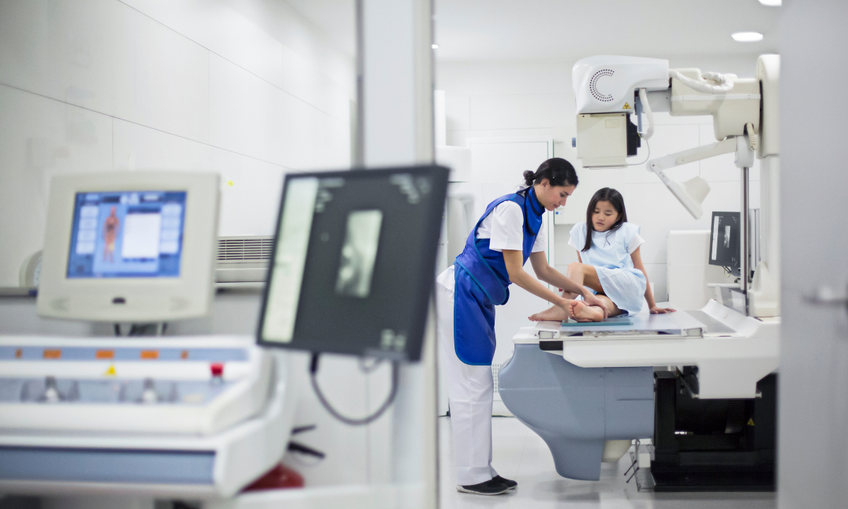
Bone and Joint X-Rays for Fractures or Pain

Bone and joint X-rays are common for injuries, fractures, or joint pain. They show any cracks or dislocations clearly.

Dental X-Ray for Tooth and Jaw Issues

Dental X-rays help your dentist check tooth decay, infections, or jaw problems before treatment.

What Happens During an X-Ray at Koshikaa?

How to Prepare Before Your Scan

You usually don’t need any special preparation. Just wear comfortable clothes and remove any jeweler or metal objects.

Quick and Painless Procedure Explained

The X-ray process is simple and painless. You will be asked to stand or lie down depending on the area to be scanned. The technician will position you correctly, and the scan takes only a few minutes.

When Will You Get Your Reports

At Koshikaa, we provide reports quickly, often on the same day, so you can consult your doctor without waiting.

Is Getting an X-Ray Safe?

Understanding Radiation and Safety

Many people worry about radiation. X-rays use very low doses, and the benefits of finding health problems early are much higher than the small risk.

Safety Measures We Follow at Koshikaa

We follow all safety guidelines to keep radiation exposure as low as possible. Our staff ensures you are safe and comfortable throughout the scan.

X-Ray Scan Cost at Koshikaa Bangalore

We offer affordable X-ray prices to make sure everyone can get the scans they need without worrying about high costs. Contact us for detailed pricing based on the scan you require.

How to Book Your X-Ray Appointment?

Walk-In Facility for Immediate Scans

You can walk into our centre anytime during working hours for your X-ray without prior booking.

Book Online for Faster Service

For quicker service, you can also book your X-ray scan online through our website.

Contact Details and Exact Location

Visit us at Koshikaa Diagnostics in Bangalore. For directions, timings, and contact numbers, check our website or call us directly.

FAQs About X-Ray Scans at Koshikaa

1. Is an X-ray scan painful?
No, it is completely painless and quick.

2. Do I need to fast before an X-ray?
Usually, no fasting is required except for certain special X-rays. Our team will guide you if needed.

3. How long does it take to get my X-ray report?
You will get your report on the same day in most cases.

4. Is an appointment needed for an X-ray?
No, you can walk in, but booking online will save you time.

Conclusion

When it comes to your health, quick and accurate diagnosis matters. Whether you’re dealing with sudden pain, an injury, or a doctor’s referral, Koshikaa is here to provide reliable, high-quality X-ray scans in Bangalore. With advanced digital machines, experienced radiologists, same-day reports, and affordable pricing, we make the entire process smooth and stress-free.

Don’t wait when your health can’t—visit Koshikaa today or book your scan online for faster service. We’re here to help you get the care you need, when you need it.

MRI of the uterus is primarily used for diagnosing and evaluating conditions such as uterine fibroids, adenomyosis, and malignancies. It is particularly useful in the planning of surgical procedures and assessing the extent of gynecological diseases. This imaging test is valuable for its ability to deliver detailed cross-sectional images of the uterus and surrounding structures.
An MRI of the spleen can help diagnose various disorders affecting the spleen including splenomegaly (enlarged spleen), tumors, and other abnormalities. It provides clear, detailed images that are crucial for assessing the health of the spleen and planning potential surgical interventions.
Prostate MRI is a non-invasive imaging technique that provides detailed pictures of the prostate gland and surrounding tissues. It helps in the detection, staging, and monitoring of prostate cancer. Prostate MRIs are increasingly used to guide biopsy procedures and assist in the planning of treatment strategies for prostate health.
An MRI of the pancreas is used to identify issues such as pancreatitis, pancreatic cancer, and other pancreatic disorders. It offers high-resolution images that help in detecting small lesions and assessing the pancreatic ducts. This imaging technique is valuable for guiding biopsies and planning surgeries involving the pancreas.
MRI scans of the liver, kidney, and bile ducts provide detailed images that help diagnose various conditions, such as tumors, cysts, and blockages in the bile ducts. They are crucial for assessing organ damage, staging cancer, and planning liver or kidney-related surgeries. This type of MRI can also evaluate the health and function of these organs comprehensively.
Breast MRI is highly sensitive for detecting breast cancer, especially in individuals with dense breast tissue or a high risk of cancer. It is often used in conjunction with mammography to provide a more comprehensive evaluation. Breast MRIs can also monitor the response to breast cancer treatments and provide detailed pre-surgical mapping.
Brain and spine MRIs are critical tools for diagnosing and monitoring neurological conditions, including tumors, spinal cord injuries, and multiple sclerosis. These MRIs can reveal abnormalities in brain tissue and spinal cord structure that might not be visible with other imaging methods. They are essential for planning surgeries and assessing the effectiveness of treatments in neurology.
PET-CT scans of transplant organs like the liver, kidney, or pancreas are performed to assess metabolic activity, detect complications, and monitor post-transplant function. These scans play a vital role in the early detection of issues such as organ rejection, recurrence of disease, or infection, guiding timely interventions and optimizing transplant outcomes for patients.
PET-CT examinations of organs such as the liver, kidney, or prostate are crucial for assessing metabolic activity, detecting abnormalities, and monitoring disease progression. These scans help diagnose conditions such as liver metastases, renal cell carcinoma, or prostate cancer, guiding treatment decisions and monitoring response to therapy over time.
PET-CT scans of soft-tissue masses are utilized to assess metabolic activity and detect malignant tumors or other abnormalities. By combining PET imaging with CT imaging, these scans provide detailed information about tumor location, size, and metabolic characteristics, aiding in accurate diagnosis and treatment planning for soft-tissue cancers.
PET-CT scans are valuable for detecting cysts or fluid collections within the body, including cysts in the liver, kidneys, or other organs. These scans help characterize the nature and metabolic activity of cysts, guiding diagnosis and treatment planning for conditions such as cystic liver disease, renal cysts, or pancreatic cysts.
PET-CT scans of muscles are performed to assess metabolic activity and detect abnormalities such as inflammation, infection, or tumors. These scans provide valuable information about muscle function and pathology, aiding in the diagnosis and management of conditions like myositis, muscular dystrophy, or soft-tissue sarcomas.
PET-CT scans of the brain are utilized to evaluate brain metabolism and detect abnormalities such as tumors, epilepsy, or neurodegenerative diseases. By combining functional PET imaging with anatomical CT imaging, these scans offer comprehensive information about brain function and structure, guiding neurosurgical interventions and treatment decisions for neurological disorders
PET-CT scans of bones are commonly used to detect bone metastases in cancer patients, assess bone health in conditions like osteoporosis, and evaluate response to treatment. By combining PET and CT imaging, these scans provide detailed information about metabolic activity and anatomical changes in bone structures, aiding in accurate diagnosis and treatment planning for various bone-related conditions.
CT scans of the spine are conducted to assess the vertebrae, spinal cord, and surrounding soft tissues for conditions like herniated discs, spinal fractures, or spinal stenosis, aiding in the diagnosis and management of spinal disorders with detailed imaging essential for surgical planning and intervention.
CT scans of the pelvis are utilized to visualize the pelvic bones, reproductive organs, bladder, and surrounding structures for conditions such as fractures, tumors, or pelvic inflammatory disease, guiding treatment plans and surgical interventions with precise anatomical information.
CT scans of the neck are performed to evaluate structures such as the throat, thyroid gland, lymph nodes, and blood vessels for abnormalities like tumors, infections, or injuries, assisting in diagnosis and management of head and neck disorders with detailed imaging.
CT scans of the extremities are employed to assess bones, joints, and soft tissues in the arms or legs, aiding in the diagnosis of fractures, arthritis, or other musculoskeletal conditions, guiding orthopedic treatment and rehabilitation plans for optimal recovery.
CT scans of the chest are utilized to examine the lungs, heart, blood vessels, and surrounding structures for conditions such as lung nodules, pneumonia, or pulmonary embolism, facilitating accurate diagnosis and management of respiratory and cardiovascular diseases.
CT scans of the brain are conducted to assess brain structures, identify abnormalities like tumors, hemorrhages, or strokes, and aid in diagnosing neurological conditions, guiding neurosurgical interventions and treatment strategies with precise imaging of the brain’s anatomy.
CT scans of the abdomen are performed to visualize and evaluate the internal organs such as the liver, kidneys, pancreas, and intestines for conditions like tumors, infections, or injuries, providing detailed images essential for diagnosis and treatment planning.
Transplant organ ultrasound scans, including those of the liver, kidney, or pancreas, are performed to assess organ viability, detect complications, and monitor post-transplant function. These scans play a vital role in the early detection of issues such as organ rejection, vascular complications, or fluid collections, guiding timely interventions and optimizing transplant outcomes for patients.
Ultrasound examinations of organs such as the liver, kidney, or prostate are crucial for assessing organ structure, detecting abnormalities, and monitoring disease progression. These scans help diagnose conditions such as liver cirrhosis, kidney stones, or prostate enlargement, guiding treatment decisions and monitoring response to therapy over time.
Soft-tissue masses can be evaluated using ultrasound to determine their size, location, and characteristics. Ultrasound imaging helps differentiate between benign and malignant masses, guiding further diagnostic evaluation and treatment planning. By providing real-time imaging, ultrasound assists in the precise localization of soft-tissue masses for biopsy or surgical removal if needed.
Ultrasound scans are valuable for detecting cysts or fluid collections within the body, including cysts in the liver, kidneys, or other organs. These scans help characterize the nature and size of cysts, aiding in diagnosis and treatment planning for conditions such as cystic liver disease or renal cysts, and guiding interventions such as cyst drainage or sclerotherapy.
Musculoskeletal ultrasound scans are used to assess the health and integrity of muscles, identifying issues such as strains, tears, or muscle inflammation. These scans provide detailed images of muscle structures, allowing healthcare providers to diagnose muscle injuries accurately and develop personalized rehabilitation plans to promote recovery and restore function.
Ultrasound examinations of joints are essential for evaluating joint health, detecting abnormalities such as inflammation, fluid accumulation, or cartilage damage. By visualizing the joint structures in real-time, ultrasound helps diagnose conditions like arthritis, bursitis, or ligament injuries, guiding treatment decisions and monitoring disease progression over time.
Ultrasound scans of tendons are performed to assess for injuries, inflammation, or tears, aiding in the diagnosis and management of conditions such as tendonitis or tendon tears. These scans provide detailed images of tendon structures, helping healthcare providers determine the extent of damage and plan appropriate treatment strategies, including physical therapy or surgical intervention if necessary.
Pelvis X-rays play a crucial role in diagnosing injuries, diseases, and developmental conditions affecting the pelvic region. They provide detailed images of the hip bones, sacrum, and coccyx, aiding in the assessment of fractures, dislocations, or signs of arthritis. Pelvic X-rays are instrumental in guiding surgical interventions, monitoring healing progress, and evaluating conditions such as osteoporosis or tumors. By providing precise anatomical information, these imaging studies enable healthcare professionals to deliver personalized care and improve patient outcomes.
Teeth X-rays, known as dental radiographs, are indispensable tools for dentists to assess oral health. They aid in diagnosing dental decay, gum disease, and abnormalities in tooth structure or alignment. These images provide valuable insights into hidden dental issues, allowing for early intervention and prevention of complications. Additionally, dental X-rays are essential for planning treatments such as orthodontics, root canals, or dental implants, ensuring optimal outcomes and patient satisfaction.
Spine X-rays are pivotal in diagnosing spinal fractures, degenerative conditions, and spinal misalignments, providing critical information for treatment planning and monitoring spinal health. They offer detailed images of the vertebrae and surrounding structures, enabling healthcare professionals to identify issues such as scoliosis, disc herniation, or spinal stenosis
Skull X-rays are valuable for detecting fractures, assessing head trauma, and evaluating the alignment of facial bones, crucial in diagnosing conditions like fractures, tumors, or developmental abnormalities in the skull. They provide detailed views of the cranial structure, aiding clinicians in determining appropriate treatment strategies and ensuring patient safety.
For the heart, X-ray solutions can aid in the detection of various cardiac problems, ranging from mild to severe, including – Heart failure, Cardiac congestion and other heart-related conditions. In addition to cardiac assessments, X-ray solutions are also used for breast examinations to identify tumors or signs of breast cancer.

Bone X-rays are essential for diagnosing fractures and assessing bone alignment, aiding doctors in developing treatment plans and monitoring healing progress. They provide detailed images of the skeletal system, helping identify abnormalities or injuries that may not be visible through other imaging techniques

Abdominal X-rays are performed when patients experience symptoms such as significant nausea, ongoing vomiting, abdominal pain, flank or back pain, or abdominal swelling. These X-rays can help diagnose various conditions.

Frequently Booked Tests

Frequently Booked Checkups