Kidney Function Test – Key Markers and What They Reveal About Renal Health

Table of Contents

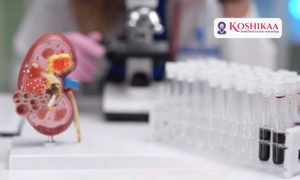
Understanding your renal health begins with a comprehensive kidney function test that measures how efficiently your kidneys filter waste, balance fluids, and regulate essential minerals in your body. 

These vital organs work silently every day, processing about 200 litres of blood and producing 1-2 litres of urine to eliminate toxins. Proactive health monitoring empowers you to catch potential kidney issues before they become serious concerns. 

Whether you’re managing existing conditions or simply prioritizing preventive care, getting a blood test in Bengaluru at a reliable facility helps you understand what’s happening inside your body and gives you the knowledge to make informed decisions about your health.

What Is Included in a Kidney Function Test

A kidney function test is actually a panel of several blood and urine tests that work together to paint a complete picture of your renal health. These tests measure different substances that indicate how well your kidneys are performing their filtering duties.

The most common kidney function blood work test includes measurements of creatinine, blood urea nitrogen (BUN), and estimated glomerular filtration rate (eGFR). Electrolyte levels, including sodium, potassium, and chloride, are also assessed. Urine tests examine protein levels and albumin.

Key Kidney Function Test Parameters Explained

Understanding kidney function tests explained helps you interpret your results with confidence. Each parameter provides unique insights into different aspects of renal health.

Serum creatinine measures a waste product from muscle metabolism that healthy kidneys filter out efficiently. Elevated levels suggest your kidneys aren’t removing waste properly.

Blood Urea Nitrogen (BUN) indicates how much urea nitrogen is in your blood. High BUN levels can signal kidney problems or dehydration.

eGFR (Estimated Glomerular Filtration Rate) is perhaps the most important marker. It estimates how much blood passes through the glomeruli (tiny filters in your kidneys) each minute.

The albumin-to-creatinine ratio (ACR) detects small amounts of albumin in urine. Even slight albumin leakage can indicate early kidney disease, especially in people with diabetes.

Kidney Function Test Normal Value Ranges

Knowing the normal ranges helps you understand where your results fall. Here’s a comprehensive table of kidney function test normal value ranges:

ParameterNormal RangeWhat It Measures
Serum Creatinine (Male)0.7 – 1.3 mg/dLWaste removal efficiency
Serum Creatinine (Female)0.6 – 1.1 mg/dLWaste removal efficiency
Blood Urea Nitrogen7 – 20 mg/dLProtein breakdown waste
eGFRAbove 60 mL/min/1.73 m²Overall kidney function
Albumin (Urine)Less than 30 mg/24 hoursProtein leakage
Sodium136 – 145 mEq/LElectrolyte balance
Potassium3.5 – 5.0 mEq/LElectrolyte balance

Remember that these ranges can vary slightly depending on the laboratory and testing methods used. Your doctor will interpret results based on your individual health profile, age, and medical history.

The Kidney Function Test Procedure

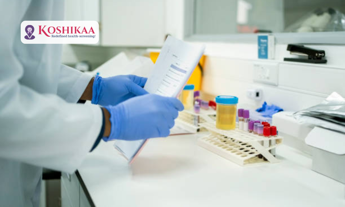
Getting tested is straightforward and minimally invasive. Most kidney function tests require a simple blood draw that takes just a few minutes.

You may be asked to fast for 8-12 hours before the test. A healthcare professional will collect blood from a vein in your arm, and the sample is then sent to a laboratory for analysis.

For those seeking convenience, scheduling a blood test in Bengaluru through a reputable diagnostic centre ensures quick, accurate results. Results typically arrive within 24-48 hours, and your doctor will review them and discuss what they mean for your health.

Understanding Kidney Function Test Results

Interpreting kidney function test results requires looking at the complete picture rather than individual numbers in isolation. We help patients understand how different markers relate to each other.

High creatinine with low eGFR strongly suggests reduced kidney function. The kidneys aren’t filtering waste as efficiently as they should be.

Elevated BUN with normal creatinine might indicate dehydration or high protein intake rather than kidney disease.

Low eGFR is categorised into stages from 1 (mild damage) to 5 (kidney failure). Early detection allows for interventions that can slow progression.

Protein in urine (proteinuria), even with normal blood tests, indicates kidney damage. This often appears before blood markers become abnormal.

Diseases and Conditions Identified Through Kidney Function Tests

These tests are invaluable for detecting various kidney-related conditions at early, treatable stages. Early diagnosis significantly improves outcomes and quality of life.

  1. Chronic Kidney Disease (CKD) is identified through persistently reduced eGFR and elevated creatinine. This progressive condition requires ongoing management to prevent kidney failure.
  2. Acute kidney injury shows sudden spikes in creatinine and BUN levels. Quick intervention can often reverse this temporary kidney damage.
  3. Diabetic nephropathy appears as protein in urine along with declining kidney function. Diabetes is a leading cause of kidney disease worldwide.
  4. Hypertensive nephrosclerosis results from long-term high blood pressure damaging the kidney blood vessels. Regular testing helps catch this before irreversible damage occurs.
  5. Glomerulonephritis involves inflammation of the kidney’s filtering units, while polycystic kidney disease shows declining function over time.

When Should You Get Tested?

Regular kidney function testing is crucial for certain groups. We recommend testing if you have diabetes, high blood pressure, heart disease, or a family history of kidney problems.

People over 60 should get tested annually, while those taking medications that affect the kidneys should have regular monitoring. Symptoms like fatigue, swelling, changes in urination, or unexplained weight changes warrant immediate testing.

Many people opt for a full body checkup in Bangalore that includes kidney function tests along with other health markers. This comprehensive approach provides a complete health snapshot.

Price of a Kidney Function Test

Understanding the cost helps you plan for regular health monitoring. The price of a kidney function test varies based on which specific tests are included in the panel.

Basic kidney function panels typically range from ₹300 to ₹800 in most diagnostic centres. 

Many health screening centre in Bangalore offers package deals where kidney tests are bundled with other important health checks. Investing in regular testing is far more economical than treating advanced kidney disease.

Tips for Maintaining Healthy Kidneys

  • Stay well-hydrated by drinking 8-10 glasses of water daily. Proper hydration helps your kidneys flush out toxins efficiently.
  • Control blood sugar if you have diabetes. High blood sugar is extremely damaging to the kidney blood vessels over time.
  • Manage blood pressure through diet, exercise, and medication if needed. Hypertension is a major cause of kidney damage.
  • Limit salt intake to reduce strain on your kidneys. Excessive sodium makes the kidneys work harder to maintain fluid balance.
  • Exercise regularly to maintain a healthy weight and circulation. Physical activity supports overall kidney health.
  • Avoid overuse of painkillers, especially NSAIDs like ibuprofen. These medications can damage the kidneys when used excessively.
  • Don’t smoke, as it reduces blood flow to the kidneys and accelerates kidney disease progression in those already affected.

Final Thoughts

Your kidneys perform hundreds of vital functions every day, and monitoring their health through regular testing is one of the most important steps you can take for long-term wellness. Understanding what kidney function tests reveal empowers you to take proactive control of your renal health.

Whether you are seeking routine screening or following up on symptoms, getting a blood test in Bangalore at a trusted diagnostic facility ensures accurate results and professional guidance. Early detection of kidney issues dramatically improves treatment outcomes and prevents progression to serious disease.

At Koshikaa, we are committed to providing comprehensive diagnostic services that help you understand and protect your health. Your kidneys deserve the attention and care that comes from regular monitoring. Schedule your kidney function test today and take an important step toward lifelong wellness.

Frequently Asked Questions

1. What does a kidney function test tell you?

A kidney function test reveals how efficiently your kidneys filter waste, balance electrolytes, and remove toxins from your blood. It measures creatinine, BUN, eGFR, and other markers to detect kidney disease, damage, or declining function early.

2. What do creatinine levels mean in pregnancy?

During pregnancy, creatinine levels typically decrease due to increased blood volume and enhanced kidney filtration. The normal range is 0.4-0.9 mg/dL. Elevated levels may indicate kidney problems, preeclampsia, or dehydration requiring immediate medical attention.

3. Which test indicates a kidney problem?

Tests indicating kidney problems include elevated serum creatinine, high BUN levels, low eGFR (below 60), protein in urine (proteinuria), abnormal albumin-to-creatinine ratio, and electrolyte imbalances. These markers reveal reduced kidney function or damage.

4. How do you tell if your kidneys are healthy?

Healthy kidneys show normal creatinine levels, eGFR above 60, no protein in urine, balanced electrolytes, and normal blood pressure. Regular kidney function tests, proper hydration, stable weight, and normal urination patterns indicate good kidney health.

5. How often should I get kidney function tests?

For healthy adults with no risk factors, every 1-2 years is sufficient. Those with diabetes, hypertension, or existing kidney issues should test every 3-6 months or as their doctor recommends.

MRI of the uterus is primarily used for diagnosing and evaluating conditions such as uterine fibroids, adenomyosis, and malignancies. It is particularly useful in the planning of surgical procedures and assessing the extent of gynecological diseases. This imaging test is valuable for its ability to deliver detailed cross-sectional images of the uterus and surrounding structures.
An MRI of the spleen can help diagnose various disorders affecting the spleen including splenomegaly (enlarged spleen), tumors, and other abnormalities. It provides clear, detailed images that are crucial for assessing the health of the spleen and planning potential surgical interventions.
Prostate MRI is a non-invasive imaging technique that provides detailed pictures of the prostate gland and surrounding tissues. It helps in the detection, staging, and monitoring of prostate cancer. Prostate MRIs are increasingly used to guide biopsy procedures and assist in the planning of treatment strategies for prostate health.
An MRI of the pancreas is used to identify issues such as pancreatitis, pancreatic cancer, and other pancreatic disorders. It offers high-resolution images that help in detecting small lesions and assessing the pancreatic ducts. This imaging technique is valuable for guiding biopsies and planning surgeries involving the pancreas.
MRI scans of the liver, kidney, and bile ducts provide detailed images that help diagnose various conditions, such as tumors, cysts, and blockages in the bile ducts. They are crucial for assessing organ damage, staging cancer, and planning liver or kidney-related surgeries. This type of MRI can also evaluate the health and function of these organs comprehensively.
Breast MRI is highly sensitive for detecting breast cancer, especially in individuals with dense breast tissue or a high risk of cancer. It is often used in conjunction with mammography to provide a more comprehensive evaluation. Breast MRIs can also monitor the response to breast cancer treatments and provide detailed pre-surgical mapping.
Brain and spine MRIs are critical tools for diagnosing and monitoring neurological conditions, including tumors, spinal cord injuries, and multiple sclerosis. These MRIs can reveal abnormalities in brain tissue and spinal cord structure that might not be visible with other imaging methods. They are essential for planning surgeries and assessing the effectiveness of treatments in neurology.
PET-CT scans of transplant organs like the liver, kidney, or pancreas are performed to assess metabolic activity, detect complications, and monitor post-transplant function. These scans play a vital role in the early detection of issues such as organ rejection, recurrence of disease, or infection, guiding timely interventions and optimizing transplant outcomes for patients.
PET-CT examinations of organs such as the liver, kidney, or prostate are crucial for assessing metabolic activity, detecting abnormalities, and monitoring disease progression. These scans help diagnose conditions such as liver metastases, renal cell carcinoma, or prostate cancer, guiding treatment decisions and monitoring response to therapy over time.
PET-CT scans of soft-tissue masses are utilized to assess metabolic activity and detect malignant tumors or other abnormalities. By combining PET imaging with CT imaging, these scans provide detailed information about tumor location, size, and metabolic characteristics, aiding in accurate diagnosis and treatment planning for soft-tissue cancers.
PET-CT scans are valuable for detecting cysts or fluid collections within the body, including cysts in the liver, kidneys, or other organs. These scans help characterize the nature and metabolic activity of cysts, guiding diagnosis and treatment planning for conditions such as cystic liver disease, renal cysts, or pancreatic cysts.
PET-CT scans of muscles are performed to assess metabolic activity and detect abnormalities such as inflammation, infection, or tumors. These scans provide valuable information about muscle function and pathology, aiding in the diagnosis and management of conditions like myositis, muscular dystrophy, or soft-tissue sarcomas.
PET-CT scans of the brain are utilized to evaluate brain metabolism and detect abnormalities such as tumors, epilepsy, or neurodegenerative diseases. By combining functional PET imaging with anatomical CT imaging, these scans offer comprehensive information about brain function and structure, guiding neurosurgical interventions and treatment decisions for neurological disorders
PET-CT scans of bones are commonly used to detect bone metastases in cancer patients, assess bone health in conditions like osteoporosis, and evaluate response to treatment. By combining PET and CT imaging, these scans provide detailed information about metabolic activity and anatomical changes in bone structures, aiding in accurate diagnosis and treatment planning for various bone-related conditions.
CT scans of the spine are conducted to assess the vertebrae, spinal cord, and surrounding soft tissues for conditions like herniated discs, spinal fractures, or spinal stenosis, aiding in the diagnosis and management of spinal disorders with detailed imaging essential for surgical planning and intervention.
CT scans of the pelvis are utilized to visualize the pelvic bones, reproductive organs, bladder, and surrounding structures for conditions such as fractures, tumors, or pelvic inflammatory disease, guiding treatment plans and surgical interventions with precise anatomical information.
CT scans of the neck are performed to evaluate structures such as the throat, thyroid gland, lymph nodes, and blood vessels for abnormalities like tumors, infections, or injuries, assisting in diagnosis and management of head and neck disorders with detailed imaging.
CT scans of the extremities are employed to assess bones, joints, and soft tissues in the arms or legs, aiding in the diagnosis of fractures, arthritis, or other musculoskeletal conditions, guiding orthopedic treatment and rehabilitation plans for optimal recovery.
CT scans of the chest are utilized to examine the lungs, heart, blood vessels, and surrounding structures for conditions such as lung nodules, pneumonia, or pulmonary embolism, facilitating accurate diagnosis and management of respiratory and cardiovascular diseases.
CT scans of the brain are conducted to assess brain structures, identify abnormalities like tumors, hemorrhages, or strokes, and aid in diagnosing neurological conditions, guiding neurosurgical interventions and treatment strategies with precise imaging of the brain’s anatomy.
CT scans of the abdomen are performed to visualize and evaluate the internal organs such as the liver, kidneys, pancreas, and intestines for conditions like tumors, infections, or injuries, providing detailed images essential for diagnosis and treatment planning.
Transplant organ ultrasound scans, including those of the liver, kidney, or pancreas, are performed to assess organ viability, detect complications, and monitor post-transplant function. These scans play a vital role in the early detection of issues such as organ rejection, vascular complications, or fluid collections, guiding timely interventions and optimizing transplant outcomes for patients.
Ultrasound examinations of organs such as the liver, kidney, or prostate are crucial for assessing organ structure, detecting abnormalities, and monitoring disease progression. These scans help diagnose conditions such as liver cirrhosis, kidney stones, or prostate enlargement, guiding treatment decisions and monitoring response to therapy over time.
Soft-tissue masses can be evaluated using ultrasound to determine their size, location, and characteristics. Ultrasound imaging helps differentiate between benign and malignant masses, guiding further diagnostic evaluation and treatment planning. By providing real-time imaging, ultrasound assists in the precise localization of soft-tissue masses for biopsy or surgical removal if needed.
Ultrasound scans are valuable for detecting cysts or fluid collections within the body, including cysts in the liver, kidneys, or other organs. These scans help characterize the nature and size of cysts, aiding in diagnosis and treatment planning for conditions such as cystic liver disease or renal cysts, and guiding interventions such as cyst drainage or sclerotherapy.
Musculoskeletal ultrasound scans are used to assess the health and integrity of muscles, identifying issues such as strains, tears, or muscle inflammation. These scans provide detailed images of muscle structures, allowing healthcare providers to diagnose muscle injuries accurately and develop personalized rehabilitation plans to promote recovery and restore function.
Ultrasound examinations of joints are essential for evaluating joint health, detecting abnormalities such as inflammation, fluid accumulation, or cartilage damage. By visualizing the joint structures in real-time, ultrasound helps diagnose conditions like arthritis, bursitis, or ligament injuries, guiding treatment decisions and monitoring disease progression over time.
Ultrasound scans of tendons are performed to assess for injuries, inflammation, or tears, aiding in the diagnosis and management of conditions such as tendonitis or tendon tears. These scans provide detailed images of tendon structures, helping healthcare providers determine the extent of damage and plan appropriate treatment strategies, including physical therapy or surgical intervention if necessary.
Pelvis X-rays play a crucial role in diagnosing injuries, diseases, and developmental conditions affecting the pelvic region. They provide detailed images of the hip bones, sacrum, and coccyx, aiding in the assessment of fractures, dislocations, or signs of arthritis. Pelvic X-rays are instrumental in guiding surgical interventions, monitoring healing progress, and evaluating conditions such as osteoporosis or tumors. By providing precise anatomical information, these imaging studies enable healthcare professionals to deliver personalized care and improve patient outcomes.
Teeth X-rays, known as dental radiographs, are indispensable tools for dentists to assess oral health. They aid in diagnosing dental decay, gum disease, and abnormalities in tooth structure or alignment. These images provide valuable insights into hidden dental issues, allowing for early intervention and prevention of complications. Additionally, dental X-rays are essential for planning treatments such as orthodontics, root canals, or dental implants, ensuring optimal outcomes and patient satisfaction.
Spine X-rays are pivotal in diagnosing spinal fractures, degenerative conditions, and spinal misalignments, providing critical information for treatment planning and monitoring spinal health. They offer detailed images of the vertebrae and surrounding structures, enabling healthcare professionals to identify issues such as scoliosis, disc herniation, or spinal stenosis
Skull X-rays are valuable for detecting fractures, assessing head trauma, and evaluating the alignment of facial bones, crucial in diagnosing conditions like fractures, tumors, or developmental abnormalities in the skull. They provide detailed views of the cranial structure, aiding clinicians in determining appropriate treatment strategies and ensuring patient safety.
For the heart, X-ray solutions can aid in the detection of various cardiac problems, ranging from mild to severe, including – Heart failure, Cardiac congestion and other heart-related conditions. In addition to cardiac assessments, X-ray solutions are also used for breast examinations to identify tumors or signs of breast cancer.

Bone X-rays are essential for diagnosing fractures and assessing bone alignment, aiding doctors in developing treatment plans and monitoring healing progress. They provide detailed images of the skeletal system, helping identify abnormalities or injuries that may not be visible through other imaging techniques

Abdominal X-rays are performed when patients experience symptoms such as significant nausea, ongoing vomiting, abdominal pain, flank or back pain, or abdominal swelling. These X-rays can help diagnose various conditions.

Frequently Booked Tests

Frequently Booked Checkups