How Ultrasound Helps in Diagnosing Liver, Kidney & Gallbladder Issues

CT scan

Table of Contents

Early diagnosis in modern society translates to a higher likelihood of successful treatment and recovery. An ultrasound scan is one of the best and non-invasive methods for identifying organ problems, particularly in the abdomen. The place of ultrasound in examining the liver, kidneys, and gallbladder is as significant as where people have heard about it, due to other reasons, like being pregnant or the heart. This article examines the process of applying abdominal ultrasound as a method to diagnose the most common problems with the given organs, its significance, and the way patients experience its use.

Understanding Ultrasound Scan: A Short Synopsis

Ultrasound scan

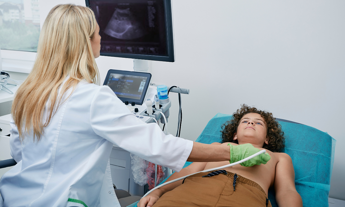
Ultrasound scan is a test whereby high-frequency sound waves are used to produce images of body organs. Compared with X-rays, they are radiation-free; thus, ultrasounds pose no harm even to individuals of any age. In the case of stomach or abdominal pain or other symptoms such as jaundice or swelling, the doctors may ask or prescribe to undergo an ultrasound scan of the stomach or a wider ultrasound scan of the abdomen and pelvis. Such procedures are used in determining the cause of symptoms without pain at all or taking days to recover.

What Makes Abdominal Ultrasound So Useful?

Abdominal ultrasound is an important diagnostic instrument. It can display the size, shape, and texture of organs, spot masses or cysts, show stones, and display blockage to blood flow. Booking an ultrasound scan in Bangalore is easy and trustworthy in towns such as Bangalore that have access to high-quality health technology.

An Investigation into the Liver: Why Ultrasound is Important to the Liver?

The liver is an important organ that filters toxins from the blood, stores nutrients, and aids in digestion. Such issues as fatty liver, hepatitis, cirrhosis, or a tumor may grow unnoticeably and develop rapidly. This is when a Liver ultrasound comes in handy. Abnormal enlargements, fatty changes, lesions, or blood vessel blockage can be found in the scan.

A Liver ultrasound is usually advised by the doctors when there are abnormalities in the blood test, which indicates a disrupted liver, or when the patient experiences certain symptoms such as constant ache on the right side of the abdomen, yellowing of the skin (jaundice), or unexplained weight loss. Since the ultrasound imaging is a real-time procedure, it is possible to see changes as the patient is undergoing the examination.

Diagnosis of Kidney Problems using Ultrasound

Kidneys maintain our body without waste and maintain our fluids. Kidney stones, cysts, infections, and even tumors are common. Symptoms like pain on your back or side, the presence of blood in urine, or frequent urinary infections may send the doctor to conduct an abdominal and pelvic ultrasound or a stomach-specific ultrasound.

The size, shape of the kidneys and their location is evident in the scan. It is particularly helpful as it may detect swelling and obstruction, or even allow tracing the progression of chronic kidney disease. In a city where you get advanced radiology, more so the ultrasound scan in Bangalore, you will find modern equipment that will detect even microscopic kidney stones.

Gallbladder: Stones and Inflammation Made Easy

The problems associated with the gall bladder are stones and infection. The bile is stored in the gallbladder and aids in fat digestion. The gallbladder may, however, become stone-like, resulting in pain when fatty foods are consumed. Obstruction of the duct by these stones may result in acute cramping and inflammation, and even jaundice. The initial test that the physicians order to inquire about the existence of gallstones or an inflamed gallbladder (cholecystitis) is an abdominal ultrasound.

In the course of ultrasound examination, the physician will be able to tell whether there are stones and their size, and the presence of swelling or infection. An early diagnosis provides time that can be used to provide the treatment earlier, before developing complications.

What to Expect: The Perspective of the Patient

It is easy, and many individuals are always scared of the first scan. During a stomach (and other conditions) ultrasound scan, you may be requested not to eat or drink for many hours to obtain the best photos. Gel is put on your belly by the technician, and a small device known as a transducer is moved about by the technician. It is pain-free, fast, and you can go home immediately.

In cities, there is no problem in getting an ultrasound scan in Bangalore with less waiting time. The reports can be prepared on the same day or the next in order to make immediate medical decisions.

Comparison of the other Diagnostic Methods to Ultrasound

Although other tests, such as blood tests and CT scans, are applicable, an ultrasound scan for the abdomen and pelvis is non-invasive, safe, cost-effective, and has no exposure to radiation. It may be used as a first step, and when problems are identified, it can be followed by more detailed imaging in case it is necessary.

The Importance of Timely Diagnosis

Psychological stubbornness to such symptoms as abdominal pains, inexplicable vomiting, yellowish color of the skin, or alterations in urine may postpone the required care. A liver ultrasound or a kidney ultrasound, or a gallbladder ultrasound may provide a rapid diagnosis of your state. The scan is so good that it finds the problems early, and it occasionally prevents a life.

People who have risk factors, such as diabetes, a family history of liver disease, or any previous problems with the kidneys or the gallbladder, are also advised to have regular check-ups using an ultrasound scan.

Conclusion: The Adoption of Safe and Effective Diagnosis

In conclusion, there are always lots of methods that can help us detect and cure our health issues at the beginning stages in the modern world of medicine, and an ultrasound examination of the stomach is one of the least demanding but most efficient. If the Liver ultrasound is required or a full ultrasound scan for abdomen and pelvis because there is a problem with the kidney or the gallbladder, this is the technology that can offer answers with little disruption. In the case of your suspicion of any problems, do not go and take an ultrasound scan in Bangalore because it is not worth your health.

Key Points Summary

  • Ultrasound scan is pain-free, non-invasive, and safe.
  • Abdominal ultrasound is effective in the detection of issues in the liver, kidney, and gallbladder.
  • The sooner the detection is made, the better the outcomes in treatment.
  • The process is very ubiquitous and fast.
  • Should you exhibit symptoms or risk factors, consult with your doctor in relation to having a scan conducted, be it a Liver ultrasound or an abdomen and pelvis ultrasound scan.

A proactive step, however, towards your health can be the difference. 

Koshikaa provides high-tech ultrasound scans in Bangalore with skilled people. We achieve proper diagnostics in a comfortable setting. Make your appointment now to have a healthier future! Call us today!

MRI of the uterus is primarily used for diagnosing and evaluating conditions such as uterine fibroids, adenomyosis, and malignancies. It is particularly useful in the planning of surgical procedures and assessing the extent of gynecological diseases. This imaging test is valuable for its ability to deliver detailed cross-sectional images of the uterus and surrounding structures.
An MRI of the spleen can help diagnose various disorders affecting the spleen including splenomegaly (enlarged spleen), tumors, and other abnormalities. It provides clear, detailed images that are crucial for assessing the health of the spleen and planning potential surgical interventions.
Prostate MRI is a non-invasive imaging technique that provides detailed pictures of the prostate gland and surrounding tissues. It helps in the detection, staging, and monitoring of prostate cancer. Prostate MRIs are increasingly used to guide biopsy procedures and assist in the planning of treatment strategies for prostate health.
An MRI of the pancreas is used to identify issues such as pancreatitis, pancreatic cancer, and other pancreatic disorders. It offers high-resolution images that help in detecting small lesions and assessing the pancreatic ducts. This imaging technique is valuable for guiding biopsies and planning surgeries involving the pancreas.
MRI scans of the liver, kidney, and bile ducts provide detailed images that help diagnose various conditions, such as tumors, cysts, and blockages in the bile ducts. They are crucial for assessing organ damage, staging cancer, and planning liver or kidney-related surgeries. This type of MRI can also evaluate the health and function of these organs comprehensively.
Breast MRI is highly sensitive for detecting breast cancer, especially in individuals with dense breast tissue or a high risk of cancer. It is often used in conjunction with mammography to provide a more comprehensive evaluation. Breast MRIs can also monitor the response to breast cancer treatments and provide detailed pre-surgical mapping.
Brain and spine MRIs are critical tools for diagnosing and monitoring neurological conditions, including tumors, spinal cord injuries, and multiple sclerosis. These MRIs can reveal abnormalities in brain tissue and spinal cord structure that might not be visible with other imaging methods. They are essential for planning surgeries and assessing the effectiveness of treatments in neurology.
PET-CT scans of transplant organs like the liver, kidney, or pancreas are performed to assess metabolic activity, detect complications, and monitor post-transplant function. These scans play a vital role in the early detection of issues such as organ rejection, recurrence of disease, or infection, guiding timely interventions and optimizing transplant outcomes for patients.
PET-CT examinations of organs such as the liver, kidney, or prostate are crucial for assessing metabolic activity, detecting abnormalities, and monitoring disease progression. These scans help diagnose conditions such as liver metastases, renal cell carcinoma, or prostate cancer, guiding treatment decisions and monitoring response to therapy over time.
PET-CT scans of soft-tissue masses are utilized to assess metabolic activity and detect malignant tumors or other abnormalities. By combining PET imaging with CT imaging, these scans provide detailed information about tumor location, size, and metabolic characteristics, aiding in accurate diagnosis and treatment planning for soft-tissue cancers.
PET-CT scans are valuable for detecting cysts or fluid collections within the body, including cysts in the liver, kidneys, or other organs. These scans help characterize the nature and metabolic activity of cysts, guiding diagnosis and treatment planning for conditions such as cystic liver disease, renal cysts, or pancreatic cysts.
PET-CT scans of muscles are performed to assess metabolic activity and detect abnormalities such as inflammation, infection, or tumors. These scans provide valuable information about muscle function and pathology, aiding in the diagnosis and management of conditions like myositis, muscular dystrophy, or soft-tissue sarcomas.
PET-CT scans of the brain are utilized to evaluate brain metabolism and detect abnormalities such as tumors, epilepsy, or neurodegenerative diseases. By combining functional PET imaging with anatomical CT imaging, these scans offer comprehensive information about brain function and structure, guiding neurosurgical interventions and treatment decisions for neurological disorders
PET-CT scans of bones are commonly used to detect bone metastases in cancer patients, assess bone health in conditions like osteoporosis, and evaluate response to treatment. By combining PET and CT imaging, these scans provide detailed information about metabolic activity and anatomical changes in bone structures, aiding in accurate diagnosis and treatment planning for various bone-related conditions.
CT scans of the spine are conducted to assess the vertebrae, spinal cord, and surrounding soft tissues for conditions like herniated discs, spinal fractures, or spinal stenosis, aiding in the diagnosis and management of spinal disorders with detailed imaging essential for surgical planning and intervention.
CT scans of the pelvis are utilized to visualize the pelvic bones, reproductive organs, bladder, and surrounding structures for conditions such as fractures, tumors, or pelvic inflammatory disease, guiding treatment plans and surgical interventions with precise anatomical information.
CT scans of the neck are performed to evaluate structures such as the throat, thyroid gland, lymph nodes, and blood vessels for abnormalities like tumors, infections, or injuries, assisting in diagnosis and management of head and neck disorders with detailed imaging.
CT scans of the extremities are employed to assess bones, joints, and soft tissues in the arms or legs, aiding in the diagnosis of fractures, arthritis, or other musculoskeletal conditions, guiding orthopedic treatment and rehabilitation plans for optimal recovery.
CT scans of the chest are utilized to examine the lungs, heart, blood vessels, and surrounding structures for conditions such as lung nodules, pneumonia, or pulmonary embolism, facilitating accurate diagnosis and management of respiratory and cardiovascular diseases.
CT scans of the brain are conducted to assess brain structures, identify abnormalities like tumors, hemorrhages, or strokes, and aid in diagnosing neurological conditions, guiding neurosurgical interventions and treatment strategies with precise imaging of the brain’s anatomy.
CT scans of the abdomen are performed to visualize and evaluate the internal organs such as the liver, kidneys, pancreas, and intestines for conditions like tumors, infections, or injuries, providing detailed images essential for diagnosis and treatment planning.
Transplant organ ultrasound scans, including those of the liver, kidney, or pancreas, are performed to assess organ viability, detect complications, and monitor post-transplant function. These scans play a vital role in the early detection of issues such as organ rejection, vascular complications, or fluid collections, guiding timely interventions and optimizing transplant outcomes for patients.
Ultrasound examinations of organs such as the liver, kidney, or prostate are crucial for assessing organ structure, detecting abnormalities, and monitoring disease progression. These scans help diagnose conditions such as liver cirrhosis, kidney stones, or prostate enlargement, guiding treatment decisions and monitoring response to therapy over time.
Soft-tissue masses can be evaluated using ultrasound to determine their size, location, and characteristics. Ultrasound imaging helps differentiate between benign and malignant masses, guiding further diagnostic evaluation and treatment planning. By providing real-time imaging, ultrasound assists in the precise localization of soft-tissue masses for biopsy or surgical removal if needed.
Ultrasound scans are valuable for detecting cysts or fluid collections within the body, including cysts in the liver, kidneys, or other organs. These scans help characterize the nature and size of cysts, aiding in diagnosis and treatment planning for conditions such as cystic liver disease or renal cysts, and guiding interventions such as cyst drainage or sclerotherapy.
Musculoskeletal ultrasound scans are used to assess the health and integrity of muscles, identifying issues such as strains, tears, or muscle inflammation. These scans provide detailed images of muscle structures, allowing healthcare providers to diagnose muscle injuries accurately and develop personalized rehabilitation plans to promote recovery and restore function.
Ultrasound examinations of joints are essential for evaluating joint health, detecting abnormalities such as inflammation, fluid accumulation, or cartilage damage. By visualizing the joint structures in real-time, ultrasound helps diagnose conditions like arthritis, bursitis, or ligament injuries, guiding treatment decisions and monitoring disease progression over time.
Ultrasound scans of tendons are performed to assess for injuries, inflammation, or tears, aiding in the diagnosis and management of conditions such as tendonitis or tendon tears. These scans provide detailed images of tendon structures, helping healthcare providers determine the extent of damage and plan appropriate treatment strategies, including physical therapy or surgical intervention if necessary.
Pelvis X-rays play a crucial role in diagnosing injuries, diseases, and developmental conditions affecting the pelvic region. They provide detailed images of the hip bones, sacrum, and coccyx, aiding in the assessment of fractures, dislocations, or signs of arthritis. Pelvic X-rays are instrumental in guiding surgical interventions, monitoring healing progress, and evaluating conditions such as osteoporosis or tumors. By providing precise anatomical information, these imaging studies enable healthcare professionals to deliver personalized care and improve patient outcomes.
Teeth X-rays, known as dental radiographs, are indispensable tools for dentists to assess oral health. They aid in diagnosing dental decay, gum disease, and abnormalities in tooth structure or alignment. These images provide valuable insights into hidden dental issues, allowing for early intervention and prevention of complications. Additionally, dental X-rays are essential for planning treatments such as orthodontics, root canals, or dental implants, ensuring optimal outcomes and patient satisfaction.
Spine X-rays are pivotal in diagnosing spinal fractures, degenerative conditions, and spinal misalignments, providing critical information for treatment planning and monitoring spinal health. They offer detailed images of the vertebrae and surrounding structures, enabling healthcare professionals to identify issues such as scoliosis, disc herniation, or spinal stenosis
Skull X-rays are valuable for detecting fractures, assessing head trauma, and evaluating the alignment of facial bones, crucial in diagnosing conditions like fractures, tumors, or developmental abnormalities in the skull. They provide detailed views of the cranial structure, aiding clinicians in determining appropriate treatment strategies and ensuring patient safety.
For the heart, X-ray solutions can aid in the detection of various cardiac problems, ranging from mild to severe, including – Heart failure, Cardiac congestion and other heart-related conditions. In addition to cardiac assessments, X-ray solutions are also used for breast examinations to identify tumors or signs of breast cancer.

Bone X-rays are essential for diagnosing fractures and assessing bone alignment, aiding doctors in developing treatment plans and monitoring healing progress. They provide detailed images of the skeletal system, helping identify abnormalities or injuries that may not be visible through other imaging techniques

Abdominal X-rays are performed when patients experience symptoms such as significant nausea, ongoing vomiting, abdominal pain, flank or back pain, or abdominal swelling. These X-rays can help diagnose various conditions.

Frequently Booked Tests

Frequently Booked Checkups