How Koshikaa’s 3D Mammography Help Women Get Clear Results

PET CT Scan in Bangalore

Table of Contents

Introduction

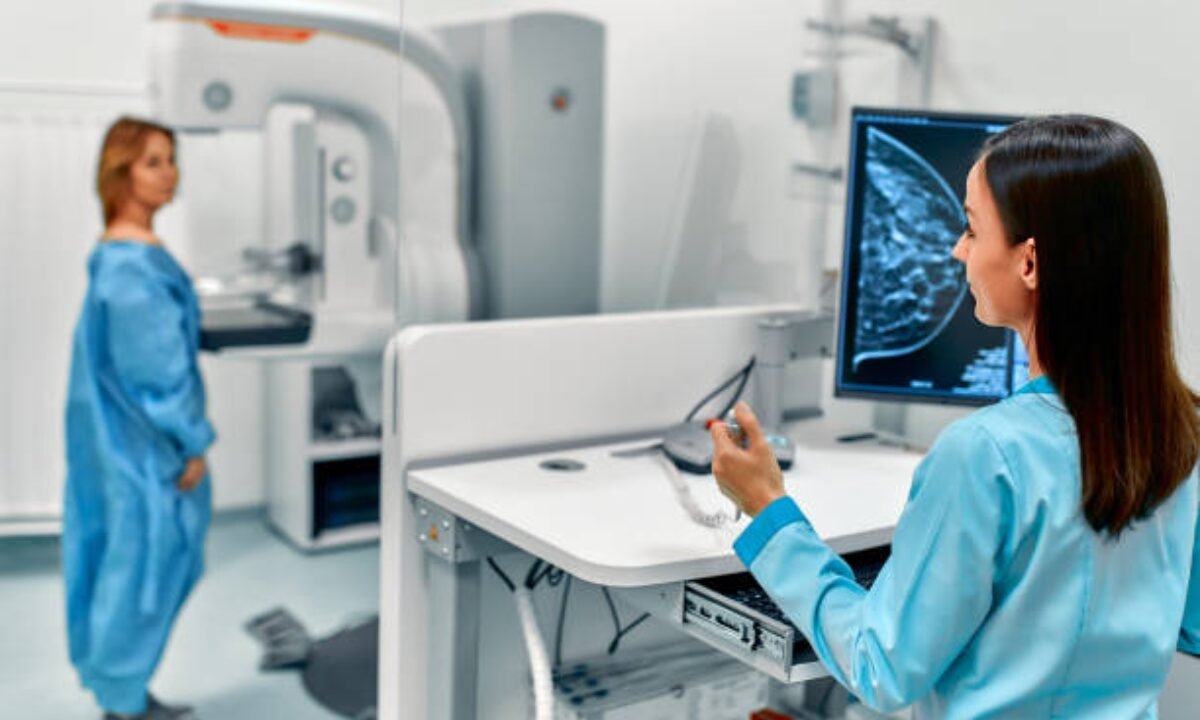
Getting a mammography can be stressful. And what’s worse than the stress? Getting a false positive result—when the test shows something suspicious, but it turns out to be nothing. For women in Bangalore, Koshikaa is changing that story.

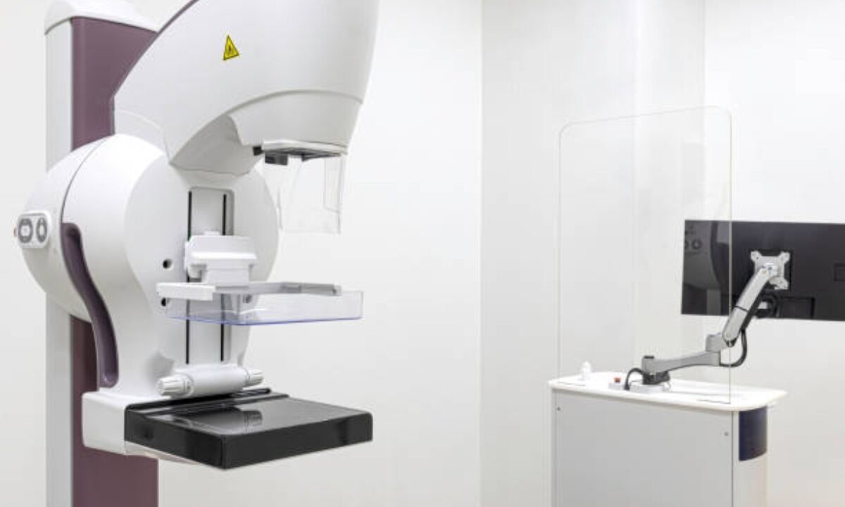
With advanced Digital Mammography with Tomosynthesis, Koshikaa is reducing the number of unnecessary scares and biopsies. If you’ve ever worried about mammogram results or felt confused by medical jargon, this blog is here to simplify it. Read on to discover how this cutting-edge technology brings peace of mind and accuracy to breast health.

What Is Digital Mammography with Tomosynthesis?

Mammography in Bangalore

Let’s break it down.

  • Digital Mammography is a modern form of breast X-ray that captures images electronically.
  • Tomosynthesis, or 3D mammography, takes a step further by capturing multiple images from different angles and creating a layered, 3D picture of the breast.

Together, they create a clear, precise image that helps doctors detect even the smallest abnormalities, without jumping to conclusions.

How Does It Reduce False Positives?

Here’s the big question. Let’s answer it with clarity and compassion.

1. Eliminates Overlapping Tissue Confusion

In traditional 2D mammography, overlapping breast tissues can look suspicious. But with tomosynthesis, doctors can look at thin “slices” of the breast, removing the confusion caused by overlapping tissue. This means:

  • Fewer callbacks
  • Fewer unnecessary biopsies
  • More accurate readings

2. Greater Visibility for Dense Breasts

Did you know that over 40% of Indian women have dense breast tissue? This makes detecting tumors harder with standard mammograms. Digital mammography tests improves visibility, especially in dense tissue, allowing doctors to spot real concerns while ignoring false alarms.

3. Proven Results from Research

A major study published in the Journal of the American Medical Association (JAMA) found that tomosynthesis reduces false positives by up to 40%. That’s a big win for emotional and financial peace of mind.

4. Better for Follow-ups and Early Detection

False positives often lead to extra testing, anxiety, and costs. At Koshikaa, tomosynthesis is used in both routine screenings and follow-ups. This ensures:

  • Faster clarity on previous findings
  • Earlier detection of real issues
  • Peace of mind for women undergoing checkups

Why Choose Koshikaa for Mammography in Bangalore?

When it comes to Mammography in Bangalore, Koshikaa stands out for several reasons:

1. State-of-the-Art Technology

Koshikaa uses premium-grade digital tomosynthesis systems trusted by radiologists worldwide. This isn’t just tech—it’s life-saving innovation tailored to Indian women.

2. Expert Radiologists

Their team includes highly trained breast imaging specialists who know how to interpret 3D mammograms with skill and accuracy.

3. Woman-Friendly Environment

Every woman deserves to feel comfortable during a mammogram. Koshikaa provides:

  • Female technicians
  • Private, clean exam rooms
  • Compassionate support throughout

4. Quick Reports with Clarity

You won’t be left waiting for results or lost in medical language. Reports are quick, clear, and focused on your well-being. Learn more about their services for Mammography in Bangalore.

FAQs About Digital Mammography with Tomosynthesis at Koshikaa

Is this technology safe?

Absolutely. It uses low-dose radiation and is FDA-approved for breast cancer screening. Tomosynthesis exposes you to only slightly more radiation than a regular mammogram, well within safety limits.

Is it more painful than a regular mammogram?

No. The experience feels nearly the same as a traditional mammogram. The machine might take a few more seconds for each image, but the comfort level is the same.

Will my insurance cover this?

Many insurance providers now recognize the value of 3D mammograms. Check with your provider—Koshikaa also offers affordable packages.

How often should I get screened?

For most women over 40, annual screening is recommended. However, if you have a family history or other risk factors, your doctor may suggest earlier.

Do I need a referral to get this done at Koshikaa?

No referral needed! You can book an appointment directly on their website or by calling the centre.

Real Stories. Real Impact.

1. Radhika, 46, from Indiranagar, shared:

“I had a scare last year. My earlier clinic asked me to come back for more scans. At Koshikaa, they used tomosynthesis and told me it was just overlapping tissue. I cried tears of relief. They were professional, quick, and kind.”

2. Anita, 52, from Whitefield, said:

“Dense breasts run in my family. Koshikaa’s 3D mammogram helped detect an early-stage lump that the old scans missed. Thanks to them, I got treatment early.”

Conclusion

If you’re frustrated by unclear mammogram results or false alarms, get clarity and peace of mind with Digital Mammography with Tomosynthesis at Koshikaa Screening Centre.

For Bangalore women, it’s more than just a test—it’s clarity, confidence, and control over your health.

Take the step today. Because when it comes to breast health, clarity is everything. Book your mammogram at Koshikaa today.

MRI of the uterus is primarily used for diagnosing and evaluating conditions such as uterine fibroids, adenomyosis, and malignancies. It is particularly useful in the planning of surgical procedures and assessing the extent of gynecological diseases. This imaging test is valuable for its ability to deliver detailed cross-sectional images of the uterus and surrounding structures.
An MRI of the spleen can help diagnose various disorders affecting the spleen including splenomegaly (enlarged spleen), tumors, and other abnormalities. It provides clear, detailed images that are crucial for assessing the health of the spleen and planning potential surgical interventions.
Prostate MRI is a non-invasive imaging technique that provides detailed pictures of the prostate gland and surrounding tissues. It helps in the detection, staging, and monitoring of prostate cancer. Prostate MRIs are increasingly used to guide biopsy procedures and assist in the planning of treatment strategies for prostate health.
An MRI of the pancreas is used to identify issues such as pancreatitis, pancreatic cancer, and other pancreatic disorders. It offers high-resolution images that help in detecting small lesions and assessing the pancreatic ducts. This imaging technique is valuable for guiding biopsies and planning surgeries involving the pancreas.
MRI scans of the liver, kidney, and bile ducts provide detailed images that help diagnose various conditions, such as tumors, cysts, and blockages in the bile ducts. They are crucial for assessing organ damage, staging cancer, and planning liver or kidney-related surgeries. This type of MRI can also evaluate the health and function of these organs comprehensively.
Breast MRI is highly sensitive for detecting breast cancer, especially in individuals with dense breast tissue or a high risk of cancer. It is often used in conjunction with mammography to provide a more comprehensive evaluation. Breast MRIs can also monitor the response to breast cancer treatments and provide detailed pre-surgical mapping.
Brain and spine MRIs are critical tools for diagnosing and monitoring neurological conditions, including tumors, spinal cord injuries, and multiple sclerosis. These MRIs can reveal abnormalities in brain tissue and spinal cord structure that might not be visible with other imaging methods. They are essential for planning surgeries and assessing the effectiveness of treatments in neurology.
PET-CT scans of transplant organs like the liver, kidney, or pancreas are performed to assess metabolic activity, detect complications, and monitor post-transplant function. These scans play a vital role in the early detection of issues such as organ rejection, recurrence of disease, or infection, guiding timely interventions and optimizing transplant outcomes for patients.
PET-CT examinations of organs such as the liver, kidney, or prostate are crucial for assessing metabolic activity, detecting abnormalities, and monitoring disease progression. These scans help diagnose conditions such as liver metastases, renal cell carcinoma, or prostate cancer, guiding treatment decisions and monitoring response to therapy over time.
PET-CT scans of soft-tissue masses are utilized to assess metabolic activity and detect malignant tumors or other abnormalities. By combining PET imaging with CT imaging, these scans provide detailed information about tumor location, size, and metabolic characteristics, aiding in accurate diagnosis and treatment planning for soft-tissue cancers.
PET-CT scans are valuable for detecting cysts or fluid collections within the body, including cysts in the liver, kidneys, or other organs. These scans help characterize the nature and metabolic activity of cysts, guiding diagnosis and treatment planning for conditions such as cystic liver disease, renal cysts, or pancreatic cysts.
PET-CT scans of muscles are performed to assess metabolic activity and detect abnormalities such as inflammation, infection, or tumors. These scans provide valuable information about muscle function and pathology, aiding in the diagnosis and management of conditions like myositis, muscular dystrophy, or soft-tissue sarcomas.
PET-CT scans of the brain are utilized to evaluate brain metabolism and detect abnormalities such as tumors, epilepsy, or neurodegenerative diseases. By combining functional PET imaging with anatomical CT imaging, these scans offer comprehensive information about brain function and structure, guiding neurosurgical interventions and treatment decisions for neurological disorders
PET-CT scans of bones are commonly used to detect bone metastases in cancer patients, assess bone health in conditions like osteoporosis, and evaluate response to treatment. By combining PET and CT imaging, these scans provide detailed information about metabolic activity and anatomical changes in bone structures, aiding in accurate diagnosis and treatment planning for various bone-related conditions.
CT scans of the spine are conducted to assess the vertebrae, spinal cord, and surrounding soft tissues for conditions like herniated discs, spinal fractures, or spinal stenosis, aiding in the diagnosis and management of spinal disorders with detailed imaging essential for surgical planning and intervention.
CT scans of the pelvis are utilized to visualize the pelvic bones, reproductive organs, bladder, and surrounding structures for conditions such as fractures, tumors, or pelvic inflammatory disease, guiding treatment plans and surgical interventions with precise anatomical information.
CT scans of the neck are performed to evaluate structures such as the throat, thyroid gland, lymph nodes, and blood vessels for abnormalities like tumors, infections, or injuries, assisting in diagnosis and management of head and neck disorders with detailed imaging.
CT scans of the extremities are employed to assess bones, joints, and soft tissues in the arms or legs, aiding in the diagnosis of fractures, arthritis, or other musculoskeletal conditions, guiding orthopedic treatment and rehabilitation plans for optimal recovery.
CT scans of the chest are utilized to examine the lungs, heart, blood vessels, and surrounding structures for conditions such as lung nodules, pneumonia, or pulmonary embolism, facilitating accurate diagnosis and management of respiratory and cardiovascular diseases.
CT scans of the brain are conducted to assess brain structures, identify abnormalities like tumors, hemorrhages, or strokes, and aid in diagnosing neurological conditions, guiding neurosurgical interventions and treatment strategies with precise imaging of the brain’s anatomy.
CT scans of the abdomen are performed to visualize and evaluate the internal organs such as the liver, kidneys, pancreas, and intestines for conditions like tumors, infections, or injuries, providing detailed images essential for diagnosis and treatment planning.
Transplant organ ultrasound scans, including those of the liver, kidney, or pancreas, are performed to assess organ viability, detect complications, and monitor post-transplant function. These scans play a vital role in the early detection of issues such as organ rejection, vascular complications, or fluid collections, guiding timely interventions and optimizing transplant outcomes for patients.
Ultrasound examinations of organs such as the liver, kidney, or prostate are crucial for assessing organ structure, detecting abnormalities, and monitoring disease progression. These scans help diagnose conditions such as liver cirrhosis, kidney stones, or prostate enlargement, guiding treatment decisions and monitoring response to therapy over time.
Soft-tissue masses can be evaluated using ultrasound to determine their size, location, and characteristics. Ultrasound imaging helps differentiate between benign and malignant masses, guiding further diagnostic evaluation and treatment planning. By providing real-time imaging, ultrasound assists in the precise localization of soft-tissue masses for biopsy or surgical removal if needed.
Ultrasound scans are valuable for detecting cysts or fluid collections within the body, including cysts in the liver, kidneys, or other organs. These scans help characterize the nature and size of cysts, aiding in diagnosis and treatment planning for conditions such as cystic liver disease or renal cysts, and guiding interventions such as cyst drainage or sclerotherapy.
Musculoskeletal ultrasound scans are used to assess the health and integrity of muscles, identifying issues such as strains, tears, or muscle inflammation. These scans provide detailed images of muscle structures, allowing healthcare providers to diagnose muscle injuries accurately and develop personalized rehabilitation plans to promote recovery and restore function.
Ultrasound examinations of joints are essential for evaluating joint health, detecting abnormalities such as inflammation, fluid accumulation, or cartilage damage. By visualizing the joint structures in real-time, ultrasound helps diagnose conditions like arthritis, bursitis, or ligament injuries, guiding treatment decisions and monitoring disease progression over time.
Ultrasound scans of tendons are performed to assess for injuries, inflammation, or tears, aiding in the diagnosis and management of conditions such as tendonitis or tendon tears. These scans provide detailed images of tendon structures, helping healthcare providers determine the extent of damage and plan appropriate treatment strategies, including physical therapy or surgical intervention if necessary.
Pelvis X-rays play a crucial role in diagnosing injuries, diseases, and developmental conditions affecting the pelvic region. They provide detailed images of the hip bones, sacrum, and coccyx, aiding in the assessment of fractures, dislocations, or signs of arthritis. Pelvic X-rays are instrumental in guiding surgical interventions, monitoring healing progress, and evaluating conditions such as osteoporosis or tumors. By providing precise anatomical information, these imaging studies enable healthcare professionals to deliver personalized care and improve patient outcomes.
Teeth X-rays, known as dental radiographs, are indispensable tools for dentists to assess oral health. They aid in diagnosing dental decay, gum disease, and abnormalities in tooth structure or alignment. These images provide valuable insights into hidden dental issues, allowing for early intervention and prevention of complications. Additionally, dental X-rays are essential for planning treatments such as orthodontics, root canals, or dental implants, ensuring optimal outcomes and patient satisfaction.
Spine X-rays are pivotal in diagnosing spinal fractures, degenerative conditions, and spinal misalignments, providing critical information for treatment planning and monitoring spinal health. They offer detailed images of the vertebrae and surrounding structures, enabling healthcare professionals to identify issues such as scoliosis, disc herniation, or spinal stenosis
Skull X-rays are valuable for detecting fractures, assessing head trauma, and evaluating the alignment of facial bones, crucial in diagnosing conditions like fractures, tumors, or developmental abnormalities in the skull. They provide detailed views of the cranial structure, aiding clinicians in determining appropriate treatment strategies and ensuring patient safety.
For the heart, X-ray solutions can aid in the detection of various cardiac problems, ranging from mild to severe, including – Heart failure, Cardiac congestion and other heart-related conditions. In addition to cardiac assessments, X-ray solutions are also used for breast examinations to identify tumors or signs of breast cancer.

Bone X-rays are essential for diagnosing fractures and assessing bone alignment, aiding doctors in developing treatment plans and monitoring healing progress. They provide detailed images of the skeletal system, helping identify abnormalities or injuries that may not be visible through other imaging techniques

Abdominal X-rays are performed when patients experience symptoms such as significant nausea, ongoing vomiting, abdominal pain, flank or back pain, or abdominal swelling. These X-rays can help diagnose various conditions.

Frequently Booked Tests

Frequently Booked Checkups