How CT Scans Work and Why They’re Important

CT Scan in Bangalore

Table of Contents

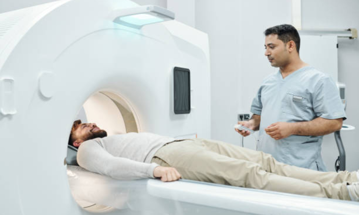
CT scans use contemporary imaging technology to produce detailed images of the body’s interior systems. Unlike traditional X-rays, CT scans combine multiple X-ray images taken from various angles around the body. These images are then interpreted by a computer, which creates more detailed cross-sectional “slices” of organs, bones, and tissues.

The process starts with the patient resting on a table that glides into a doughnut-shaped CT scanner. Inside, an X-ray tube spins around the subject, taking a sequence of photographs as it goes. A detector on the opposite side detects X-rays as they pass through the body. The computer then converts the measurements into detailed 3D images. Many clinics in Bangalore provide CT Scan, a contemporary diagnostic tool that allows doctors to precisely identify and treat a wide range of medical conditions.

CT scans Play a Significant Role in Medical Diagnosis.

CT scan in Bangalore

CT scans are essential tools in modern medical diagnostics, offering significant benefits over traditional X-rays and other imaging modalities. Here is why they matter:

CT scans provide exceptionally detailed images of soft tissues, bones, and blood vessels, which improves diagnostic accuracy. This level of information is critical for detecting tiny irregularities that other methods might overlook.

CT scanning is quick and non-invasive, making it ideal for emergency situations that require immediate, precise imaging. This efficiency allows for faster decision-making and treatment.

CT scans can diagnose a variety of problems, including tumors, internal bleeding, fractures, and infections. They provide essential information for more effective treatment planning.

A respected health screening center in Bangalore provides CT scans, which allow for precise diagnosis and timely medical treatment.

Advantages of CT Scans

CT scans offer numerous significant advantages, making them indispensable in medical imaging.

High Resolution: They produce ultra-high-resolution images capable of detecting even the most minute details in the body. This level of precision is required for an accurate diagnosis and treatment strategy.

Speed: The CT scanning procedure is short, generally taking only a few minutes. This rapid approach is especially important in emergencies because prompt imaging can have a significant impact on patient outcomes.

Versatility: CT scans are versatile and can be used to examine a variety of body parts, such as the head, chest, belly, and pelvis. Because of their common use, they are critical for identifying a wide range of illnesses.

Guidance for Treatment: CT scans can aid in diagnosis and treatment. They help with the planning and monitoring of actions, such as inserting biopsy needles or evaluating the efficacy of ongoing treatments.

Understanding the Many Types of CT scans.

CT scans are classified into several types, each of which is intended to meet distinct diagnostic purposes.

Standard CT Scan: This type produces basic cross-sectional images of the body and is commonly used for diagnostic purposes. It offers a broad perspective that can aid in the diagnosis of a variety of diseases.

CT Angiography: CT Angiography which focuses on imaging blood vessels, is critical for diagnosing vascular problems such aneurysms, blockages, and other anomalies. It generates detailed images of the blood arteries, which aids treatment planning.

CT Enterography: CT enterography examines the gastrointestinal tract and creates comprehensive images of the intestines. It is especially useful for detecting and monitoring gastrointestinal issues, such as inflammatory bowel disease.

High-Resolution CT Scan: This type generates images that are even more detailed than standard CT scans. It is frequently used to evaluate lung disorders such as chronic obstructive pulmonary disease (COPD) or interstitial lung disease when precise data are required for correct diagnosis.

The Importance of CT Scans in Early Detection and Prevention of Diseases

Early identification is critical for effective treatment and better health outcomes, and CT scans play an important part in this process.

Cancer Detection: CT scans are useful in detecting malignancies in their early stages. By detecting malignant growths early, these scans enable earlier intervention, which is crucial for improving prognosis and treatment success. CT imaging delivers detailed information to patients undergoing cancer diagnosis and treatment, which can have a significant impact on treatment planning.

Trauma Assessment: CT scans are crucial for swiftly detecting internal injuries in emergencies such as car accidents. They produce clear images that clinicians may use to promptly assess and treat interior damage, which is essential for timely and effective therapy.

Chronic Condition Monitoring: CT scans can also be used to monitor chronic conditions such as chronic obstructive pulmonary disease (COPD) and inflammatory bowel disease (IBD). They enable continuous examination of critical circumstances, ensuring that any changes are detected early and addressed effectively.

Overall, CT scans are useful tools for early disease detection and continuing monitoring, resulting in better diagnosis and treatment outcomes.

Conclusion: The Use of CT Scans in Modern Healthcare

CT scans have become an essential component of modern healthcare, producing precise and accurate images of the human body. Their capacity to diagnose a wide range of disorders, acute and chronic, makes them extremely helpful in medical practice. CT scans generate high-resolution images that aid in precise diagnosis and treatment planning, hence improving patient care and results. As technology advances, CT scans become more accurate, resulting in better healthcare solutions.

To summarize, the significance of understanding how CT scans function and its role in healthcare cannot be overemphasized. CT scans, whether used for routine diagnostics or emergency medical treatments, provide critical information that allows healthcare providers to make informed decisions. Individuals wanting advanced imaging services can get a CT Scan in Bangalore, which provides access to these vital diagnostic tools and ensures complete and effective patient treatment.

Koshikaa Screening Centre offers a wide range of comprehensive cancer screening services, ensuring accurate results and personalized health care. Contact us today at +91 9019800764

MRI of the uterus is primarily used for diagnosing and evaluating conditions such as uterine fibroids, adenomyosis, and malignancies. It is particularly useful in the planning of surgical procedures and assessing the extent of gynecological diseases. This imaging test is valuable for its ability to deliver detailed cross-sectional images of the uterus and surrounding structures.
An MRI of the spleen can help diagnose various disorders affecting the spleen including splenomegaly (enlarged spleen), tumors, and other abnormalities. It provides clear, detailed images that are crucial for assessing the health of the spleen and planning potential surgical interventions.
Prostate MRI is a non-invasive imaging technique that provides detailed pictures of the prostate gland and surrounding tissues. It helps in the detection, staging, and monitoring of prostate cancer. Prostate MRIs are increasingly used to guide biopsy procedures and assist in the planning of treatment strategies for prostate health.
An MRI of the pancreas is used to identify issues such as pancreatitis, pancreatic cancer, and other pancreatic disorders. It offers high-resolution images that help in detecting small lesions and assessing the pancreatic ducts. This imaging technique is valuable for guiding biopsies and planning surgeries involving the pancreas.
MRI scans of the liver, kidney, and bile ducts provide detailed images that help diagnose various conditions, such as tumors, cysts, and blockages in the bile ducts. They are crucial for assessing organ damage, staging cancer, and planning liver or kidney-related surgeries. This type of MRI can also evaluate the health and function of these organs comprehensively.
Breast MRI is highly sensitive for detecting breast cancer, especially in individuals with dense breast tissue or a high risk of cancer. It is often used in conjunction with mammography to provide a more comprehensive evaluation. Breast MRIs can also monitor the response to breast cancer treatments and provide detailed pre-surgical mapping.
Brain and spine MRIs are critical tools for diagnosing and monitoring neurological conditions, including tumors, spinal cord injuries, and multiple sclerosis. These MRIs can reveal abnormalities in brain tissue and spinal cord structure that might not be visible with other imaging methods. They are essential for planning surgeries and assessing the effectiveness of treatments in neurology.
PET-CT scans of transplant organs like the liver, kidney, or pancreas are performed to assess metabolic activity, detect complications, and monitor post-transplant function. These scans play a vital role in the early detection of issues such as organ rejection, recurrence of disease, or infection, guiding timely interventions and optimizing transplant outcomes for patients.
PET-CT examinations of organs such as the liver, kidney, or prostate are crucial for assessing metabolic activity, detecting abnormalities, and monitoring disease progression. These scans help diagnose conditions such as liver metastases, renal cell carcinoma, or prostate cancer, guiding treatment decisions and monitoring response to therapy over time.
PET-CT scans of soft-tissue masses are utilized to assess metabolic activity and detect malignant tumors or other abnormalities. By combining PET imaging with CT imaging, these scans provide detailed information about tumor location, size, and metabolic characteristics, aiding in accurate diagnosis and treatment planning for soft-tissue cancers.
PET-CT scans are valuable for detecting cysts or fluid collections within the body, including cysts in the liver, kidneys, or other organs. These scans help characterize the nature and metabolic activity of cysts, guiding diagnosis and treatment planning for conditions such as cystic liver disease, renal cysts, or pancreatic cysts.
PET-CT scans of muscles are performed to assess metabolic activity and detect abnormalities such as inflammation, infection, or tumors. These scans provide valuable information about muscle function and pathology, aiding in the diagnosis and management of conditions like myositis, muscular dystrophy, or soft-tissue sarcomas.
PET-CT scans of the brain are utilized to evaluate brain metabolism and detect abnormalities such as tumors, epilepsy, or neurodegenerative diseases. By combining functional PET imaging with anatomical CT imaging, these scans offer comprehensive information about brain function and structure, guiding neurosurgical interventions and treatment decisions for neurological disorders
PET-CT scans of bones are commonly used to detect bone metastases in cancer patients, assess bone health in conditions like osteoporosis, and evaluate response to treatment. By combining PET and CT imaging, these scans provide detailed information about metabolic activity and anatomical changes in bone structures, aiding in accurate diagnosis and treatment planning for various bone-related conditions.
CT scans of the spine are conducted to assess the vertebrae, spinal cord, and surrounding soft tissues for conditions like herniated discs, spinal fractures, or spinal stenosis, aiding in the diagnosis and management of spinal disorders with detailed imaging essential for surgical planning and intervention.
CT scans of the pelvis are utilized to visualize the pelvic bones, reproductive organs, bladder, and surrounding structures for conditions such as fractures, tumors, or pelvic inflammatory disease, guiding treatment plans and surgical interventions with precise anatomical information.
CT scans of the neck are performed to evaluate structures such as the throat, thyroid gland, lymph nodes, and blood vessels for abnormalities like tumors, infections, or injuries, assisting in diagnosis and management of head and neck disorders with detailed imaging.
CT scans of the extremities are employed to assess bones, joints, and soft tissues in the arms or legs, aiding in the diagnosis of fractures, arthritis, or other musculoskeletal conditions, guiding orthopedic treatment and rehabilitation plans for optimal recovery.
CT scans of the chest are utilized to examine the lungs, heart, blood vessels, and surrounding structures for conditions such as lung nodules, pneumonia, or pulmonary embolism, facilitating accurate diagnosis and management of respiratory and cardiovascular diseases.
CT scans of the brain are conducted to assess brain structures, identify abnormalities like tumors, hemorrhages, or strokes, and aid in diagnosing neurological conditions, guiding neurosurgical interventions and treatment strategies with precise imaging of the brain’s anatomy.
CT scans of the abdomen are performed to visualize and evaluate the internal organs such as the liver, kidneys, pancreas, and intestines for conditions like tumors, infections, or injuries, providing detailed images essential for diagnosis and treatment planning.
Transplant organ ultrasound scans, including those of the liver, kidney, or pancreas, are performed to assess organ viability, detect complications, and monitor post-transplant function. These scans play a vital role in the early detection of issues such as organ rejection, vascular complications, or fluid collections, guiding timely interventions and optimizing transplant outcomes for patients.
Ultrasound examinations of organs such as the liver, kidney, or prostate are crucial for assessing organ structure, detecting abnormalities, and monitoring disease progression. These scans help diagnose conditions such as liver cirrhosis, kidney stones, or prostate enlargement, guiding treatment decisions and monitoring response to therapy over time.
Soft-tissue masses can be evaluated using ultrasound to determine their size, location, and characteristics. Ultrasound imaging helps differentiate between benign and malignant masses, guiding further diagnostic evaluation and treatment planning. By providing real-time imaging, ultrasound assists in the precise localization of soft-tissue masses for biopsy or surgical removal if needed.
Ultrasound scans are valuable for detecting cysts or fluid collections within the body, including cysts in the liver, kidneys, or other organs. These scans help characterize the nature and size of cysts, aiding in diagnosis and treatment planning for conditions such as cystic liver disease or renal cysts, and guiding interventions such as cyst drainage or sclerotherapy.
Musculoskeletal ultrasound scans are used to assess the health and integrity of muscles, identifying issues such as strains, tears, or muscle inflammation. These scans provide detailed images of muscle structures, allowing healthcare providers to diagnose muscle injuries accurately and develop personalized rehabilitation plans to promote recovery and restore function.
Ultrasound examinations of joints are essential for evaluating joint health, detecting abnormalities such as inflammation, fluid accumulation, or cartilage damage. By visualizing the joint structures in real-time, ultrasound helps diagnose conditions like arthritis, bursitis, or ligament injuries, guiding treatment decisions and monitoring disease progression over time.
Ultrasound scans of tendons are performed to assess for injuries, inflammation, or tears, aiding in the diagnosis and management of conditions such as tendonitis or tendon tears. These scans provide detailed images of tendon structures, helping healthcare providers determine the extent of damage and plan appropriate treatment strategies, including physical therapy or surgical intervention if necessary.
Pelvis X-rays play a crucial role in diagnosing injuries, diseases, and developmental conditions affecting the pelvic region. They provide detailed images of the hip bones, sacrum, and coccyx, aiding in the assessment of fractures, dislocations, or signs of arthritis. Pelvic X-rays are instrumental in guiding surgical interventions, monitoring healing progress, and evaluating conditions such as osteoporosis or tumors. By providing precise anatomical information, these imaging studies enable healthcare professionals to deliver personalized care and improve patient outcomes.
Teeth X-rays, known as dental radiographs, are indispensable tools for dentists to assess oral health. They aid in diagnosing dental decay, gum disease, and abnormalities in tooth structure or alignment. These images provide valuable insights into hidden dental issues, allowing for early intervention and prevention of complications. Additionally, dental X-rays are essential for planning treatments such as orthodontics, root canals, or dental implants, ensuring optimal outcomes and patient satisfaction.
Spine X-rays are pivotal in diagnosing spinal fractures, degenerative conditions, and spinal misalignments, providing critical information for treatment planning and monitoring spinal health. They offer detailed images of the vertebrae and surrounding structures, enabling healthcare professionals to identify issues such as scoliosis, disc herniation, or spinal stenosis
Skull X-rays are valuable for detecting fractures, assessing head trauma, and evaluating the alignment of facial bones, crucial in diagnosing conditions like fractures, tumors, or developmental abnormalities in the skull. They provide detailed views of the cranial structure, aiding clinicians in determining appropriate treatment strategies and ensuring patient safety.
For the heart, X-ray solutions can aid in the detection of various cardiac problems, ranging from mild to severe, including – Heart failure, Cardiac congestion and other heart-related conditions. In addition to cardiac assessments, X-ray solutions are also used for breast examinations to identify tumors or signs of breast cancer.
Bone X-rays are essential for diagnosing fractures and assessing bone alignment, aiding doctors in developing treatment plans and monitoring healing progress. They provide detailed images of the skeletal system, helping identify abnormalities or injuries that may not be visible through other imaging techniques
Abdominal X-rays are performed when patients experience symptoms such as significant nausea, ongoing vomiting, abdominal pain, flank or back pain, or abdominal swelling. These X-rays can help diagnose various conditions.
Search Bar

Frequently Booked Tests

Frequently Booked Checkups