Everything You Need to Know About Doppler Scan During Pregnancy

Table of Contents

Have you ever wondered how doctors ensure your baby is receiving enough oxygen and nutrients inside the womb? During my pregnancy journey, one test that often comes up, especially in critical situations, is the Doppler scan during pregnancy. If you are planning an ultrasound scan in Bangalore, understanding this advanced scan can make you feel more confident and prepared.

Unlike routine ultrasounds, this scan goes deeper, giving real-time insights into your baby’s blood flow and overall well-being. Curious to know when, why, and how this scan can safeguard your baby’s health? Let’s explore everything step by step.

Medical Disclaimer:

This blog is intended for informational purposes only and should not be considered medical advice. Always consult a qualified healthcare professional or your doctor for diagnosis, recommendations, and treatment related to your pregnancy.

Key Points at a Glance

  • A Doppler scan pregnancy test measures blood flow in the fetus, placenta, and umbilical cord
  • It is non-invasive, safe, and radiation-free
  • Commonly recommended in high-risk pregnancies
  • Usually performed between 24 and 40 weeks, often around 32 weeks
  • Helps detect growth issues, placental problems, and oxygen supply concerns
  • Results are quick and help doctors take timely action

What is a Doppler Scan in Pregnancy?

Doppler Scan in Pregnancy Explained

If you are asking, ‘What is a Doppler scan in pregnancy?’ let me simplify it for you. It’s an advanced ultrasound technique that uses sound waves to study the speed and direction of blood flow in your baby’s body.

Unlike regular scans that show images, this one shows how well blood circulates through critical areas like the placenta and umbilical cord. This makes it incredibly valuable in assessing whether your baby is developing safely.

Why the Doppler Test Is Done in Pregnancy

Why Doppler Scan Is Done

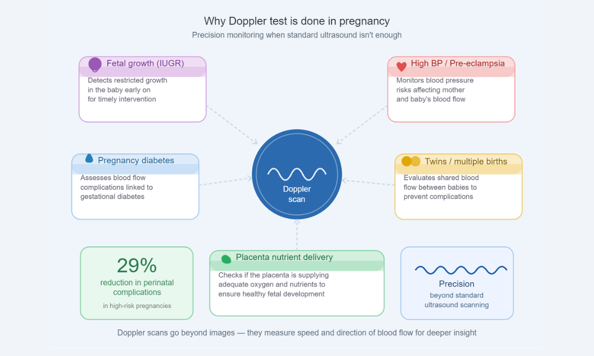
Now you might be wondering, why is a Doppler test done in pregnancy when regular ultrasounds already exist? The answer lies in precision and prevention.

Doctors recommend this scan when they need a deeper understanding of your baby’s condition. It helps in:

  • Detecting restricted fetal growth (IUGR)
  • Monitoring high blood pressure or pre-eclampsia
  • Assessing pregnancies with diabetes
  • Evaluating twins or multiple pregnancies
  • Checking if the placenta is delivering enough nutrients

In fact, studies suggest that Doppler scans can reduce perinatal complications by up to 29% in high-risk pregnancies, making them a crucial diagnostic tool.

When a Doppler Scan Is Done in Pregnancy

Timing of Doppler Scan

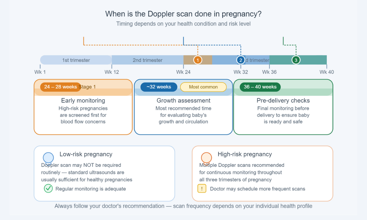
Timing plays a key role. So, when a Doppler scan is done in pregnancy, it depends on your health condition.

Here’s a quick breakdown:

Stage of PregnancyPurpose
24–28 weeksEarly monitoring in high-risk cases
Around 32 weeksThe most common time for growth assessment
36–40 weeksFinal checks before delivery

For low-risk pregnancies, it may not be required routinely. However, in high-risk cases, your doctor may recommend multiple scans for continuous monitoring.

What Does the Doppler Scan Measure?

What Does Doppler Measure

This scan focuses on three vital blood flow areas that directly impact your baby’s health and overall development:

  • Umbilical Artery – Ensures nutrients and oxygen reach the baby. It also helps detect if the placenta is functioning properly or if there is any resistance in blood flow, which could indicate growth concerns.
  • Uterine Artery – Checks blood flow from the mother to the placenta. Abnormal flow here may signal conditions like pre-eclampsia or reduced placental efficiency.
  • Middle Cerebral Artery (MCA) – Evaluates the blood supply to the baby’s brain. Increased blood flow in this artery can sometimes indicate that the baby is compensating for low oxygen levels.

Each of these measurements provides crucial insights, helping doctors identify potential complications early and take timely steps to ensure a safe and healthy pregnancy.

Types of Doppler Scans

Types of Doppler Scans

There are mainly two types used during pregnancy, and each serves a slightly different purpose in assessing your baby’s well-being:
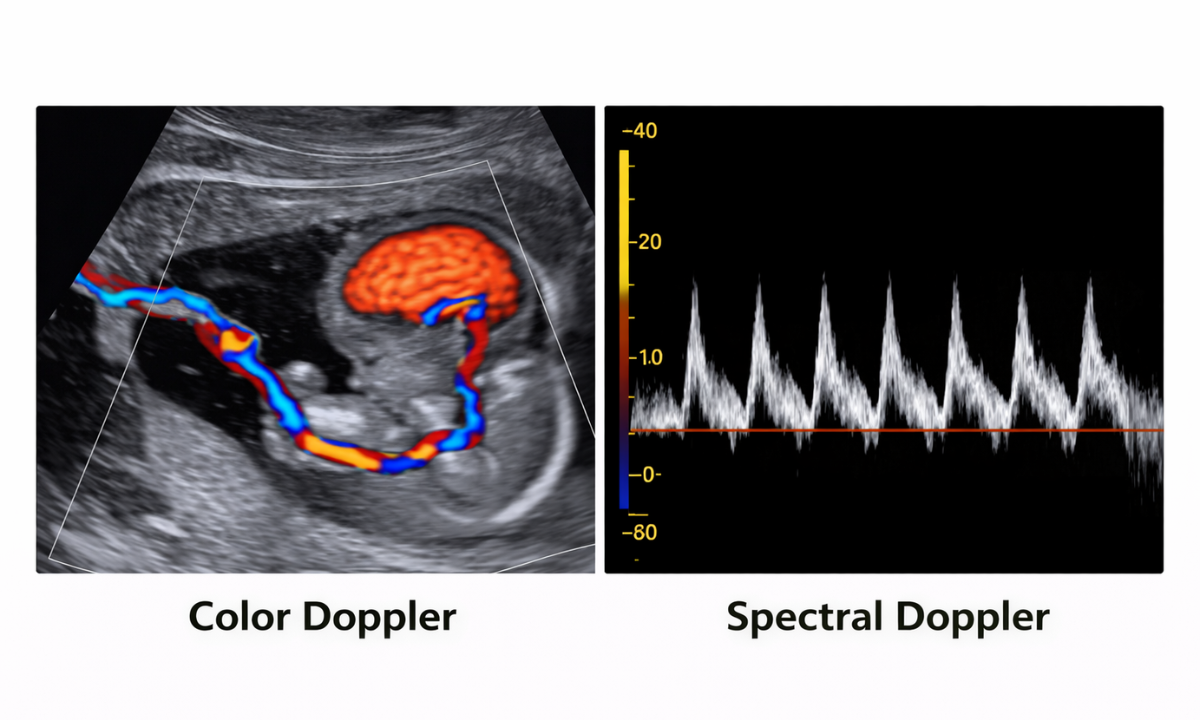
Color Doppler

  • Shows blood flow using different colours (usually red and blue)
  • Helps visualise circulation patterns clearly in real time
  • Makes it easier for doctors to identify blockages, reduced flow, or abnormal circulation in the placenta and umbilical cord

Spectral Doppler

  • Displays blood flow as waveforms on a graph
  • Helps measure the speed, direction, and resistance of blood flow
  • Provides detailed numerical values that doctors use to assess whether the blood supply is normal or needs closer monitoring

Both are equally safe and non-invasive, and doctors often use them together for a more comprehensive evaluation, depending on what needs to be assessed in your pregnancy.

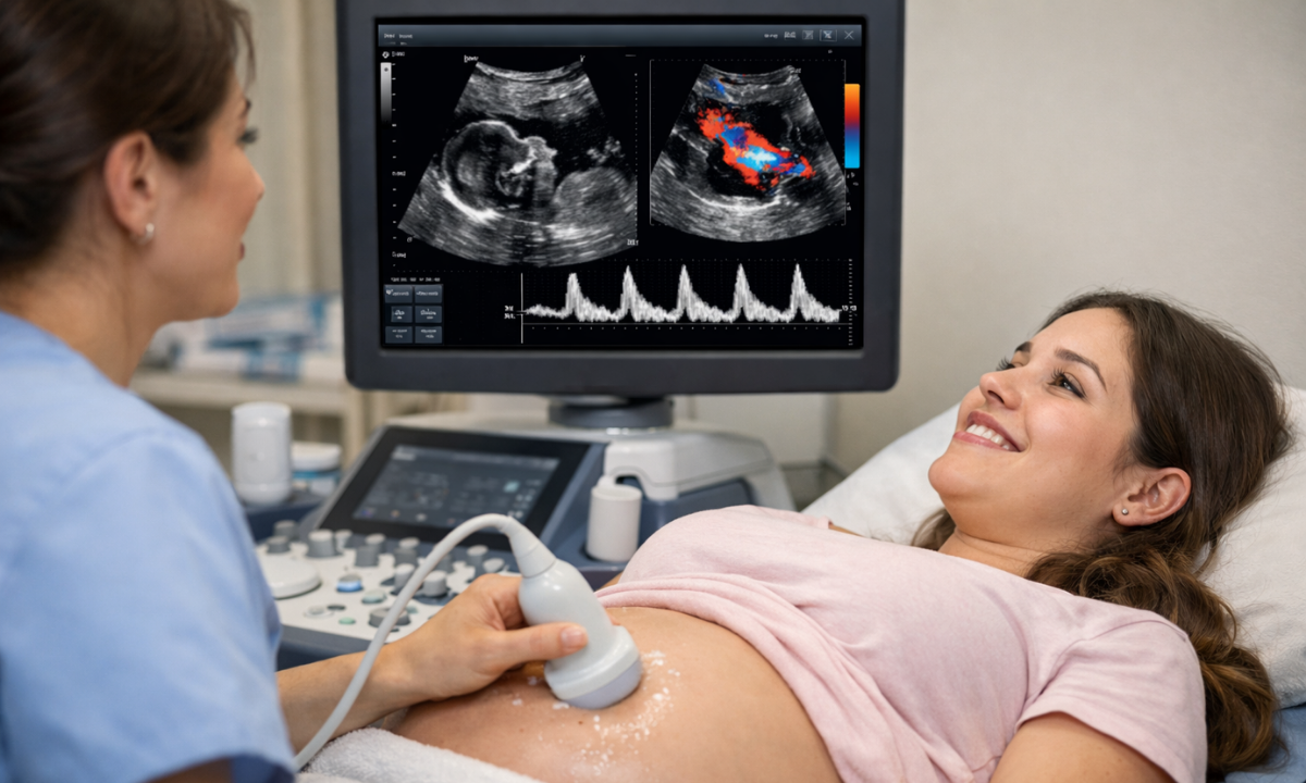
What Happens During the Procedure?

If you are feeling anxious, let me reassure you; the procedure is simple, safe, and completely painless. It is very similar to a regular ultrasound, with a slightly more detailed focus on blood flow.

  • A clear gel is applied to your abdomen to help transmit sound waves effectively
  • A handheld device (transducer) is gently moved across the skin
  • Sound waves create detailed images along with colour or waveform patterns showing blood flow
  • You may hear a soft “whooshing” sound, which represents the baby’s blood circulation

Duration: 20–60 minutes, depending on how detailed the assessment needs to be
Results: Usually available within 15–60 minutes, and your doctor will explain them to you

In most cases, no special preparation is needed, and you can eat and drink normally before the scan. You can walk in and out without any discomfort, which makes it a stress-free and reassuring experience for expectant mothers.

Understanding Doppler Scan Pregnancy Results

The next big question is about Doppler scan pregnancy results: what do they really indicate, and how should you interpret them?

Doctors carefully analyse blood flow patterns and specific indices (like resistance and pulsatility) to understand how well oxygen and nutrients are reaching your baby. Based on this, results are typically interpreted as follows:

  • Normal blood flow: Indicates healthy fetal development and efficient placental function. This means your baby is receiving adequate oxygen and nutrients, and routine monitoring will usually continue.
  • Reduced flow: May signal placental insufficiency, where the placenta is not delivering enough nutrients. In such cases, doctors may recommend more frequent scans, rest, or medication to support better outcomes.
  • Absent or reversed flow: This is a serious finding that suggests significant compromise in blood circulation. It requires immediate medical attention, close monitoring, and sometimes early delivery to protect the baby’s health.

Doctors may also compare these results with your baby’s growth, heartbeat, and other ultrasound findings to get a complete picture. These insights help them decide whether to continue monitoring, adjust treatment, or take timely action, ensuring the safest possible outcome for both mother and baby.

Is There Any Doppler Scan Pregnancy Risk?

One of the most common concerns I hear is about the Doppler scan pregnancy risk.

Here’s the truth:

  • It uses sound waves, not radiation
  • It is completely non-invasive
  • It is considered safe for both mother and baby

However, it is only recommended when medically necessary, ensuring there’s no overuse.

Who Should Definitely Get a Doppler Scan?

While not mandatory for everyone, some pregnancies benefit greatly from this scan:

  • Mothers with high blood pressure or diabetes
  • Cases of slow fetal growth
  • Multiple pregnancies (twins/triplets)
  • Previous history of pregnancy complications

If you fall into any of these categories, your doctor may strongly recommend it.

Tips to Prepare for Your Doppler Scan

To make the most of your ultrasound Doppler scan, here are a few simple tips I always suggest:

  • Wear comfortable, loose clothing
  • Stay hydrated before the test
  • Carry your previous reports
  • Ask questions and clarify doubts immediately

Being prepared helps you feel more relaxed and informed.

Final Thoughts

A Doppler scan during pregnancy is more than just a test; it’s a powerful tool that ensures your baby’s safety by monitoring vital blood flow. If you are planning an ultrasound scan in Bangalore, choosing a trusted health screening centre in Bangalore can make all the difference in accuracy and care.

At Koshikaa, the focus is always on combining advanced technology with compassionate care, ensuring every mother feels informed, reassured, and supported throughout her pregnancy journey.

FAQs

1. Is a Doppler scan painful?

No, a Doppler scan is completely painless and non-invasive. It feels very similar to a regular ultrasound, where a gel is applied, and a device is moved over your abdomen. There are no needles, injections, or discomfort involved, making it a safe and reassuring experience for expectant mothers.

2. How long does the scan take?

A Doppler scan usually takes between 20 and 60 minutes, depending on the level of detail required. In some cases, it may take slightly longer if the baby’s position makes imaging difficult. However, the procedure is generally quick, smooth, and completed within a single outpatient visit.

3. Can it harm my baby?

No, a Doppler scan is considered completely safe for your baby when performed under medical supervision. It uses sound waves, not radiation, to assess blood flow. Doctors recommend it only when necessary, ensuring that both mother and baby benefit from accurate monitoring without any known risks.

4. Do all pregnant women need it?

Not all pregnant women require a Doppler scan. It is usually recommended for high-risk pregnancies, such as those involving high blood pressure, diabetes, or concerns about the baby’s growth. Your doctor will decide if it is necessary based on your medical condition and overall pregnancy progress.

5. How soon will I get the results?

In most cases, Doppler scan results are available within 30 to 60 minutes after the procedure. A radiologist analyses the blood flow patterns and shares the findings with your doctor. Your doctor will then explain the results and guide you on any next steps if required.

Reference

1. From Google

2. https://www.medanta.org/patient-education-blog/doppler-ultrasound-in-pregnancy-assessing-blood-flow-for-babys-health

MRI of the uterus is primarily used for diagnosing and evaluating conditions such as uterine fibroids, adenomyosis, and malignancies. It is particularly useful in the planning of surgical procedures and assessing the extent of gynecological diseases. This imaging test is valuable for its ability to deliver detailed cross-sectional images of the uterus and surrounding structures.
An MRI of the spleen can help diagnose various disorders affecting the spleen including splenomegaly (enlarged spleen), tumors, and other abnormalities. It provides clear, detailed images that are crucial for assessing the health of the spleen and planning potential surgical interventions.
Prostate MRI is a non-invasive imaging technique that provides detailed pictures of the prostate gland and surrounding tissues. It helps in the detection, staging, and monitoring of prostate cancer. Prostate MRIs are increasingly used to guide biopsy procedures and assist in the planning of treatment strategies for prostate health.
An MRI of the pancreas is used to identify issues such as pancreatitis, pancreatic cancer, and other pancreatic disorders. It offers high-resolution images that help in detecting small lesions and assessing the pancreatic ducts. This imaging technique is valuable for guiding biopsies and planning surgeries involving the pancreas.
MRI scans of the liver, kidney, and bile ducts provide detailed images that help diagnose various conditions, such as tumors, cysts, and blockages in the bile ducts. They are crucial for assessing organ damage, staging cancer, and planning liver or kidney-related surgeries. This type of MRI can also evaluate the health and function of these organs comprehensively.
Breast MRI is highly sensitive for detecting breast cancer, especially in individuals with dense breast tissue or a high risk of cancer. It is often used in conjunction with mammography to provide a more comprehensive evaluation. Breast MRIs can also monitor the response to breast cancer treatments and provide detailed pre-surgical mapping.
Brain and spine MRIs are critical tools for diagnosing and monitoring neurological conditions, including tumors, spinal cord injuries, and multiple sclerosis. These MRIs can reveal abnormalities in brain tissue and spinal cord structure that might not be visible with other imaging methods. They are essential for planning surgeries and assessing the effectiveness of treatments in neurology.
PET-CT scans of transplant organs like the liver, kidney, or pancreas are performed to assess metabolic activity, detect complications, and monitor post-transplant function. These scans play a vital role in the early detection of issues such as organ rejection, recurrence of disease, or infection, guiding timely interventions and optimizing transplant outcomes for patients.
PET-CT examinations of organs such as the liver, kidney, or prostate are crucial for assessing metabolic activity, detecting abnormalities, and monitoring disease progression. These scans help diagnose conditions such as liver metastases, renal cell carcinoma, or prostate cancer, guiding treatment decisions and monitoring response to therapy over time.
PET-CT scans of soft-tissue masses are utilized to assess metabolic activity and detect malignant tumors or other abnormalities. By combining PET imaging with CT imaging, these scans provide detailed information about tumor location, size, and metabolic characteristics, aiding in accurate diagnosis and treatment planning for soft-tissue cancers.
PET-CT scans are valuable for detecting cysts or fluid collections within the body, including cysts in the liver, kidneys, or other organs. These scans help characterize the nature and metabolic activity of cysts, guiding diagnosis and treatment planning for conditions such as cystic liver disease, renal cysts, or pancreatic cysts.
PET-CT scans of muscles are performed to assess metabolic activity and detect abnormalities such as inflammation, infection, or tumors. These scans provide valuable information about muscle function and pathology, aiding in the diagnosis and management of conditions like myositis, muscular dystrophy, or soft-tissue sarcomas.
PET-CT scans of the brain are utilized to evaluate brain metabolism and detect abnormalities such as tumors, epilepsy, or neurodegenerative diseases. By combining functional PET imaging with anatomical CT imaging, these scans offer comprehensive information about brain function and structure, guiding neurosurgical interventions and treatment decisions for neurological disorders
PET-CT scans of bones are commonly used to detect bone metastases in cancer patients, assess bone health in conditions like osteoporosis, and evaluate response to treatment. By combining PET and CT imaging, these scans provide detailed information about metabolic activity and anatomical changes in bone structures, aiding in accurate diagnosis and treatment planning for various bone-related conditions.
CT scans of the spine are conducted to assess the vertebrae, spinal cord, and surrounding soft tissues for conditions like herniated discs, spinal fractures, or spinal stenosis, aiding in the diagnosis and management of spinal disorders with detailed imaging essential for surgical planning and intervention.
CT scans of the pelvis are utilized to visualize the pelvic bones, reproductive organs, bladder, and surrounding structures for conditions such as fractures, tumors, or pelvic inflammatory disease, guiding treatment plans and surgical interventions with precise anatomical information.
CT scans of the neck are performed to evaluate structures such as the throat, thyroid gland, lymph nodes, and blood vessels for abnormalities like tumors, infections, or injuries, assisting in diagnosis and management of head and neck disorders with detailed imaging.
CT scans of the extremities are employed to assess bones, joints, and soft tissues in the arms or legs, aiding in the diagnosis of fractures, arthritis, or other musculoskeletal conditions, guiding orthopedic treatment and rehabilitation plans for optimal recovery.
CT scans of the chest are utilized to examine the lungs, heart, blood vessels, and surrounding structures for conditions such as lung nodules, pneumonia, or pulmonary embolism, facilitating accurate diagnosis and management of respiratory and cardiovascular diseases.
CT scans of the brain are conducted to assess brain structures, identify abnormalities like tumors, hemorrhages, or strokes, and aid in diagnosing neurological conditions, guiding neurosurgical interventions and treatment strategies with precise imaging of the brain’s anatomy.
CT scans of the abdomen are performed to visualize and evaluate the internal organs such as the liver, kidneys, pancreas, and intestines for conditions like tumors, infections, or injuries, providing detailed images essential for diagnosis and treatment planning.
Transplant organ ultrasound scans, including those of the liver, kidney, or pancreas, are performed to assess organ viability, detect complications, and monitor post-transplant function. These scans play a vital role in the early detection of issues such as organ rejection, vascular complications, or fluid collections, guiding timely interventions and optimizing transplant outcomes for patients.
Ultrasound examinations of organs such as the liver, kidney, or prostate are crucial for assessing organ structure, detecting abnormalities, and monitoring disease progression. These scans help diagnose conditions such as liver cirrhosis, kidney stones, or prostate enlargement, guiding treatment decisions and monitoring response to therapy over time.
Soft-tissue masses can be evaluated using ultrasound to determine their size, location, and characteristics. Ultrasound imaging helps differentiate between benign and malignant masses, guiding further diagnostic evaluation and treatment planning. By providing real-time imaging, ultrasound assists in the precise localization of soft-tissue masses for biopsy or surgical removal if needed.
Ultrasound scans are valuable for detecting cysts or fluid collections within the body, including cysts in the liver, kidneys, or other organs. These scans help characterize the nature and size of cysts, aiding in diagnosis and treatment planning for conditions such as cystic liver disease or renal cysts, and guiding interventions such as cyst drainage or sclerotherapy.
Musculoskeletal ultrasound scans are used to assess the health and integrity of muscles, identifying issues such as strains, tears, or muscle inflammation. These scans provide detailed images of muscle structures, allowing healthcare providers to diagnose muscle injuries accurately and develop personalized rehabilitation plans to promote recovery and restore function.
Ultrasound examinations of joints are essential for evaluating joint health, detecting abnormalities such as inflammation, fluid accumulation, or cartilage damage. By visualizing the joint structures in real-time, ultrasound helps diagnose conditions like arthritis, bursitis, or ligament injuries, guiding treatment decisions and monitoring disease progression over time.
Ultrasound scans of tendons are performed to assess for injuries, inflammation, or tears, aiding in the diagnosis and management of conditions such as tendonitis or tendon tears. These scans provide detailed images of tendon structures, helping healthcare providers determine the extent of damage and plan appropriate treatment strategies, including physical therapy or surgical intervention if necessary.
Pelvis X-rays play a crucial role in diagnosing injuries, diseases, and developmental conditions affecting the pelvic region. They provide detailed images of the hip bones, sacrum, and coccyx, aiding in the assessment of fractures, dislocations, or signs of arthritis. Pelvic X-rays are instrumental in guiding surgical interventions, monitoring healing progress, and evaluating conditions such as osteoporosis or tumors. By providing precise anatomical information, these imaging studies enable healthcare professionals to deliver personalized care and improve patient outcomes.
Teeth X-rays, known as dental radiographs, are indispensable tools for dentists to assess oral health. They aid in diagnosing dental decay, gum disease, and abnormalities in tooth structure or alignment. These images provide valuable insights into hidden dental issues, allowing for early intervention and prevention of complications. Additionally, dental X-rays are essential for planning treatments such as orthodontics, root canals, or dental implants, ensuring optimal outcomes and patient satisfaction.
Spine X-rays are pivotal in diagnosing spinal fractures, degenerative conditions, and spinal misalignments, providing critical information for treatment planning and monitoring spinal health. They offer detailed images of the vertebrae and surrounding structures, enabling healthcare professionals to identify issues such as scoliosis, disc herniation, or spinal stenosis
Skull X-rays are valuable for detecting fractures, assessing head trauma, and evaluating the alignment of facial bones, crucial in diagnosing conditions like fractures, tumors, or developmental abnormalities in the skull. They provide detailed views of the cranial structure, aiding clinicians in determining appropriate treatment strategies and ensuring patient safety.
For the heart, X-ray solutions can aid in the detection of various cardiac problems, ranging from mild to severe, including – Heart failure, Cardiac congestion and other heart-related conditions. In addition to cardiac assessments, X-ray solutions are also used for breast examinations to identify tumors or signs of breast cancer.

Bone X-rays are essential for diagnosing fractures and assessing bone alignment, aiding doctors in developing treatment plans and monitoring healing progress. They provide detailed images of the skeletal system, helping identify abnormalities or injuries that may not be visible through other imaging techniques

Abdominal X-rays are performed when patients experience symptoms such as significant nausea, ongoing vomiting, abdominal pain, flank or back pain, or abdominal swelling. These X-rays can help diagnose various conditions.

Frequently Booked Tests

Frequently Booked Checkups