
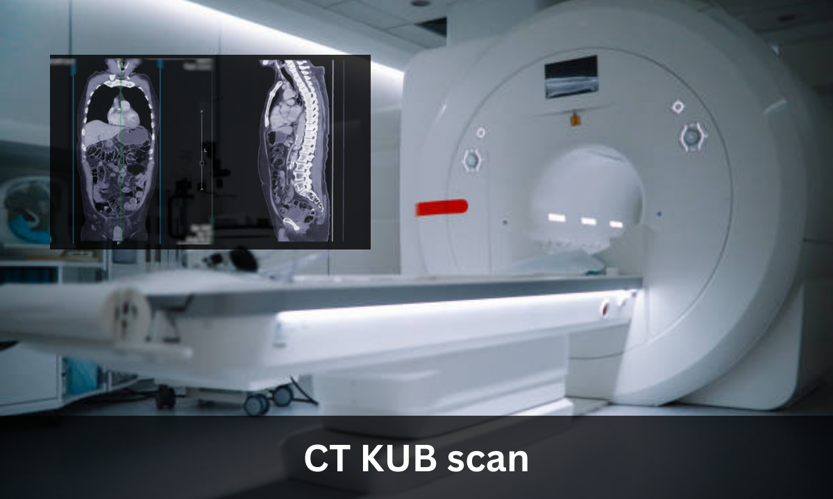
Patients who experience abdominal pains, unexplained urinary issues, and have possible kidney stones are likely to be referred to a CT KUB exam. This type of imaging test is a non-invasive test that delivers intricate cross-sectional images of pelvic organs. Being aware of the functioning of the exam, the reasons why a doctor prescribes this test, and the things you might anticipate may give you a feeling of comfort before your visit. This article describes what a CT KUB scan is used to do, how it is done, and the patient experience.
What is CT KUB?
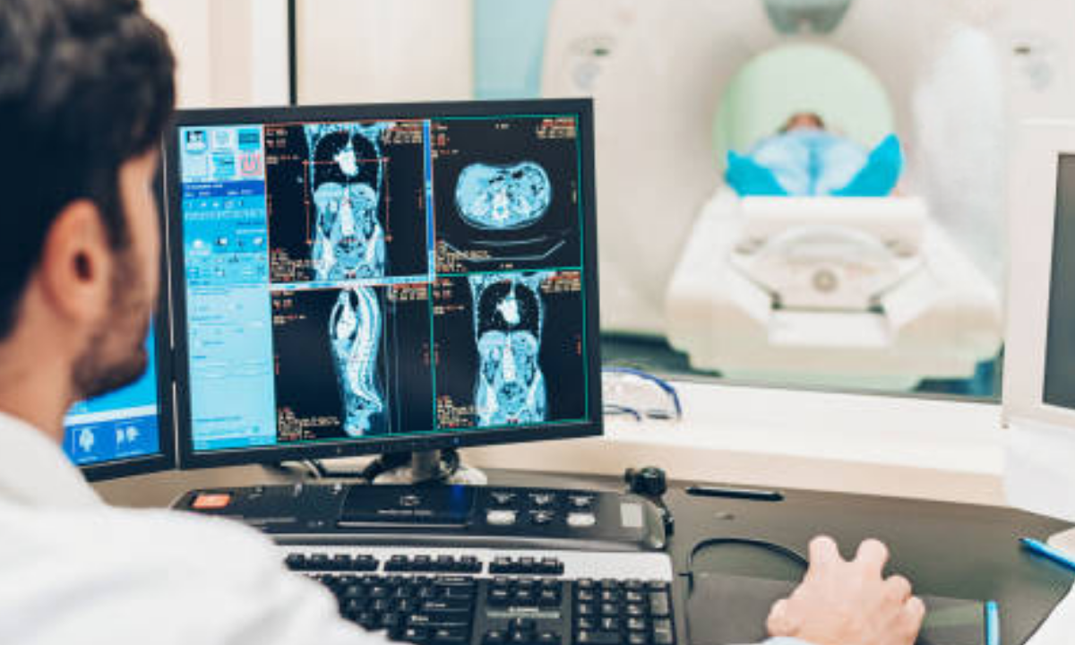
A CT KUB is a type of computerized tomographic (CT) scan that specifically examines the Kidneys, Ureters, and Bladder (KUB). Computerized tomography uses a series of X-ray images taken from different angles and computer processing to create detailed pictures of these organs.
A CT KUB scan is a type of X-ray scan that uses computer technology to create detailed images of the urinary tract, which includes the kidneys, ureters, and bladder. It is non-invasive, meaning it does not require surgery or entering the body. The scan can help identify kidney stones, blockages, tumors, or any unusual structures within these organs.
CT KUB Scan Uses
Before booking, many patients want to know about the CT KUB scan uses. This test is especially helpful for:
- Diagnosing kidney stones, including their size and location
- Identifying blockages or narrowing (strictures) in the urinary tract
- Evaluating unexplained blood in the urine (also called hematuria)
- Examining the kidneys and surrounding tissue for tumors or abnormal growths
A CT KUB scan can determine treatment options and plan surgery because it gives a clear visual of the soft-tissue structures through its high-contrast resolution.
What does CT KUB show
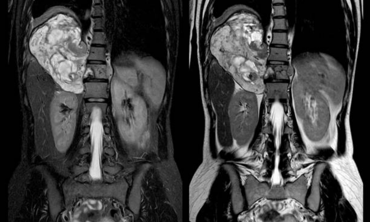
When it comes to what does CT KUB shows, keep in mind that it shows:
- The cortex, medulla and the kidney.
- Pathways of the ureters and narrowing/broadening
- The organization of the bladder ventricles, the Thickness of the walls, and possible lesions
- Around the structures, there are lymph nodes or vascular structures.
- Such data will allow clinicians to differentiate between the stone disease, infections, vascular problems, and malignancies, which will guarantee a correct diagnosis.
CT Scan KUB Procedure
The CT scan KUB procedure typically follows these steps:
1. Positioning
You lie down on a motorized table, which is shifted inside a CT gantry.
2. Contrast Administration
A contrast may be administered by an intravenous (IV) line in order to demonstrate blood vessels and enhance tissue detail.
3. Image acquisition
After you get inside, the scanner rotates around you, taking numerous pictures within seconds.
4. Completion
Picture quality is also checked by the technologist before you go; this scan takes around 15 minutes or less.
CT KUB Scan Preparation
Follow these pointers to escape blurred shots:
- When there is the use of contrast dye, fast for 4- 6 hours before an exam.
- Drink a lot of water prior to a ride.
- Avoid caffeine or diuretics that can impair the functioning of the kidneys.
- Dress loosely and wear no metal, and take off the jewelry at home.
CT KUB Scan Cost
When patients plan their expenses, many of them inquire about the CT KUB scan cost. Prices differ, depending on:
1. Geographic – nomenclature option (free-standing hospital vs outpatient center)
2. IV contrast application that can increase the total billing.
3. Insurance cover, insured deductibles and co-payments.
Radiologist’s fee for interpretation and additional post-processing.
CT Scan in Bangalore
Suppose you need a CT scan in Bangalore, you will find a number of hospitals and diagnostic centers with modern CT devices. The facilities include:
- High-speed, resolution scanners with their high-slice.
- Radiologists had expertise in the processing of the urological images.
- Low-priced packages that usually carry contrast and reporting charges.
- Checking each center: Accreditation, experience of the technicians, and turnaround or cycle time should be checked when booking a scan.
Selecting the Right Center in Bangalore
As far as booking a CT KUB scan in Bangalore goes, you need to ensure that the place where it is to happen is appropriately accredited, inquire about the generation and maintenance of the scanners and go through the qualification and experience of radiologists. At Koshikaa, the patients undergo a comprehensive process of CT KUB imaging with minimal waiting time and comprehensive and absolutely readable reports. You can also access your images and completed report digitally at many of the centers within 24 to 48 hours of completion, so that you can share your results with your healthcare provider immediately.
Conclusion: The Expectation from CT KUB Scan
Kidney stones, a condition of recurrent kidney or flank pain, urinary tract issues, and abdominal masses, may be detected accurately by using a CT KUB scan. Being able to know what is going to happen ahead of time is beneficial in limiting the feelings of anxiety. Prepare as instructed by your clinician, and feel free to enquire about cost and venues, such as a CT scan in Bangalore. The CT KUB process is generally a comfortable, informative, and fast process with proper communication and a slight level of planning.
Koshikaa provides high-end CT scan in Bangalore with a skilled team, high-quality reporting and an approach towards the patients. Reach out to the team directly for your requirements or in case of an emergency.