Common Signs that indicate the need for whole body CT scan

cost of ct scan in bangalore

Table of Contents

In recent years, the use of imaging technology, particularly CT (Computed Tomography) scans, has improved tremendously in the diagnosis and treatment of many health problems. Among them, the whole-body CT scan is a very powerful tool that allows healthcare professionals to identify potential issues in a variety of areas of the body at the same time. 

This article will help you determine when you may need a whole-body CT scan, and why you would want to see a healthcare provider.

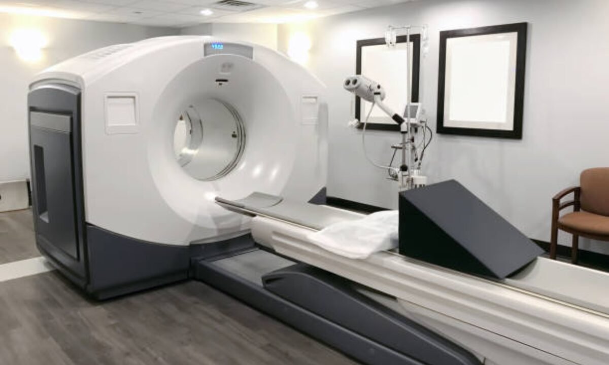
Understanding Whole Body CT Scan

Before diving into the signs, understanding what a whole-body CT scan is is important. A whole-body CT scan is a diagnostic imaging technique involving X-rays and computer technology that helps take detailed cross-sectional images of a person’s body. This scan is different from the individual scans that look at one specific area of the body, such as the head, chest, abdomen, and pelvis.

The scan is usually used for early detection of diseases such as cancer, internal injuries, or abnormalities. It’s not performed on healthy people very often but can be critical to evaluating conditions for patients with certain symptoms.

Common Signs for a Whole Body CT Scan

whole body ct scan

Below are some common reasons a doctor may decide to perform this comprehensive scan.

Persistent, Unexplained Pain

A whole body CT scan may be helpful if you have pain that persistently, and cannot be attributed to an easily identifiable cause. It’s also often used when the pain persists despite medication, physical therapy, or other treatment. This scan provides detailed images of bones, tissues, and organs, helping doctors identify underlying issues such as:

  • Tumors: But there may be tumors that don’t show any signs in the early stages and persistent pain could be a sign of their presence.
  • Internal Inflammations or Infections: This scan, however, may reveal infections that cannot be spotted with simple blood tests or external checks. This could mean, for example, spots in the abdominal area which might indicate abscesses.

After an Accident — Traumatic Injuries

Traumatic injury, e.g. after a car accident or fall, is complex and it may not always present obvious external signs. A whole body CT scan can help assess internal injuries, such as:

  • Internal organs can be damaged, such as the liver, spleen, or kidneys.
  • An inability to notice fractures or breaks in the spinal cord or bones — with X-rays.
  • Brain injuries or bleeding.

Cancer Screening or Suspicion of Cancer

A whole-body CT scan is very important to determine and monitor cancer. Symptoms such as unexplained weight loss, prolonged cough, or fatigue might indicate a patient is at high risk due to family history, or environmental exposure and this scan might be recommended. It helps in:

  • The ability to recognize different types of cancerous growths throughout different parts of the body.
  • Determine which cancer has spread (metastasis).
  • Checking what cancer treatment works.

Yet, routine scans in the absence of specific symptoms or risk factors are not routinely recommended for cancer screening, because overuse of CT scans exposes people to potentially unnecessary radiation.

Breathing Problems or Persistent Chest Pain Require Immediate Attention

Symptoms like shortness of breath and chest pain could point to severe underlying conditions, such as:

  • Pulmonary Embolism: An MCA, which can be detected between a blood clot in the lungs and cause sudden chest pain or difficulty breathing.
  • Heart Conditions: Other methods of testing, such as an echocardiogram, may be used to begin with while a more complete evaluation can be done with a whole-body CT scan to look at issues with blood flow or damage to blood vessels.
  •  Lung Diseases: They can even evaluate conditions such as infections, chronic obstructive pulmonary disease (COPD), and lung cancer.

No Clear Cause for Recurrent Infections

A whole-body CT scan may also be warranted if there are recurrent, unexplained infections. For example:

  • Respiratory infections or urinary tract infections that keep coming back even though they are being treated over and over may mean there is a problem.
  • If further evaluation is necessary, infections secondary to compromised immunity, e.g. in HIV/AIDS, should be considered.

With its in-depth look into internal organs and systems, this comprehensive scan allows pinpointing of causes that wouldn’t otherwise be detectable.

Appetite Changes or Unexplained Weight Loss

Changes in appetite or sudden unexplained weight loss can not only indicate any number of medical conditions but may also call for urgent medical attention. This may be indicative of thyroid disorders, digestive disorders, or cancers, such as stomach or pancreatic cancer. Whole-body CT scans help doctors find places where these symptoms could be coming from.

Chronic Fatigue or Weakness

Chronic fatigue, even if you can’t blame it on fatigue from lifestyle factors like stress or sleep deprivation, might be a telltale sign of a more serious health condition. However, a whole-body CT scan might find abnormalities in the blood vessels, hidden infections, or damage to several systems, including the nervous or circulatory system.

Blood in Stool, Urine, or Cough

Never is ‘bleeding’ in stool, urine, or sputum a symptom to ignore. That is exactly why it is called a sign. These could be indicative of:

  •  Internal bleeding.
  • Infections.
  • Cancers of the digestive tract, urinary tract, or lungs.

A whole-body CT scan is often a valuable step in finding out where in the body the bleeding is coming from and where.

Fevers, or sweating, without an easily explained cause

Long periods of varying temperatures of high fevers or night sweats that can’t be explained can sometimes indicate infections or diseases of the immune system. In some cases, they also can be early cues to cancer, such as lymphoma. A whole-body CT scan allows doctors to see all of the problems that could cause these symptoms from a top-down view.

After Treatment or Surgery Follow Up

If your cancer has been treated or if you had a major surgery for a serious injury and need to have follow-up imaging. A whole-body CT scan is often used to:

  •  Follow up to see if treatments such as chemotherapy or radiation succeeded.
  • It will also help to detect any signs of recurrence of the disease or new complications.
  • Check to see how well organs heal and how their overall condition is after surgery.

What Does a CT Scan Reveal?

CT scan produces a more detailed picture inside the body of bones, organs, blood vessels, and tissues. Standard X-rays or physical exams might miss fractures, tumors, infections, internal bleeding, and other abnormalities. 

Conclusion

The CT scan of a whole body, sometimes called a body CT scan, can be a lifesaver in identifying serious health problems, such as cancer, traumatic injury, or something that doesn’t make sense. The full body CT scan price can vary depending on several factors, including the location, the healthcare facility, the technology used, and whether the scan is covered by insurance. 

These are common signs to speak with your doctor about, such as persistent pain, difficulty breathing, unexplained fevers, or recurrent infections.

If you believe there’s any issue regarding health, please speak to a medical professional. They can also decide if this is appropriate for your condition and get you an accurate diagnosis. 

CT scan in Bangalore is one of the advanced services offered by Koshikaa. It is an incredibly trusted provider of CT scans, using the latest technology and expert interpretation for accurate diagnoses, with exceptional care given to patients.

MRI of the uterus is primarily used for diagnosing and evaluating conditions such as uterine fibroids, adenomyosis, and malignancies. It is particularly useful in the planning of surgical procedures and assessing the extent of gynecological diseases. This imaging test is valuable for its ability to deliver detailed cross-sectional images of the uterus and surrounding structures.
An MRI of the spleen can help diagnose various disorders affecting the spleen including splenomegaly (enlarged spleen), tumors, and other abnormalities. It provides clear, detailed images that are crucial for assessing the health of the spleen and planning potential surgical interventions.
Prostate MRI is a non-invasive imaging technique that provides detailed pictures of the prostate gland and surrounding tissues. It helps in the detection, staging, and monitoring of prostate cancer. Prostate MRIs are increasingly used to guide biopsy procedures and assist in the planning of treatment strategies for prostate health.
An MRI of the pancreas is used to identify issues such as pancreatitis, pancreatic cancer, and other pancreatic disorders. It offers high-resolution images that help in detecting small lesions and assessing the pancreatic ducts. This imaging technique is valuable for guiding biopsies and planning surgeries involving the pancreas.
MRI scans of the liver, kidney, and bile ducts provide detailed images that help diagnose various conditions, such as tumors, cysts, and blockages in the bile ducts. They are crucial for assessing organ damage, staging cancer, and planning liver or kidney-related surgeries. This type of MRI can also evaluate the health and function of these organs comprehensively.
Breast MRI is highly sensitive for detecting breast cancer, especially in individuals with dense breast tissue or a high risk of cancer. It is often used in conjunction with mammography to provide a more comprehensive evaluation. Breast MRIs can also monitor the response to breast cancer treatments and provide detailed pre-surgical mapping.
Brain and spine MRIs are critical tools for diagnosing and monitoring neurological conditions, including tumors, spinal cord injuries, and multiple sclerosis. These MRIs can reveal abnormalities in brain tissue and spinal cord structure that might not be visible with other imaging methods. They are essential for planning surgeries and assessing the effectiveness of treatments in neurology.
PET-CT scans of transplant organs like the liver, kidney, or pancreas are performed to assess metabolic activity, detect complications, and monitor post-transplant function. These scans play a vital role in the early detection of issues such as organ rejection, recurrence of disease, or infection, guiding timely interventions and optimizing transplant outcomes for patients.
PET-CT examinations of organs such as the liver, kidney, or prostate are crucial for assessing metabolic activity, detecting abnormalities, and monitoring disease progression. These scans help diagnose conditions such as liver metastases, renal cell carcinoma, or prostate cancer, guiding treatment decisions and monitoring response to therapy over time.
PET-CT scans of soft-tissue masses are utilized to assess metabolic activity and detect malignant tumors or other abnormalities. By combining PET imaging with CT imaging, these scans provide detailed information about tumor location, size, and metabolic characteristics, aiding in accurate diagnosis and treatment planning for soft-tissue cancers.
PET-CT scans are valuable for detecting cysts or fluid collections within the body, including cysts in the liver, kidneys, or other organs. These scans help characterize the nature and metabolic activity of cysts, guiding diagnosis and treatment planning for conditions such as cystic liver disease, renal cysts, or pancreatic cysts.
PET-CT scans of muscles are performed to assess metabolic activity and detect abnormalities such as inflammation, infection, or tumors. These scans provide valuable information about muscle function and pathology, aiding in the diagnosis and management of conditions like myositis, muscular dystrophy, or soft-tissue sarcomas.
PET-CT scans of the brain are utilized to evaluate brain metabolism and detect abnormalities such as tumors, epilepsy, or neurodegenerative diseases. By combining functional PET imaging with anatomical CT imaging, these scans offer comprehensive information about brain function and structure, guiding neurosurgical interventions and treatment decisions for neurological disorders
PET-CT scans of bones are commonly used to detect bone metastases in cancer patients, assess bone health in conditions like osteoporosis, and evaluate response to treatment. By combining PET and CT imaging, these scans provide detailed information about metabolic activity and anatomical changes in bone structures, aiding in accurate diagnosis and treatment planning for various bone-related conditions.
CT scans of the spine are conducted to assess the vertebrae, spinal cord, and surrounding soft tissues for conditions like herniated discs, spinal fractures, or spinal stenosis, aiding in the diagnosis and management of spinal disorders with detailed imaging essential for surgical planning and intervention.
CT scans of the pelvis are utilized to visualize the pelvic bones, reproductive organs, bladder, and surrounding structures for conditions such as fractures, tumors, or pelvic inflammatory disease, guiding treatment plans and surgical interventions with precise anatomical information.
CT scans of the neck are performed to evaluate structures such as the throat, thyroid gland, lymph nodes, and blood vessels for abnormalities like tumors, infections, or injuries, assisting in diagnosis and management of head and neck disorders with detailed imaging.
CT scans of the extremities are employed to assess bones, joints, and soft tissues in the arms or legs, aiding in the diagnosis of fractures, arthritis, or other musculoskeletal conditions, guiding orthopedic treatment and rehabilitation plans for optimal recovery.
CT scans of the chest are utilized to examine the lungs, heart, blood vessels, and surrounding structures for conditions such as lung nodules, pneumonia, or pulmonary embolism, facilitating accurate diagnosis and management of respiratory and cardiovascular diseases.
CT scans of the brain are conducted to assess brain structures, identify abnormalities like tumors, hemorrhages, or strokes, and aid in diagnosing neurological conditions, guiding neurosurgical interventions and treatment strategies with precise imaging of the brain’s anatomy.
CT scans of the abdomen are performed to visualize and evaluate the internal organs such as the liver, kidneys, pancreas, and intestines for conditions like tumors, infections, or injuries, providing detailed images essential for diagnosis and treatment planning.
Transplant organ ultrasound scans, including those of the liver, kidney, or pancreas, are performed to assess organ viability, detect complications, and monitor post-transplant function. These scans play a vital role in the early detection of issues such as organ rejection, vascular complications, or fluid collections, guiding timely interventions and optimizing transplant outcomes for patients.
Ultrasound examinations of organs such as the liver, kidney, or prostate are crucial for assessing organ structure, detecting abnormalities, and monitoring disease progression. These scans help diagnose conditions such as liver cirrhosis, kidney stones, or prostate enlargement, guiding treatment decisions and monitoring response to therapy over time.
Soft-tissue masses can be evaluated using ultrasound to determine their size, location, and characteristics. Ultrasound imaging helps differentiate between benign and malignant masses, guiding further diagnostic evaluation and treatment planning. By providing real-time imaging, ultrasound assists in the precise localization of soft-tissue masses for biopsy or surgical removal if needed.
Ultrasound scans are valuable for detecting cysts or fluid collections within the body, including cysts in the liver, kidneys, or other organs. These scans help characterize the nature and size of cysts, aiding in diagnosis and treatment planning for conditions such as cystic liver disease or renal cysts, and guiding interventions such as cyst drainage or sclerotherapy.
Musculoskeletal ultrasound scans are used to assess the health and integrity of muscles, identifying issues such as strains, tears, or muscle inflammation. These scans provide detailed images of muscle structures, allowing healthcare providers to diagnose muscle injuries accurately and develop personalized rehabilitation plans to promote recovery and restore function.
Ultrasound examinations of joints are essential for evaluating joint health, detecting abnormalities such as inflammation, fluid accumulation, or cartilage damage. By visualizing the joint structures in real-time, ultrasound helps diagnose conditions like arthritis, bursitis, or ligament injuries, guiding treatment decisions and monitoring disease progression over time.
Ultrasound scans of tendons are performed to assess for injuries, inflammation, or tears, aiding in the diagnosis and management of conditions such as tendonitis or tendon tears. These scans provide detailed images of tendon structures, helping healthcare providers determine the extent of damage and plan appropriate treatment strategies, including physical therapy or surgical intervention if necessary.
Pelvis X-rays play a crucial role in diagnosing injuries, diseases, and developmental conditions affecting the pelvic region. They provide detailed images of the hip bones, sacrum, and coccyx, aiding in the assessment of fractures, dislocations, or signs of arthritis. Pelvic X-rays are instrumental in guiding surgical interventions, monitoring healing progress, and evaluating conditions such as osteoporosis or tumors. By providing precise anatomical information, these imaging studies enable healthcare professionals to deliver personalized care and improve patient outcomes.
Teeth X-rays, known as dental radiographs, are indispensable tools for dentists to assess oral health. They aid in diagnosing dental decay, gum disease, and abnormalities in tooth structure or alignment. These images provide valuable insights into hidden dental issues, allowing for early intervention and prevention of complications. Additionally, dental X-rays are essential for planning treatments such as orthodontics, root canals, or dental implants, ensuring optimal outcomes and patient satisfaction.
Spine X-rays are pivotal in diagnosing spinal fractures, degenerative conditions, and spinal misalignments, providing critical information for treatment planning and monitoring spinal health. They offer detailed images of the vertebrae and surrounding structures, enabling healthcare professionals to identify issues such as scoliosis, disc herniation, or spinal stenosis
Skull X-rays are valuable for detecting fractures, assessing head trauma, and evaluating the alignment of facial bones, crucial in diagnosing conditions like fractures, tumors, or developmental abnormalities in the skull. They provide detailed views of the cranial structure, aiding clinicians in determining appropriate treatment strategies and ensuring patient safety.
For the heart, X-ray solutions can aid in the detection of various cardiac problems, ranging from mild to severe, including – Heart failure, Cardiac congestion and other heart-related conditions. In addition to cardiac assessments, X-ray solutions are also used for breast examinations to identify tumors or signs of breast cancer.

Bone X-rays are essential for diagnosing fractures and assessing bone alignment, aiding doctors in developing treatment plans and monitoring healing progress. They provide detailed images of the skeletal system, helping identify abnormalities or injuries that may not be visible through other imaging techniques

Abdominal X-rays are performed when patients experience symptoms such as significant nausea, ongoing vomiting, abdominal pain, flank or back pain, or abdominal swelling. These X-rays can help diagnose various conditions.
Search Bar

Frequently Booked Tests

Frequently Booked Checkups