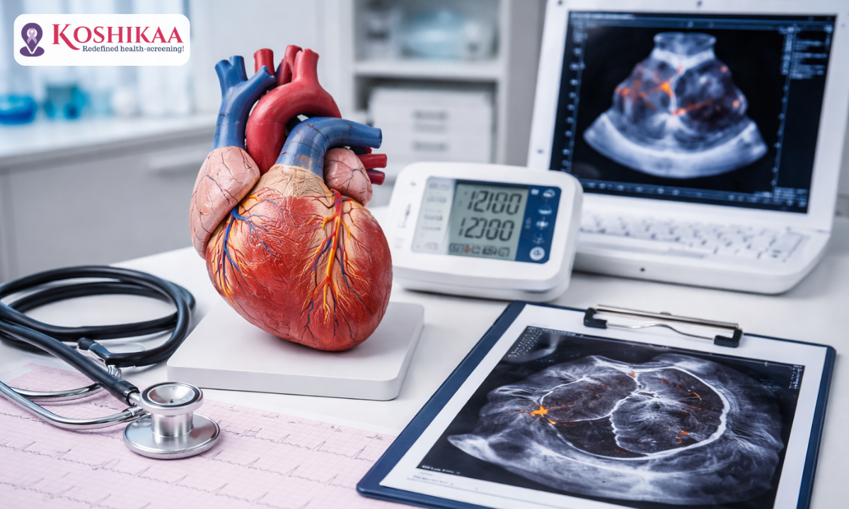
When it comes to your cardiovascular health, proactive diagnosis literally saves lives. Finding a highly reliable Health screening centre in Bangalore is the first vital step toward protecting your heart. At Koshikaa, widely recognized as the Best Health screening centre in Bangalore, we offer a comprehensive suite of cardiovascular diagnostics under one roof.
Whether you need a routine ECG Test in Bangalore, highly specialized ECG services in Bangalore, or an advanced high-resolution CT scan in Bangalore, our facility is equipped with state-of-the-art technology to capture every detail.
Patients frequently visit our clinic seeking the best available heart blockage test, often confused by the complicated medical terminology surrounding coronary health.
Understanding your own cardiovascular system can often feel completely overwhelming. Many patients use the terms “heart block” and “heart blockage” interchangeably, but they actually refer to two entirely different biological emergencies.
One is a strict electrical signalling problem, while the other involves the buildup of plaque in your major arteries. At Koshikaa, we believe that patient education is just as important as the diagnostic tests themselves.
In this comprehensive guide, we will break down the exact biological differences between these two conditions and explain the highly precise imaging tools our specialists use to detect them long before they become life-threatening.
Physical Blockages vs. Electrical Blocks
When patients ask our specialists at Koshikaa to check their cardiovascular health, they are usually worried about clogged arteries. However, in clinical cardiology, a physical blockage and an electrical block mean entirely different things. To understand exactly what test you need, you must first understand the two distinct systems that keep your heart functioning smoothly.
Physical Blockages
Coronary Artery Disease: A physical heart blockage, officially known as Coronary Artery Disease (CAD), occurs within your blood vessels. Over time, cholesterol and fat build up into hardened plaque inside the walls of your coronary arteries.
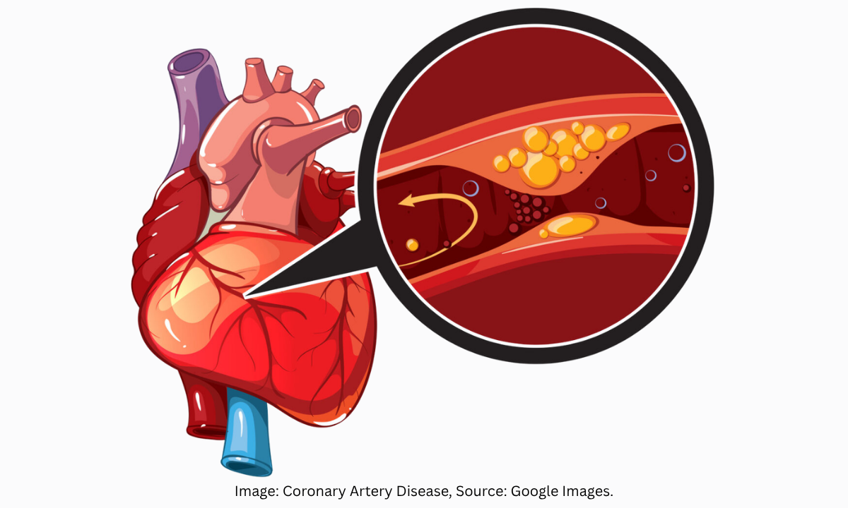
This plaque physically narrows the blood vessel, directly restricting the flow of oxygen-rich blood to your heart muscle. This physical obstruction is the condition most people are referring to when they seek a test to prevent a heart attack.
Electrical Blocks: Conduction System Disorders
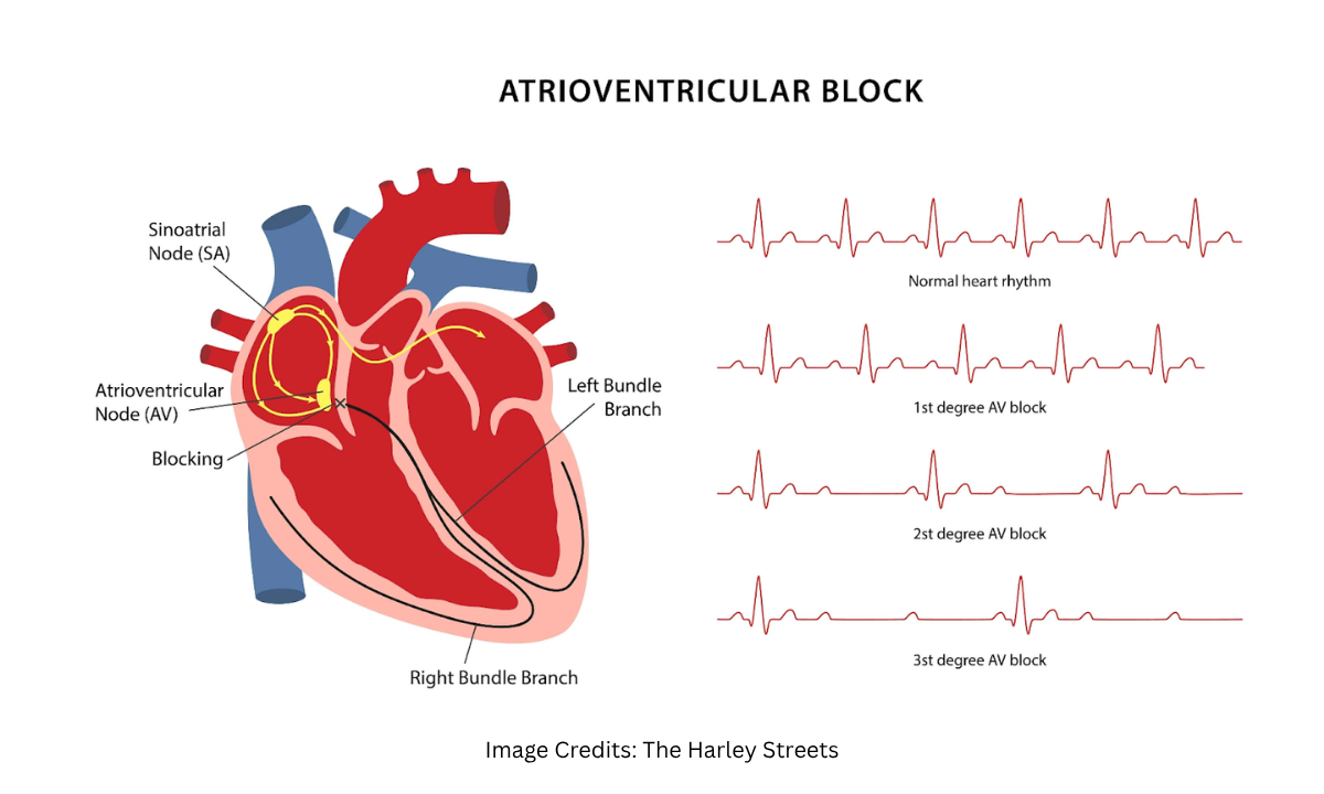
Conversely, an electrical heart block is a communication error within the heart’s internal pacemaker system. Your heart beats in a perfect, synchronized rhythm because an electrical signal travels from the upper chambers down to the lower chambers.
When this specific signal is delayed or completely obstructed, your heart beats irregularly or far too slowly. The American Heart Association categorizes these electrical disruptions into three severity levels.
Clinical Classification of Electrical Heart Blocks
| Diagnosis | Biological Mechanism | Clinical Impact |
|---|---|---|
| Heart block, first degree | The electrical signal slows down significantly as it moves through the heart, but every signal eventually reaches the lower chambers. | This is the mildest form. Patients are rarely aware they have it; it causes zero noticeable symptoms, and it generally requires no treatment. |
| Heart block 2nd degree | Not all electrical signals successfully reach the lower chambers. The signals get progressively slower until one drops completely. | Patients often feel a skipped beat, sudden dizziness, or fatigue. This condition requires careful monitoring by a cardiologist to prevent it from worsening. |
| Heart block 3rd degree | Also known as a complete heart block. No electrical signals reach the lower chambers. The ventricles are forced to rely on a dangerously slow backup pacemaker to survive. | This is a severe medical emergency. It drastically reduces the amount of blood pumped to the brain and body, almost always requiring the surgical implantation of an artificial pacemaker. |
By separating physical plaque buildup from electrical signalling errors, we can pinpoint exactly which diagnostic tools are required. An electrical issue requires tracking the signal, while a physical plaque issue requires visualizing the blood vessels.
Non-Invasive Screening
When patients visit Koshikaa with chest pain or palpitations, they immediately want to know which heart blockage test to take first.
The diagnostic journey always begins with the least invasive options available. These frontline screening tools are entirely painless, take only a few minutes to complete, and provide our cardiologists with a crucial baseline understanding of your cardiovascular health.
The most common initial screening tool is the Electrocardiogram (ECG). This simple test records the electrical signals travelling through your heart muscle.
This is what an ECG detects:
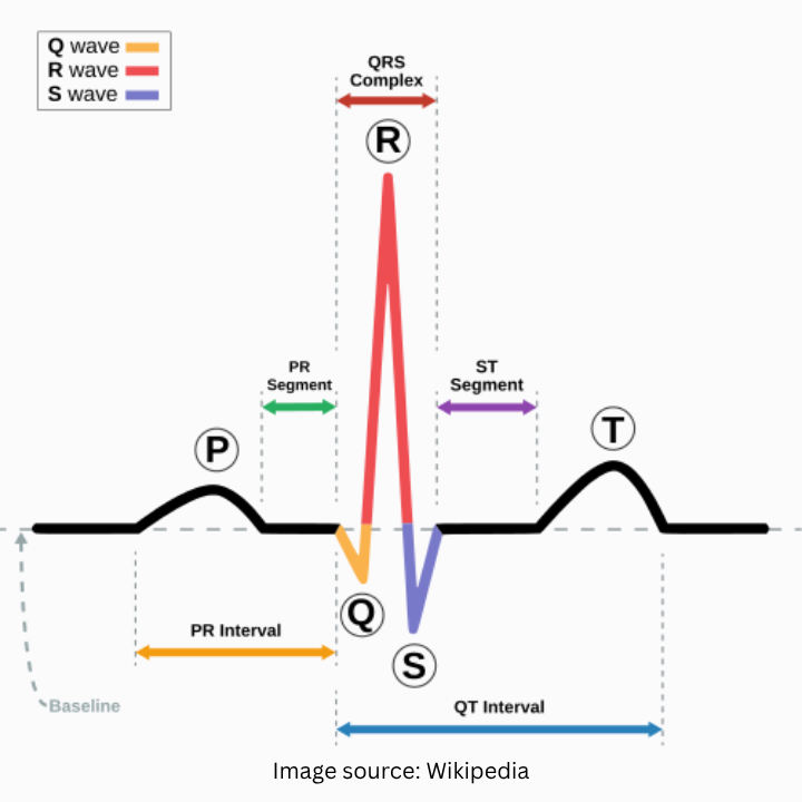
While an ECG is the ideal tool for diagnosing electrical conduction blocks, as discussed in the previous section, it has specific limitations when it comes to physical plaque.
An ECG cannot actually look inside your blood vessels. However, if a physical blockage is severe enough to restrict blood flow or cause a mild heart attack, the resulting muscle stress will alter the electrical signal. This allows the ECG to detect a major cardiac event that has occurred or is occurring.
Following an abnormal ECG, patients frequently ask about scheduling an echocardiogram to evaluate heart blockage. An echocardiogram utilizes high-frequency sound waves to create a live, moving video of your heart. Here is what it looks like:
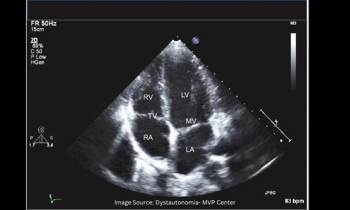
It allows our specialists to view the physical structure of your heart valves and measure exactly how forcefully your heart muscle pumps blood with every single beat.
Comparing Frontline Cardiac Screening Tools
| Diagnostic Tool | How It Works | What It Can Detect | Limitations for Plaque |
|---|---|---|---|
| Electrocardiogram (ECG) | Uses small adhesive sensors attached to the chest to record electrical wave patterns. | Rapidly diagnoses electrical blocks, irregular heartbeats (arrhythmias), and signs of previous muscle damage. | It cannot directly visualize plaque buildup or show the physical narrowing of the coronary arteries. |
| Echocardiogram (Echo) | Uses a handheld ultrasound wand to create a real-time structural video of the beating heart. | Identifies weak pumping action, valve abnormalities, and areas of the heart muscle that are lacking oxygen. | It cannot see inside the tiny coronary arteries to measure the exact percentage of a physical blockage. |
While these non-invasive tests are fantastic for assessing overall heart function and electrical stability, they are usually just the beginning of the diagnostic process.
If your ECG or Echo shows signs that your heart muscle is struggling to get enough oxygen, our specialists at Koshikaa will recommend advanced imaging to look directly inside your blood vessels.
Non-Invasive CT Scans
If your frontline screenings indicate a potential problem, or if you simply possess a strong family history of cardiovascular disease, our cardiologists will want to look directly at your coronary arteries.
For patients seeking the most comprehensive heart blockage check test without undergoing a surgical procedure, the CT Coronary Angiogram is the absolute gold standard in non-invasive imaging.
How the CT Coronary Angiogram Works
During this advanced scan, a specialized contrast dye is injected through a simple intravenous (IV) line in your arm. As this dye safely travels into your chest and illuminates your blood vessels, our state-of-the-art CT scanner rapidly captures thousands of high-resolution X-ray images in mere seconds.
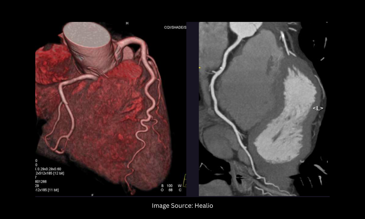
A powerful computer then compiles these cross-sectional images to construct a remarkably detailed, three-dimensional model of your beating heart and its entire plumbing network.
The Clinical Advantage of 3D Visualization
The clarity provided by modern CT technology is truly revolutionary. Our specialists at Koshikaa can virtually navigate through your arteries on a computer monitor to pinpoint the exact location, density, and severity of any calcified plaque buildup. This test offers several distinct advantages over traditional surgical methods:
- Zero Surgical Risk: Because the procedure does not require threading a physical catheter tube through the arteries in your groin or wrist, the risk of complications is practically non-existent.
- Rapid Results: The entire scanning process usually takes less than fifteen minutes from start to finish.
- Immediate Recovery: No downtime required. You simply walk into our clinic, complete your scan, and walk right back out to resume your normal daily activities.
For the vast majority of patients looking to rule out severe coronary artery disease, this non-invasive scan provides all the necessary answers.
However, if the CT scan reveals a critical physical blockage that requires immediate intervention, our team will escalate your care to the final therapeutic tier of cardiac diagnostics.
Traditional Angiography
When a non-invasive CT scan reveals a severe physical obstruction, or if a patient is actively experiencing a heart attack, cardiologists must immediately pivot from simply taking pictures to actively treating the problem. This is where traditional angiography for heart blockage, also known as cardiac catheterization, serves as the absolute gold standard in cardiovascular medicine.
The Invasive Diagnostic Process
Unlike a CT scan, where you simply lie on a table, traditional angiography is an invasive surgical procedure performed in a specialized hospital room called a Cath Lab.
A cardiologist makes a small incision in your wrist or groin and inserts a thin, flexible tube known as a catheter. This tube is carefully threaded through your blood vessels all the way up to your heart.
Once it reaches the coronary arteries, a special contrast dye is injected directly into the vessels. Real-time X-ray cameras capture exactly how the dye flows, instantly highlighting the precise location and severity of any plaque buildup.
Diagnostic and Therapeutic Power
The primary reason traditional angiography remains the ultimate medical tool is its dual capability. It is not just a test. It is a potential cure.
- Immediate Intervention: If the cardiologist spots a critical, life-threatening blockage on the X-ray, they can immediately inflate a tiny balloon inside the same catheter and guide it directly to the site of the plaque.
- Stent Placement: They inflate a balloon to crush the plaque against the arterial wall physically, then place a tiny wire-mesh tube, called a stent, to keep the blood vessel permanently open.
This seamless transition from precise diagnosis to life-saving treatment is exactly why traditional angiography is reserved exclusively for high-risk patients who require immediate physical intervention.
Why Choose Koshikaa for Your Cardiac Diagnostics?
When it comes to the complex physical and electrical systems of your heart, diagnostic precision is simply non-negotiable. As a premier Health screening centre in Bangalore, Koshikaa stands out by combining cutting-edge technology with deeply personalized patient care.
Here is exactly what makes our approach to cardiac care entirely different:
- State-of-the-Art Imaging: We utilise the latest high-resolution 3D CT scanners and advanced echocardiography machines to capture crystal-clear images of your heart, leaving absolutely no room for diagnostic guesswork.
- Rapid Digital Reporting: We understand that waiting for cardiac results is often the most stressful part of the process. Our streamlined system delivers highly accurate, smart reports directly to your digital devices within 24 to 48 hours.
- Expert Clinical Oversight: A machine is only as good as the doctor reading the results. Our dedicated team of top-tier cardiologists and skilled radiologists ensures every scan is interpreted with absolute clinical perfection.
- Accessible Convenience: With multiple state-of-the-art centres across Bangalore, including specialized diagnostic hubs in HSR Layout and Banashankari, scheduling your vital screenings is completely effortless.
Over 10,000 patients have already trusted our NABH-accredited facilities to protect their cardiovascular health and prevent life-threatening emergencies.
Conclusion
Understanding the critical biological difference between an electrical conduction error and a physical coronary blockage is your first and most powerful defense against cardiovascular disease.
You do not need to wait for a terrifying medical emergency to find out what is actually happening inside your chest. Whether you simply need a baseline ECG Test in Bangalore to monitor your internal pacemaker or a highly advanced CT scan in Bangalore to clearly visualize the health of your coronary arteries, early detection provides the ultimate clinical peace of mind.
Do not leave your most vital organ to chance. Take total control of your cardiovascular future by booking a comprehensive health package today.
Visit Koshikaa to experience the very best in preventive cardiac care, and let our expert medical team guide you toward a much longer, healthier life.