Annual Health Screening Tests You Should Not Ignore

Table of Contents

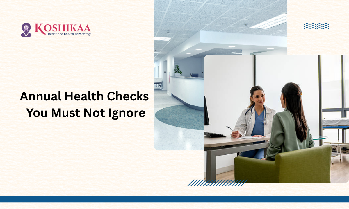
At Koshikaa, we believe that regular health screening tests are one of the most powerful ways to safeguard your well-being. These are not just routine check-ups; they are essential tools to detect early changes in your body before they turn into serious health issues. Understanding the importance of timely screenings can help you stay proactive and enjoy lasting peace of mind.

Many people are surprised to learn that even when they feel completely healthy, preventive screenings can uncover hidden health issues early on. That’s why doctors recommend scheduling a full-body checkup in Bangalore before any symptoms arise. Being proactive about your health today can help prevent complications tomorrow. Wondering which medical tests can give you valuable insights into your overall well-being? Let’s take a closer look.

Why Health Screening Matters

When we talk about normal health check-ups, we often think of simple blood work or a doctor’s visit. But a comprehensive screening covers multiple systems—heart, liver, kidney, bones, hormones, and more. Research shows that proactive screening helps in early detection of non-communicable diseases (NCDs) such as diabetes, hypertension, dyslipidaemia and thyroid disorders.

At Koshikaa, we see the value: a small abnormality caught early often means simpler treatment, better outcomes and less stress. The concept of “we’ll wait till it hurts” is outdated for modern healthcare. Prevention is far better.

Key Screening Tests We Recommend

While the exact list of tests may vary depending on your age, gender, family history and lifestyle, here are important ones we routinely include at Koshikaa:

  • Complete Blood Count (CBC) to check for anaemia, infections and basic blood health.
  • Blood sugar tests, including fasting glucose and HbA1c, are used to screen for diabetes or pre-diabetes.
  • Lipid Profile (total cholesterol, LDL, HDL, triglycerides) to assess cardiovascular risk.
  • Liver Function Test (LFT) and Kidney Function Test (KFT) to check the function of major organs.
  • Thyroid Function Test (TSH, T3, T4), because hormonal imbalances often go unnoticed for years.
  • ECG and in some cases Echocardiogram or Treadmill Test for heart screening—especially in men and in people over age 40.
  • Cancer screening tests appropriate to age and gender (for example, mammography, Pap smear for women over 40).
  • Urinalysis and other basic screenings like eye exam, dental check, and bone density in older age.

Health Screening for Women Over 40

For women over 40, the screening focus shifts slightly. We still emphasize many of the tests listed above—but we also add gender-specific screening:

  • Mammography and clinical breast examinations to detect early breast changes.
  • Pap smear and HPV testing as needed for cervical cancer risk.
  • Bone density (osteoporosis screening) because bone health becomes more critical with age.
  • Hormonal panels (if symptoms suggest thyroid changes or menopausal transition).
  • Continue regular lipid, liver, kidney, thyroid, and sugar screenings as standard.

As a radiologist here at Koshikaa, I emphasize that combining gender-specific tests with full-body screenings gives the best defence.

Health Screening for Men

Men also have specific screening needs. In addition to general tests, our focus at Koshikaa includes:

  • Prostate health screening (PSA test and digital rectal exam – depending on risk profile).
  • Colon cancer screening from around age 45–50 (for example, stool tests, colonoscopy).
  • Cardiac screening: considering the higher risk of heart disease in men, we emphasise ECG, lipid profile, and, if indicated, stress tests.
  • Testosterone/hormonal screening if symptoms arise (low energy, mood changes).
  • All the standard screening tests: CBC, sugar, thyroid, liver, kidney, etc.

Health Screening for Ages 65 and Older

Once you cross age 65, health screening becomes even more essential. Our protocol at Koshikaa includes:

  • Continued monitoring of all routine tests (CBC, lipid, sugar, liver, and kidney).
  • More frequent cardiac checks are necessary because heart disease risk rises with age.
  • Bone density tests (osteoporosis risk increases).
  • Vision and hearing checks to maintain quality of life.
  • Fall-risk assessment and screening for cognitive changes (memory, etc).
  • More intensive cancer screening, depending on previous history and family risk.

At this stage, we shift from just detection to also focusing on maintaining independence, mobility, and quality of life.

How to Choose the Right Health Screening Centre in Bangalore

Here at Koshikaa, we believe that selecting the best health screening centre in Bangalore matters as much as choosing the tests themselves. Here are my tips:

  1. Ensure the centre offers both diagnostic tests (blood, imaging) and expert consultation (radiology, pathology).
  2. Check for accreditation, up-to-date equipment, and experienced specialists.
  3. Look for a centre that adapts screening packages to your age, gender, risk profile and family history rather than a one-size-fits-all.
  4. Choose convenience: location, appointment ease, report delivery on time, and clear advice on follow-up.
  5. Ask about the clarity of pricing, what’s included, and how the results are communicated (in person or digital).

At Koshikaa, we emphasize all of these, so you get more than just a test; you get actionable insight and continuity of care.

Preparing for Your Health Screening

Because the accuracy of your screening depends on preparing well, as your radiologist, I’m advising:

  • Fast overnight if required (many blood tests ask for 8-12 hour fasting).
  • Avoid heavy meals, alcohol or nicotine 24 hours before tests.
  • Bring your previous medical records, medications list, and family history summary.
  • Wear comfortable clothing, especially if you might require an ECG or a treadmill test.
  • Inform the team of any existing conditions (diabetes, heart disease, kidney issues) so we can plan accordingly.

What Happens After the Screening?

After your tests at Koshikaa, we review everything together. If we identify any deviations from the “normal health check-up tests” baseline, we discuss:

  • What does the deviation mean (risk factor vs disease)
  • Possible follow-up investigations (imaging, specialist consult)
  • Lifestyle, diet, exercise or medication changes required
  • A plan for repeat screening, monitoring or intervention

We aim to turn screening from a checkbox activity into a meaningful health action.

Final Thoughts

When I reflect on my career as a radiologist at Koshikaa, one truth stands out: health screening tests are your best shot at staying ahead of disease and protecting your future quality of life. Whether you’re a woman over 40 navigating hormonal shifts, a man looking to manage heart risk, or someone above 65 ensuring your health remains robust, there is no substitute for a thoughtful, comprehensive screening plan.

At Koshikaa, the best health screening centre in Bangalore, we don’t just offer tests; we offer assurance, clarity and a pathway to well-being. We believe you deserve more than just results; you deserve a health roadmap tailored to you. And because we recognise how busy life in Bangalore is, we’ve built our screening process to be efficient, clear and patient-focused.

If you’ve been putting off a check-up, please don’t wait until something pops up. Let’s act early together. Let’s get ahead.

FAQs

Q1: At what age should I start routine health screenings?

I recommend starting annual or biennial health screenings from your mid-30s if you’re healthy, or earlier if you have a family history of chronic disease.

Q2: How often should key tests like blood sugar or lipid profile be repeated?

If you’re in good health and low risk, annually is acceptable. If you have risk factors, more frequent review may be needed.

Q3: Are the tests the same for men and women?

The core tests overlap (CBC, sugar, lipids, and organ function). But women over 40 and men over a certain age have additional gender-specific screening requirements (breast/mammogram and Pap smear for women; prostate and colon screening for men).

Q4: What if my screenings come back “normal”?

That’s great news. But “normal” does not mean you pause vigilance. We still recommend annual or age‐appropriate screening plus healthy lifestyle habits to maintain those normal results.

Q5: Why should I choose a specialised screening centre in Bangalore rather than my regular lab?

Specialized centres like Koshikaa integrate diagnostics, expert consultation, tailored packages, follow-up planning and patient support. That holistic model is what makes screening truly effective.

MRI of the uterus is primarily used for diagnosing and evaluating conditions such as uterine fibroids, adenomyosis, and malignancies. It is particularly useful in the planning of surgical procedures and assessing the extent of gynecological diseases. This imaging test is valuable for its ability to deliver detailed cross-sectional images of the uterus and surrounding structures.
An MRI of the spleen can help diagnose various disorders affecting the spleen including splenomegaly (enlarged spleen), tumors, and other abnormalities. It provides clear, detailed images that are crucial for assessing the health of the spleen and planning potential surgical interventions.
Prostate MRI is a non-invasive imaging technique that provides detailed pictures of the prostate gland and surrounding tissues. It helps in the detection, staging, and monitoring of prostate cancer. Prostate MRIs are increasingly used to guide biopsy procedures and assist in the planning of treatment strategies for prostate health.
An MRI of the pancreas is used to identify issues such as pancreatitis, pancreatic cancer, and other pancreatic disorders. It offers high-resolution images that help in detecting small lesions and assessing the pancreatic ducts. This imaging technique is valuable for guiding biopsies and planning surgeries involving the pancreas.
MRI scans of the liver, kidney, and bile ducts provide detailed images that help diagnose various conditions, such as tumors, cysts, and blockages in the bile ducts. They are crucial for assessing organ damage, staging cancer, and planning liver or kidney-related surgeries. This type of MRI can also evaluate the health and function of these organs comprehensively.
Breast MRI is highly sensitive for detecting breast cancer, especially in individuals with dense breast tissue or a high risk of cancer. It is often used in conjunction with mammography to provide a more comprehensive evaluation. Breast MRIs can also monitor the response to breast cancer treatments and provide detailed pre-surgical mapping.
Brain and spine MRIs are critical tools for diagnosing and monitoring neurological conditions, including tumors, spinal cord injuries, and multiple sclerosis. These MRIs can reveal abnormalities in brain tissue and spinal cord structure that might not be visible with other imaging methods. They are essential for planning surgeries and assessing the effectiveness of treatments in neurology.
PET-CT scans of transplant organs like the liver, kidney, or pancreas are performed to assess metabolic activity, detect complications, and monitor post-transplant function. These scans play a vital role in the early detection of issues such as organ rejection, recurrence of disease, or infection, guiding timely interventions and optimizing transplant outcomes for patients.
PET-CT examinations of organs such as the liver, kidney, or prostate are crucial for assessing metabolic activity, detecting abnormalities, and monitoring disease progression. These scans help diagnose conditions such as liver metastases, renal cell carcinoma, or prostate cancer, guiding treatment decisions and monitoring response to therapy over time.
PET-CT scans of soft-tissue masses are utilized to assess metabolic activity and detect malignant tumors or other abnormalities. By combining PET imaging with CT imaging, these scans provide detailed information about tumor location, size, and metabolic characteristics, aiding in accurate diagnosis and treatment planning for soft-tissue cancers.
PET-CT scans are valuable for detecting cysts or fluid collections within the body, including cysts in the liver, kidneys, or other organs. These scans help characterize the nature and metabolic activity of cysts, guiding diagnosis and treatment planning for conditions such as cystic liver disease, renal cysts, or pancreatic cysts.
PET-CT scans of muscles are performed to assess metabolic activity and detect abnormalities such as inflammation, infection, or tumors. These scans provide valuable information about muscle function and pathology, aiding in the diagnosis and management of conditions like myositis, muscular dystrophy, or soft-tissue sarcomas.
PET-CT scans of the brain are utilized to evaluate brain metabolism and detect abnormalities such as tumors, epilepsy, or neurodegenerative diseases. By combining functional PET imaging with anatomical CT imaging, these scans offer comprehensive information about brain function and structure, guiding neurosurgical interventions and treatment decisions for neurological disorders
PET-CT scans of bones are commonly used to detect bone metastases in cancer patients, assess bone health in conditions like osteoporosis, and evaluate response to treatment. By combining PET and CT imaging, these scans provide detailed information about metabolic activity and anatomical changes in bone structures, aiding in accurate diagnosis and treatment planning for various bone-related conditions.
CT scans of the spine are conducted to assess the vertebrae, spinal cord, and surrounding soft tissues for conditions like herniated discs, spinal fractures, or spinal stenosis, aiding in the diagnosis and management of spinal disorders with detailed imaging essential for surgical planning and intervention.
CT scans of the pelvis are utilized to visualize the pelvic bones, reproductive organs, bladder, and surrounding structures for conditions such as fractures, tumors, or pelvic inflammatory disease, guiding treatment plans and surgical interventions with precise anatomical information.
CT scans of the neck are performed to evaluate structures such as the throat, thyroid gland, lymph nodes, and blood vessels for abnormalities like tumors, infections, or injuries, assisting in diagnosis and management of head and neck disorders with detailed imaging.
CT scans of the extremities are employed to assess bones, joints, and soft tissues in the arms or legs, aiding in the diagnosis of fractures, arthritis, or other musculoskeletal conditions, guiding orthopedic treatment and rehabilitation plans for optimal recovery.
CT scans of the chest are utilized to examine the lungs, heart, blood vessels, and surrounding structures for conditions such as lung nodules, pneumonia, or pulmonary embolism, facilitating accurate diagnosis and management of respiratory and cardiovascular diseases.
CT scans of the brain are conducted to assess brain structures, identify abnormalities like tumors, hemorrhages, or strokes, and aid in diagnosing neurological conditions, guiding neurosurgical interventions and treatment strategies with precise imaging of the brain’s anatomy.
CT scans of the abdomen are performed to visualize and evaluate the internal organs such as the liver, kidneys, pancreas, and intestines for conditions like tumors, infections, or injuries, providing detailed images essential for diagnosis and treatment planning.
Transplant organ ultrasound scans, including those of the liver, kidney, or pancreas, are performed to assess organ viability, detect complications, and monitor post-transplant function. These scans play a vital role in the early detection of issues such as organ rejection, vascular complications, or fluid collections, guiding timely interventions and optimizing transplant outcomes for patients.
Ultrasound examinations of organs such as the liver, kidney, or prostate are crucial for assessing organ structure, detecting abnormalities, and monitoring disease progression. These scans help diagnose conditions such as liver cirrhosis, kidney stones, or prostate enlargement, guiding treatment decisions and monitoring response to therapy over time.
Soft-tissue masses can be evaluated using ultrasound to determine their size, location, and characteristics. Ultrasound imaging helps differentiate between benign and malignant masses, guiding further diagnostic evaluation and treatment planning. By providing real-time imaging, ultrasound assists in the precise localization of soft-tissue masses for biopsy or surgical removal if needed.
Ultrasound scans are valuable for detecting cysts or fluid collections within the body, including cysts in the liver, kidneys, or other organs. These scans help characterize the nature and size of cysts, aiding in diagnosis and treatment planning for conditions such as cystic liver disease or renal cysts, and guiding interventions such as cyst drainage or sclerotherapy.
Musculoskeletal ultrasound scans are used to assess the health and integrity of muscles, identifying issues such as strains, tears, or muscle inflammation. These scans provide detailed images of muscle structures, allowing healthcare providers to diagnose muscle injuries accurately and develop personalized rehabilitation plans to promote recovery and restore function.
Ultrasound examinations of joints are essential for evaluating joint health, detecting abnormalities such as inflammation, fluid accumulation, or cartilage damage. By visualizing the joint structures in real-time, ultrasound helps diagnose conditions like arthritis, bursitis, or ligament injuries, guiding treatment decisions and monitoring disease progression over time.
Ultrasound scans of tendons are performed to assess for injuries, inflammation, or tears, aiding in the diagnosis and management of conditions such as tendonitis or tendon tears. These scans provide detailed images of tendon structures, helping healthcare providers determine the extent of damage and plan appropriate treatment strategies, including physical therapy or surgical intervention if necessary.
Pelvis X-rays play a crucial role in diagnosing injuries, diseases, and developmental conditions affecting the pelvic region. They provide detailed images of the hip bones, sacrum, and coccyx, aiding in the assessment of fractures, dislocations, or signs of arthritis. Pelvic X-rays are instrumental in guiding surgical interventions, monitoring healing progress, and evaluating conditions such as osteoporosis or tumors. By providing precise anatomical information, these imaging studies enable healthcare professionals to deliver personalized care and improve patient outcomes.
Teeth X-rays, known as dental radiographs, are indispensable tools for dentists to assess oral health. They aid in diagnosing dental decay, gum disease, and abnormalities in tooth structure or alignment. These images provide valuable insights into hidden dental issues, allowing for early intervention and prevention of complications. Additionally, dental X-rays are essential for planning treatments such as orthodontics, root canals, or dental implants, ensuring optimal outcomes and patient satisfaction.
Spine X-rays are pivotal in diagnosing spinal fractures, degenerative conditions, and spinal misalignments, providing critical information for treatment planning and monitoring spinal health. They offer detailed images of the vertebrae and surrounding structures, enabling healthcare professionals to identify issues such as scoliosis, disc herniation, or spinal stenosis
Skull X-rays are valuable for detecting fractures, assessing head trauma, and evaluating the alignment of facial bones, crucial in diagnosing conditions like fractures, tumors, or developmental abnormalities in the skull. They provide detailed views of the cranial structure, aiding clinicians in determining appropriate treatment strategies and ensuring patient safety.
For the heart, X-ray solutions can aid in the detection of various cardiac problems, ranging from mild to severe, including – Heart failure, Cardiac congestion and other heart-related conditions. In addition to cardiac assessments, X-ray solutions are also used for breast examinations to identify tumors or signs of breast cancer.

Bone X-rays are essential for diagnosing fractures and assessing bone alignment, aiding doctors in developing treatment plans and monitoring healing progress. They provide detailed images of the skeletal system, helping identify abnormalities or injuries that may not be visible through other imaging techniques

Abdominal X-rays are performed when patients experience symptoms such as significant nausea, ongoing vomiting, abdominal pain, flank or back pain, or abdominal swelling. These X-rays can help diagnose various conditions.

Frequently Booked Tests

Frequently Booked Checkups