The Ultimate Guide to MRI Scans: Procedures, Benefits and, More

Health Screening Centre in Bangalore

Table of Contents

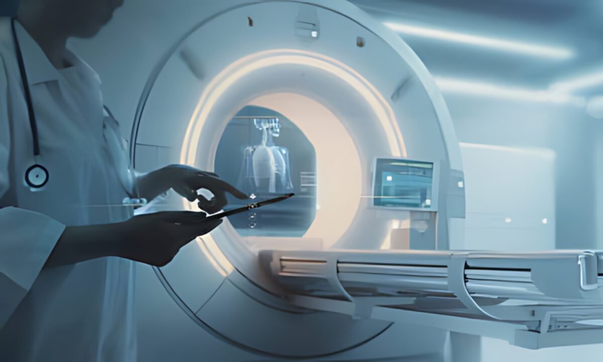
MRI Scan or Magnetic Resonance Imaging Scan is a medical technology in the assessment of organs, tissues, bones, and other body structures in different hospitals and clinics. An MRI essentially is an imaging technique that entails employing strong magnetic fields as well as radio waves to produce imagery of the interior of the human body. In this ultimate guide, we will discuss what an MRI scan means, its advantages, and some things you should know about this frequent type of examination.

What is an MRI Scan?

MRI scan in Bangalore

MRI stands for magnetic resonance imaging and it is a painless test used by medical practitioners to help in analyzing the results of various illnesses. In an MRI, you recline on a flattish platform that then moves into the huge circular scanner that does not use X-rays in taking pictures of your body but instead uses magnets and computers. 

The MRI scanner which is also known as magnetic resonance imaging utilizes magnetic fields and radiofrequency pulses to align hydrogen atoms in your body. This in turn makes the atoms emit radio signals which are read by the computer used in the method. This way, the computer is capable of producing cross-sectional pictures or sections, also referred to as slices of the part of the body under scan.

What does an MRI Procedure Entail?

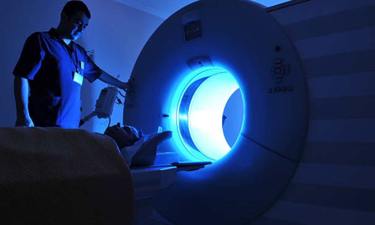
When you come for an MRI scan you have to dress according to the scan type selected and remove all metals such as; jewellery, glasses, and hair clips among others as they may hamper the strong magnetic force used. You may be asked to change into a hospital gown if you are going to be staying at the hospital for a long time. The technician will assist you arrange yourself on the table that moves into the MR machine. 

You will be completely on your own with nothing but the scanner surrounding you, however, the technician will be able to see, hear you, and communicate with you at the same time. The scanner is also bound to produce rather loud banging and humming noises during the scan. You must remain very still as movement can cause blurred images. The actual scan takes 15-90 minutes depending on the body area imaged.

Preparation Steps for an MRI

Before getting your MRI scan, there are some matters of importance you have to first accomplish. The technician or the doctor wants to know if the patient has any medical implant or metal on him/her. You may require an indication if you have a cardiac pacemaker or some other implants. Eat and drink normally unless asked to avoid foods containing dyes or iron which may blur the images.

For safety, do not wear any metal including jewellery and other metallic items. Some places may require you to put on a hospital gown if one is to be provided. When you are making the booking let them know if you are claustrophobic or if you have any other condition that may make it difficult to be enclosed.

Benefits of MRI over Other Scans

Compared to X-rays, CT scans, or ultrasound, MRI has some unique advantages:

  • MRI uses strong magnetic fields rather than ionizing radiation making it safer. It does not use any damaging radiation.
  • MRI is efficient in providing contrast between tissues and muscles, ligaments tendons, etc.
  • MRI makes it possible to get multi-planar or cross-sectional images in any plane and therefore the body structures can be viewed in different planes.
  • It has high resolution and clarity allowing the detection of even small abnormalities, lesions, or tumors.
  • MRI is very good for imaging internal organs, the brain, spinal cord, and joints where other tests may have limitations.
  • Some scans like MRI angiography can study blood vessel structure and flow non-invasively.
  • Contrast dyes, if used during MRI, provide even better visualization of targeted tissues and structures.

Important Things to Note

This is because other important information that the doctor needs to be aware of includes a history of allergy to contrast dyes if they intend to use one during an MRI.

  • Avoid bringing along items containing metal such as jewellery, watches, hair clips, and credit cards among others when going for the MRI scan.
  • You might be requested to wear a hospital gown to your operation dress depending on the regulatory measures on the wearing of metallic items such as clothes with fasteners or zippers near the MRI equipment.
  • If a person has claustrophobia or fears small enclosed spaces, he/she should inform the technicians before the procedure or operation. Medications can be given to help relax. 
  • Expect to hold very still while images are being captured as movement causes blurring artifacts. You’ll be able to breathe normally though.
  • The loud banging, thumping, and vibrations from the MRI scanner are normal. Earplugs or headphones with music are provided to some patients.
  • Wait for the radiologist’s evaluation and diagnosis. Do not self-diagnose based on the pictures which are reviewed alongside your symptoms and medical history.

MRI Areas and Applications

Some common types of MRI scans and the body areas or conditions they are used to study include:

  1. MRI brain and spinal cord – Head injuries, intracranial space-occupying lesions, infections, demyelinating diseases, spine disorders, etc.
  2. Joint MRI – Injuries to ligaments, tendons, cartilage-like torn meniscus, tendinitis and arthritis. 
  3. MRI of the abdomen and Pelvis – organs in the abdomen and pelvis, tumors, any disorders in the liver, kidney, ovary, and so on.
  4. Breast MRI – Diagnostic tool used in cases where breast cancer is suspected including on high-risk women and in cases where a mammogram is ambiguous.
  5. Conventional angiography MRI – Blood vessels and structure of the brain vessels, heart, kidney as well as the extremities.
  6. Cardiac MRI – Heart muscle, valves, and arteries assessing conditions like cardiomyopathy or blockages.

In Conclusion

MRI scan is a noninvasive technique of medical imaging that has found broad application in diagnosis and intervention in present-day practice. This guide entails details on what patients need to know in case they are scheduled for an MRI, why MRI is preferred over other scans, and places MRI can focus on. Proper preparation and communication of medical history ensures safe, quality images are obtained for accurate diagnosis. Contact your doctor from Koshikaa or a radiologist if you need any clarification on your scheduled MRI scan.

MRI of the uterus is primarily used for diagnosing and evaluating conditions such as uterine fibroids, adenomyosis, and malignancies. It is particularly useful in the planning of surgical procedures and assessing the extent of gynecological diseases. This imaging test is valuable for its ability to deliver detailed cross-sectional images of the uterus and surrounding structures.
An MRI of the spleen can help diagnose various disorders affecting the spleen including splenomegaly (enlarged spleen), tumors, and other abnormalities. It provides clear, detailed images that are crucial for assessing the health of the spleen and planning potential surgical interventions.
Prostate MRI is a non-invasive imaging technique that provides detailed pictures of the prostate gland and surrounding tissues. It helps in the detection, staging, and monitoring of prostate cancer. Prostate MRIs are increasingly used to guide biopsy procedures and assist in the planning of treatment strategies for prostate health.
An MRI of the pancreas is used to identify issues such as pancreatitis, pancreatic cancer, and other pancreatic disorders. It offers high-resolution images that help in detecting small lesions and assessing the pancreatic ducts. This imaging technique is valuable for guiding biopsies and planning surgeries involving the pancreas.
MRI scans of the liver, kidney, and bile ducts provide detailed images that help diagnose various conditions, such as tumors, cysts, and blockages in the bile ducts. They are crucial for assessing organ damage, staging cancer, and planning liver or kidney-related surgeries. This type of MRI can also evaluate the health and function of these organs comprehensively.
Breast MRI is highly sensitive for detecting breast cancer, especially in individuals with dense breast tissue or a high risk of cancer. It is often used in conjunction with mammography to provide a more comprehensive evaluation. Breast MRIs can also monitor the response to breast cancer treatments and provide detailed pre-surgical mapping.
Brain and spine MRIs are critical tools for diagnosing and monitoring neurological conditions, including tumors, spinal cord injuries, and multiple sclerosis. These MRIs can reveal abnormalities in brain tissue and spinal cord structure that might not be visible with other imaging methods. They are essential for planning surgeries and assessing the effectiveness of treatments in neurology.
PET-CT scans of transplant organs like the liver, kidney, or pancreas are performed to assess metabolic activity, detect complications, and monitor post-transplant function. These scans play a vital role in the early detection of issues such as organ rejection, recurrence of disease, or infection, guiding timely interventions and optimizing transplant outcomes for patients.
PET-CT examinations of organs such as the liver, kidney, or prostate are crucial for assessing metabolic activity, detecting abnormalities, and monitoring disease progression. These scans help diagnose conditions such as liver metastases, renal cell carcinoma, or prostate cancer, guiding treatment decisions and monitoring response to therapy over time.
PET-CT scans of soft-tissue masses are utilized to assess metabolic activity and detect malignant tumors or other abnormalities. By combining PET imaging with CT imaging, these scans provide detailed information about tumor location, size, and metabolic characteristics, aiding in accurate diagnosis and treatment planning for soft-tissue cancers.
PET-CT scans are valuable for detecting cysts or fluid collections within the body, including cysts in the liver, kidneys, or other organs. These scans help characterize the nature and metabolic activity of cysts, guiding diagnosis and treatment planning for conditions such as cystic liver disease, renal cysts, or pancreatic cysts.
PET-CT scans of muscles are performed to assess metabolic activity and detect abnormalities such as inflammation, infection, or tumors. These scans provide valuable information about muscle function and pathology, aiding in the diagnosis and management of conditions like myositis, muscular dystrophy, or soft-tissue sarcomas.
PET-CT scans of the brain are utilized to evaluate brain metabolism and detect abnormalities such as tumors, epilepsy, or neurodegenerative diseases. By combining functional PET imaging with anatomical CT imaging, these scans offer comprehensive information about brain function and structure, guiding neurosurgical interventions and treatment decisions for neurological disorders
PET-CT scans of bones are commonly used to detect bone metastases in cancer patients, assess bone health in conditions like osteoporosis, and evaluate response to treatment. By combining PET and CT imaging, these scans provide detailed information about metabolic activity and anatomical changes in bone structures, aiding in accurate diagnosis and treatment planning for various bone-related conditions.
CT scans of the spine are conducted to assess the vertebrae, spinal cord, and surrounding soft tissues for conditions like herniated discs, spinal fractures, or spinal stenosis, aiding in the diagnosis and management of spinal disorders with detailed imaging essential for surgical planning and intervention.
CT scans of the pelvis are utilized to visualize the pelvic bones, reproductive organs, bladder, and surrounding structures for conditions such as fractures, tumors, or pelvic inflammatory disease, guiding treatment plans and surgical interventions with precise anatomical information.
CT scans of the neck are performed to evaluate structures such as the throat, thyroid gland, lymph nodes, and blood vessels for abnormalities like tumors, infections, or injuries, assisting in diagnosis and management of head and neck disorders with detailed imaging.
CT scans of the extremities are employed to assess bones, joints, and soft tissues in the arms or legs, aiding in the diagnosis of fractures, arthritis, or other musculoskeletal conditions, guiding orthopedic treatment and rehabilitation plans for optimal recovery.
CT scans of the chest are utilized to examine the lungs, heart, blood vessels, and surrounding structures for conditions such as lung nodules, pneumonia, or pulmonary embolism, facilitating accurate diagnosis and management of respiratory and cardiovascular diseases.
CT scans of the brain are conducted to assess brain structures, identify abnormalities like tumors, hemorrhages, or strokes, and aid in diagnosing neurological conditions, guiding neurosurgical interventions and treatment strategies with precise imaging of the brain’s anatomy.
CT scans of the abdomen are performed to visualize and evaluate the internal organs such as the liver, kidneys, pancreas, and intestines for conditions like tumors, infections, or injuries, providing detailed images essential for diagnosis and treatment planning.
Transplant organ ultrasound scans, including those of the liver, kidney, or pancreas, are performed to assess organ viability, detect complications, and monitor post-transplant function. These scans play a vital role in the early detection of issues such as organ rejection, vascular complications, or fluid collections, guiding timely interventions and optimizing transplant outcomes for patients.
Ultrasound examinations of organs such as the liver, kidney, or prostate are crucial for assessing organ structure, detecting abnormalities, and monitoring disease progression. These scans help diagnose conditions such as liver cirrhosis, kidney stones, or prostate enlargement, guiding treatment decisions and monitoring response to therapy over time.
Soft-tissue masses can be evaluated using ultrasound to determine their size, location, and characteristics. Ultrasound imaging helps differentiate between benign and malignant masses, guiding further diagnostic evaluation and treatment planning. By providing real-time imaging, ultrasound assists in the precise localization of soft-tissue masses for biopsy or surgical removal if needed.
Ultrasound scans are valuable for detecting cysts or fluid collections within the body, including cysts in the liver, kidneys, or other organs. These scans help characterize the nature and size of cysts, aiding in diagnosis and treatment planning for conditions such as cystic liver disease or renal cysts, and guiding interventions such as cyst drainage or sclerotherapy.
Musculoskeletal ultrasound scans are used to assess the health and integrity of muscles, identifying issues such as strains, tears, or muscle inflammation. These scans provide detailed images of muscle structures, allowing healthcare providers to diagnose muscle injuries accurately and develop personalized rehabilitation plans to promote recovery and restore function.
Ultrasound examinations of joints are essential for evaluating joint health, detecting abnormalities such as inflammation, fluid accumulation, or cartilage damage. By visualizing the joint structures in real-time, ultrasound helps diagnose conditions like arthritis, bursitis, or ligament injuries, guiding treatment decisions and monitoring disease progression over time.
Ultrasound scans of tendons are performed to assess for injuries, inflammation, or tears, aiding in the diagnosis and management of conditions such as tendonitis or tendon tears. These scans provide detailed images of tendon structures, helping healthcare providers determine the extent of damage and plan appropriate treatment strategies, including physical therapy or surgical intervention if necessary.
Pelvis X-rays play a crucial role in diagnosing injuries, diseases, and developmental conditions affecting the pelvic region. They provide detailed images of the hip bones, sacrum, and coccyx, aiding in the assessment of fractures, dislocations, or signs of arthritis. Pelvic X-rays are instrumental in guiding surgical interventions, monitoring healing progress, and evaluating conditions such as osteoporosis or tumors. By providing precise anatomical information, these imaging studies enable healthcare professionals to deliver personalized care and improve patient outcomes.
Teeth X-rays, known as dental radiographs, are indispensable tools for dentists to assess oral health. They aid in diagnosing dental decay, gum disease, and abnormalities in tooth structure or alignment. These images provide valuable insights into hidden dental issues, allowing for early intervention and prevention of complications. Additionally, dental X-rays are essential for planning treatments such as orthodontics, root canals, or dental implants, ensuring optimal outcomes and patient satisfaction.
Spine X-rays are pivotal in diagnosing spinal fractures, degenerative conditions, and spinal misalignments, providing critical information for treatment planning and monitoring spinal health. They offer detailed images of the vertebrae and surrounding structures, enabling healthcare professionals to identify issues such as scoliosis, disc herniation, or spinal stenosis
Skull X-rays are valuable for detecting fractures, assessing head trauma, and evaluating the alignment of facial bones, crucial in diagnosing conditions like fractures, tumors, or developmental abnormalities in the skull. They provide detailed views of the cranial structure, aiding clinicians in determining appropriate treatment strategies and ensuring patient safety.
For the heart, X-ray solutions can aid in the detection of various cardiac problems, ranging from mild to severe, including – Heart failure, Cardiac congestion and other heart-related conditions. In addition to cardiac assessments, X-ray solutions are also used for breast examinations to identify tumors or signs of breast cancer.
Bone X-rays are essential for diagnosing fractures and assessing bone alignment, aiding doctors in developing treatment plans and monitoring healing progress. They provide detailed images of the skeletal system, helping identify abnormalities or injuries that may not be visible through other imaging techniques
Abdominal X-rays are performed when patients experience symptoms such as significant nausea, ongoing vomiting, abdominal pain, flank or back pain, or abdominal swelling. These X-rays can help diagnose various conditions.
Search Bar

Frequently Booked Tests

Frequently Booked Checkups