
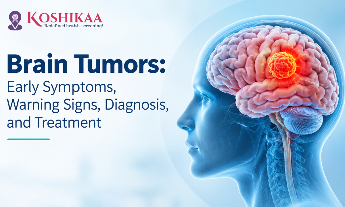
Brain health plays a vital role in overall well-being. The brain controls essential functions such as movement, memory, speech, emotions, and coordination. When abnormal cells grow in or around the brain, they can form a brain tumor. While the word “brain tumor” may sound frightening, not all brain tumors are cancerous, and many treatment options are available depending on the type, size, and location of the tumor.
Understanding the early symptoms, warning signs, diagnostic methods, and treatment options can help individuals seek timely medical attention. In this article, explores the key facts about brain tumors and explains why early evaluation at a health screening centre in Bangalore is important.
What Is a Brain Tumor?
A brain tumor is an abnormal growth of cells within the brain or surrounding tissues. Brain tumors are generally classified into two main categories: benign and malignant.
Benign brain tumors are non-cancerous and usually grow more slowly. However, they can still cause serious problems if they press against important brain structures.
Malignant brain tumors are cancerous and may grow rapidly, invade nearby tissues, or spread within the central nervous system.
Brain tumors may also be classified as primary or secondary. Primary tumors begin in the brain, while secondary tumors, also known as brain metastases, develop when cancer spreads to the brain from another part of the body.
Early Symptoms of Brain Tumors
Brain tumor symptoms vary depending on the tumor’s size, location, and growth rate. Some symptoms develop gradually, while others may appear more suddenly.
Common early symptoms include:
1. Persistent or Worsening Headaches
Headaches are common and usually have causes unrelated to brain tumors. However, a new headache pattern, persistent headaches that progressively worsen, or headaches accompanied by neurological symptoms should be evaluated by a healthcare professional.
Some people may experience chronic headaches that are worse in the morning or become more noticeable with coughing or straining.
2. Seizures
A seizure can sometimes be the first noticeable sign of a brain tumor, even in someone who has never experienced seizures before. Seizures may involve uncontrolled movements, temporary loss of awareness, unusual sensations, or brief episodes of confusion.
A first-time seizure requires urgent medical assessment.
3. Vision Problems
Tumors affecting areas of the brain responsible for vision can cause blurred vision, double vision, or loss of part of the visual field. Persistent or unexplained visual changes should not be ignored.
4. Memory and Concentration Difficulties
Some individuals may notice difficulty concentrating, forgetfulness, confusion, or changes in their ability to perform everyday tasks. These symptoms can have many causes, but persistent or progressive changes warrant medical evaluation.
5. Weakness or Balance Problems
A brain tumor may affect the nerves or brain regions responsible for movement and coordination. Symptoms can include weakness in an arm or leg, difficulty walking, poor balance, or unusual clumsiness.
6. Nausea and Vomiting
Unexplained, persistent nausea or vomiting, particularly when associated with worsening headaches or other neurological symptoms, may require further investigation.
Warning Signs That Need Immediate Medical Attention
Certain symptoms should never be ignored. Seek emergency medical care for a sudden severe headache, a first-time seizure, new weakness on one side of the body, sudden difficulty speaking, loss of consciousness, or rapidly worsening confusion.
These symptoms do not automatically mean a brain tumor is present. They can also indicate other serious conditions, including a stroke or bleeding in the brain, which require prompt assessment.
How Are Brain Tumors Diagnosed?
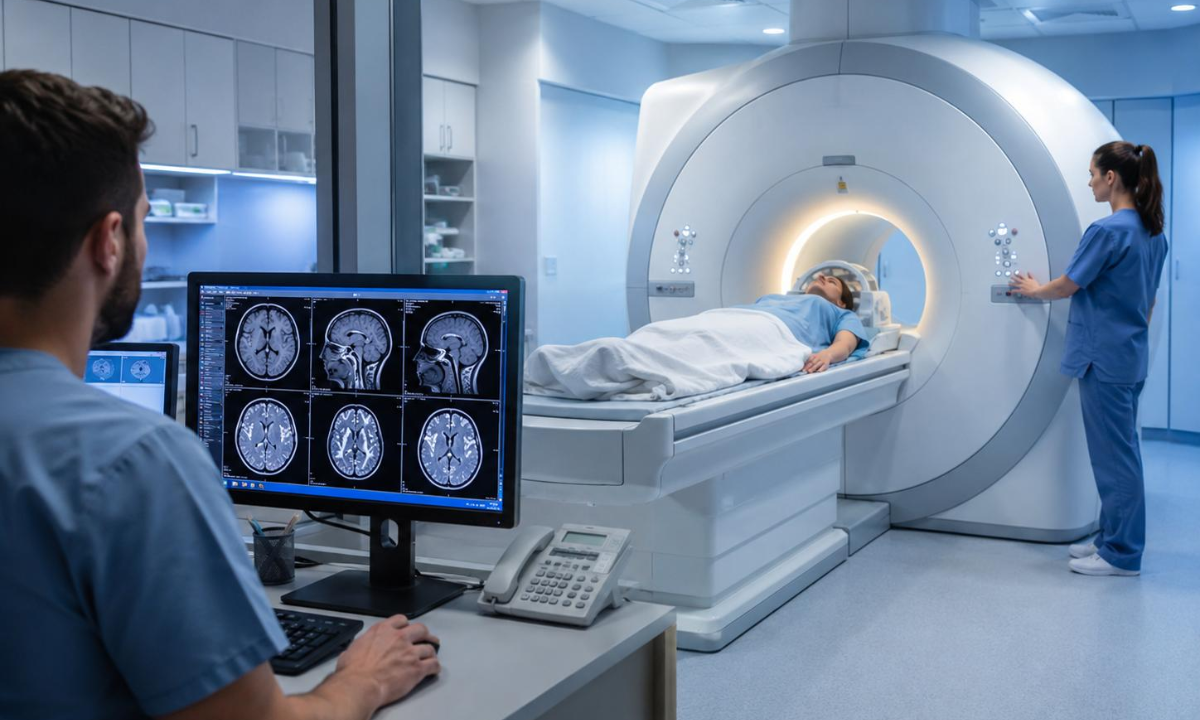
Diagnosing a brain tumor involves evaluating symptoms, performing a neurological examination, and using appropriate imaging or other tests.
1. Medical History and Neurological Examination
A doctor may ask about headaches, seizures, changes in vision, memory problems, and other symptoms. A neurological examination can assess muscle strength, reflexes, coordination, balance, hearing, and vision.
2. MRI Scan
Magnetic resonance imaging (MRI) is commonly used to examine the brain in detail. It can help identify abnormal growths and show their size, location, and relationship to surrounding structures. Contrast-enhanced MRI may provide additional information.
3. CT Scan
A computed tomography (CT) scan uses X-rays to create detailed images of the brain. It may be used in emergency situations or when doctors need to identify bleeding, swelling, or other abnormalities quickly.
4. PET Scan
A positron emission tomography (PET) scan evaluates metabolic activity or other biological processes using a small amount of a radioactive tracer. In selected cases, PET imaging can help doctors assess certain brain tumors, distinguish active tumor tissue from treatment-related changes, or investigate cancer elsewhere in the body.
PET scans are not required for every suspected brain tumor. The choice of imaging depends on the clinical situation and the treating specialist’s recommendation.
Patients and caregivers looking for advanced imaging services can explore PET scan in Bangalore to learn more about available PET/CT diagnostic services.
5. Biopsy and Additional Tests
In many cases, a tissue sample is needed to establish the exact tumor type and grade. A biopsy or surgical removal may allow specialists to examine tumor cells and perform molecular tests that help guide treatment. Not every patient requires a biopsy, and the decision depends on the tumor’s location and clinical circumstances.
Treatment Options for Brain Tumors
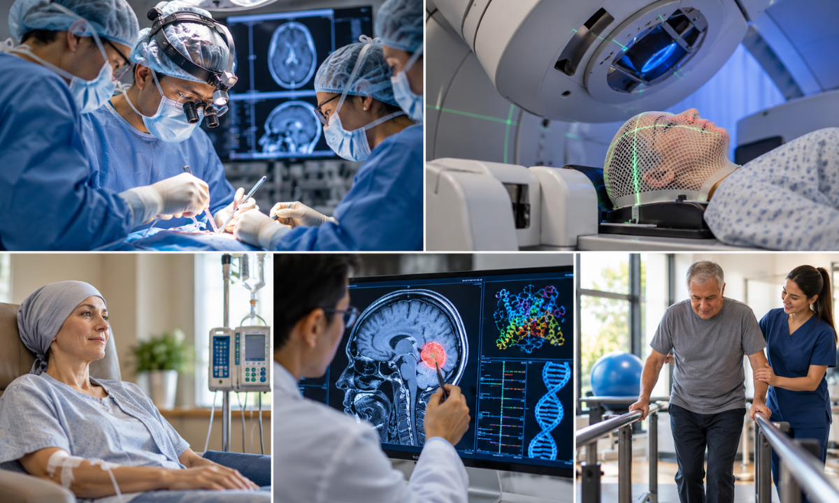
Treatment is personalized according to the tumor type, grade, location, size, symptoms, and the patient’s overall health.
1. Surgery
Surgery may be recommended to remove as much of the tumor as safely possible. It can also help relieve pressure on the brain and provide tissue for diagnosis. Some tumors cannot be completely removed because they are close to critical brain structures.
2. Radiation Therapy
Radiation therapy uses targeted radiation to destroy tumor cells or slow their growth. It may be used after surgery, instead of surgery in selected cases, or to treat tumors that have returned.
3. Chemotherapy
Chemotherapy uses medicines to destroy or control cancer cells. It is used for certain types of brain tumors, depending on their biological characteristics and treatment plan.
4. Targeted Therapy and Other Treatments
Some tumors have specific molecular features that can be treated with targeted medicines. Depending on the diagnosis, doctors may also consider other specialized therapies or clinical trials.
5. Monitoring and Supportive Care
Certain slow-growing tumors may be monitored with regular examinations and imaging rather than treated immediately. Supportive care, rehabilitation, seizure management, and medicines to control symptoms may also form part of the treatment plan.
Can Brain Tumors Be Prevented?
Most brain tumors do not have a clearly identifiable preventable cause. Although certain rare inherited conditions can increase risk, many people diagnosed with brain tumors have no known risk factors.
Maintaining a healthy lifestyle supports general well-being, but there is no proven lifestyle routine or dietary supplement that can guarantee prevention. Routine brain scans are not generally recommended for people without symptoms or specific medical indications.
Conclusion
Brain tumors can cause a range of symptoms, from persistent headaches and seizures to vision changes, weakness, and difficulties with memory or coordination. Because these symptoms can also result from other conditions, professional evaluation is essential for an accurate diagnosis.
MRI and CT scans, selected PET imaging, and sometimes biopsy help doctors understand the nature of an abnormality and plan appropriate treatment. Depending on the diagnosis, care may include surgery, radiation therapy, chemotherapy, targeted treatment, or regular monitoring.
Recognizing warning signs and seeking timely medical advice can make a meaningful difference in diagnosis and treatment planning. If you or a loved one experiences persistent or worsening neurological symptoms, consult a qualified healthcare professional rather than relying on self-diagnosis.
Disclaimer: This article is intended for general educational purposes only and should not replace professional medical advice, diagnosis, or treatment.