
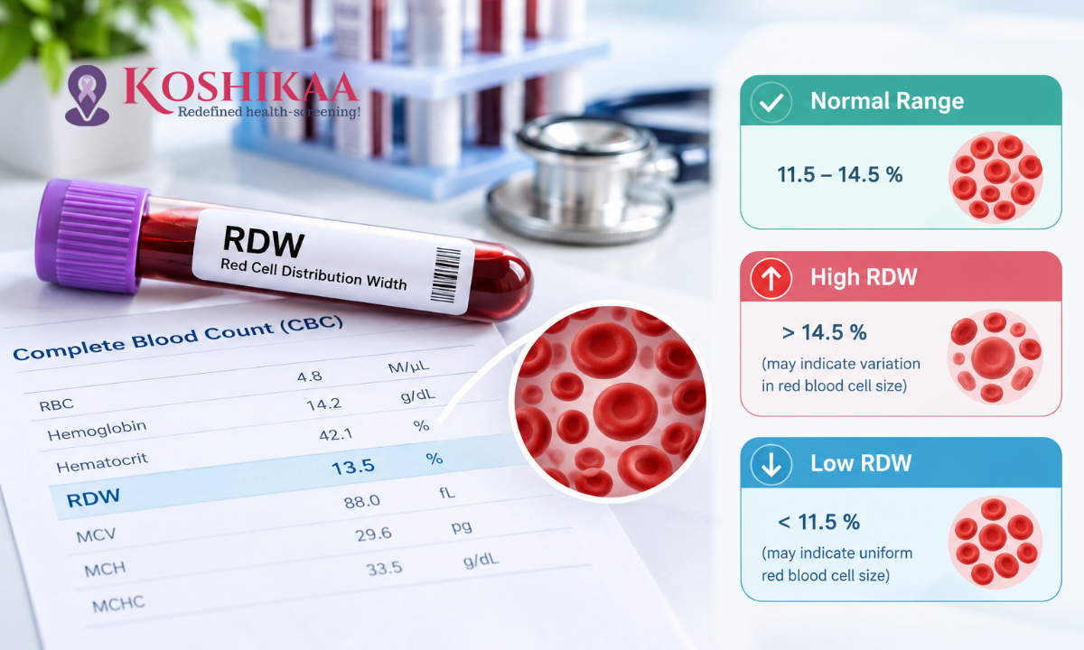
If you have recently had a complete blood count (CBC), you may have noticed a line on your report labelled RDW. For many people, it is one of the less familiar values, and seeing it marked as high or low can be worrying.
The RDW blood test measures how much your red blood cells vary in size. It gives doctors a useful clue about how your body is producing red blood cells and can help point towards possible causes of anaemia, such as iron, vitamin B12 or folate deficiency. For a clear interpretation of your results, it is best to consult a trusted healthcare provider like Koshikaa Group.
However, RDW is only one part of the picture. On its own, it cannot diagnose anaemia or identify the exact cause of an abnormal result. Doctors always interpret it alongside haemoglobin, MCV and other blood count values, as well as your symptoms and medical history. This guide explains what RDW means, what the normal range looks like, what high and low results may indicate and what to do next. For a wider overview, see our guide to common types of blood tests.
What Is an RDW Blood Test?
What Does RDW Mean in a Blood Test?
RDW stands for red cell distribution width. It measures the variation in the size of your red blood cells, which carry oxygen from your lungs to the rest of your body.
Healthy red blood cells are usually fairly similar in size. When they vary more than expected, with a mix of larger and smaller cells, the RDW value rises. The medical term for this variation in size is anisocytosis.
Why Is RDW Included in a CBC Test?
RDW is calculated automatically as part of a complete blood count, so it usually appears on your report without being requested separately. It helps clinicians assess red blood cell patterns, particularly when investigating possible anaemia or nutritional deficiencies.
Because RDW describes variation in size rather than the size itself, it is most useful when read alongside other values, especially haemoglobin, which shows how much oxygen-carrying protein is in your blood, and MCV (mean corpuscular volume), which shows the average size of your red blood cells. Learn more in our guide to understanding common blood tests.
What Is the Normal Range for an RDW Blood Test?
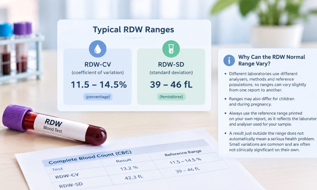
Typical RDW-CV and RDW-SD Ranges
RDW is reported in one of two ways, and some reports show both:
•RDW-CV (coefficient of variation): reported as a percentage. A commonly cited adult range is approximately 11.5 to 14.5 per cent.
•RDW-SD (standard deviation): reported in femtolitres (fL). A commonly cited adult range is approximately 39 to 46 fL.
Why Can the RDW Normal Range Vary?
Different laboratories use different analysers, methods and reference populations, so reference ranges can vary slightly from one report to another. Ranges may also differ for children and during pregnancy.
Always use the reference range printed on your own report, as it reflects the laboratory and analyser used for your sample. It is also worth knowing that a result just outside the range does not automatically mean a serious health problem. Small variations are common and are often not clinically significant on their own.
What Does a High RDW Blood Test Mean?
A high RDW means your red blood cells vary more in size than usual. This can happen for a number of reasons, some of which are common and easily treatable.
Common Causes of High RDW Levels
•Iron deficiency: one of the most common causes. Iron deficiency anaemia is particularly common in India, especially among women. When iron is low, the body produces smaller red blood cells, which creates a mix of sizes.
•Vitamin B12 or folate deficiency: these can lead to the production of larger red blood cells, again increasing variation.
•Certain types of anaemia: including mixed deficiencies, where more than one nutrient is low.
•Recovery after blood loss or treatment: when the body produces new red blood cells quickly, for example after bleeding or when starting iron treatment, newer and older cells may differ in size.
•Other conditions: some chronic illnesses, liver disease and certain bone marrow conditions may also be associated with a raised RDW, depending on the wider clinical picture.
Researchers have also studied RDW in relation to heart disease and other long-term conditions. These findings are interesting, but RDW is not used to diagnose or predict these conditions in routine practice, so a high result should not be read as a sign of heart disease.
Can High RDW Mean Anaemia?
A high RDW can occur with anaemia, but it does not confirm anaemia by itself. Anaemia is diagnosed when haemoglobin is below the expected range. A person may have a high RDW with normal haemoglobin, particularly in the early stages of iron deficiency, before anaemia has fully developed.
To understand a raised RDW, a clinician will look at haemoglobin, MCV, red blood cell count and your medical history. Depending on the findings, they may recommend further tests to identify the cause.
What Does a Low RDW Blood Test Mean?
Is Low RDW a Cause for Concern?
A low RDW usually means your red blood cells are very similar in size. On its own, a low RDW is generally not considered clinically significant and is much less concerning than many other abnormal blood count findings.
That said, RDW should never be read in isolation. Your complete blood count and any symptoms still matter.
When Should You Discuss a Low RDW Result?
Speak to your doctor if a low RDW appears alongside abnormal haemoglobin, MCV or other blood count values, or if you have symptoms such as persistent tiredness, breathlessness or unexplained weight loss.
RDW-CV vs RDW-SD: What Is the Difference?
RDW-CV and RDW-SD both describe variation in red blood cell size, but they are calculated differently.
•RDW-CV compares the spread of red blood cell sizes with the average cell size and is expressed as a percentage. Because it is related to the average size, it can be influenced by MCV.
•RDW-SD is a direct measurement of the width of the size distribution and is expressed in femtolitres. It is less affected by the average cell size.
Because they use different units and methods, the two values should not be compared as if they were the same measurement. Always interpret each one against the reference range provided by your laboratory, and follow your clinician’s interpretation.
How Is RDW Related to MCV and Other CBC Results?
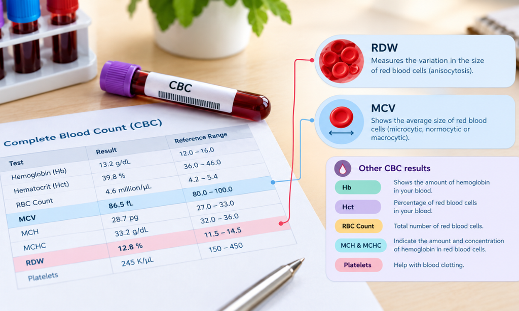
RDW tells doctors how varied your red blood cells are, while MCV tells them the average size. Looking at the two together can provide helpful clues.
| RDW | MCV | What the Pattern May Suggest |
| High | Low | Can occur with iron deficiency, among other causes |
| High | High | Can occur with vitamin B12 or folate deficiency or some liver conditions |
| High | Normal | May be seen in early nutritional deficiencies or mixed deficiencies |
| Normal | Low | May be seen in beta-thalassaemia trait or anaemia of chronic disease |
| Normal | Normal | Common in healthy individuals, but does not rule out all blood conditions |
These patterns are clues, not diagnoses. Your doctor will consider them alongside your full report, symptoms and medical history.
High RDW With Low MCV
This pattern is commonly associated with iron deficiency, but it can have other causes and needs clinical interpretation, often with iron studies.
High RDW With Normal MCV
This can occur in the early stages of a nutritional deficiency or when two deficiencies are present together, for example low iron alongside low vitamin B12, which may balance out the average cell size. In these situations, MCV can look normal even though the blood contains both unusually small and unusually large cells.
Normal RDW Does Not Rule Out Anaemia
Anaemia can occur with a normal RDW. Thalassaemia trait, which is relatively common in India, often shows small red blood cells with a normal or low-normal RDW. This is why haemoglobin and other values remain important.
What Symptoms May Be Associated With Abnormal RDW?

RDW itself does not cause symptoms. However, an underlying condition linked to an abnormal RDW, such as anaemia, may cause:
•Tiredness or weakness
•Dizziness or light-headedness
•Pale skin
•Shortness of breath, particularly on exertion
•Headaches or difficulty concentrating
These symptoms can have many causes unrelated to anaemia, such as poor sleep, stress, thyroid problems or infections, so they should always be assessed by a doctor rather than attributed to RDW alone. See a doctor if symptoms persist, and seek urgent medical care for severe breathing difficulty, chest pain or fainting.
How Can You Prepare for an RDW Blood Test?
RDW is measured as part of a complete blood count, which requires a small blood sample taken from a vein in your arm. The process takes only a few minutes.
Fasting is generally not needed for a standalone CBC. However, if other tests, such as blood sugar or a lipid profile, are being done at the same time, you may need to fast. Tell the team about any medicines or supplements you take, especially iron or vitamin supplements, and follow the specific instructions provided by your doctor or laboratory.
What Should You Do If Your RDW Is Abnormal?
•Check the reference range: compare your result with the range printed on your report.
•Look at the full CBC: consider haemoglobin, MCV and other values rather than focusing on one marker.
•Speak to your doctor: depending on the findings, they may recommend iron studies, vitamin B12, folate or other tests.
•Avoid self-treatment: do not start iron or vitamin supplements without proper assessment, as taking the wrong supplement can mask a deficiency or cause side effects.
•Follow up: repeat testing may be advised to see how your results change over time. Bring previous CBC reports to your appointment, as trends over time can be more informative than a single result.
Our guide to how to understand your blood test report explains the next steps after a health check-up in more detail.
Frequently Asked Questions About the RDW Blood Test
Is a high RDW always dangerous?
No. Its significance depends on how high it is, your other blood count results, symptoms and medical history. A mildly raised RDW is often linked to common, treatable causes such as iron deficiency.
Can RDW detect iron deficiency?
RDW can provide a useful clue, especially when combined with a low MCV. However, iron deficiency cannot be confirmed by RDW alone. Serum ferritin and iron studies are commonly used to assess iron deficiency, alongside the clinical context.
Can RDW be normal when haemoglobin is low?
Yes. A normal RDW does not exclude anaemia or other blood conditions. For example, thalassaemia trait and anaemia of chronic disease may show a normal RDW.
Does the RDW blood test require fasting?
Usually not for a standalone CBC. However, other tests collected at the same time may have different requirements, so follow the instructions you are given.
Which test should be done after an abnormal RDW?
The next test depends on your other CBC findings and the suspected cause. A clinician may recommend iron studies, vitamin B12, folate or other investigations when appropriate.
Conclusion
The RDW blood test measures how much your red blood cells vary in size, and it helps doctors interpret your complete blood count. A high RDW can point towards causes such as iron, vitamin B12 or folate deficiency, while a low RDW is usually not a concern on its own. In every case, RDW should be read alongside haemoglobin, MCV and your overall health, rather than as a diagnosis by itself.
If you need a complete blood count or want to understand your results, explore Koshikaa’s blood test services in Bengaluru or call our Banashankari centre on +91 7996666106.
References:
MedlinePlus: Red Cell Distribution Width (RDW) Test