
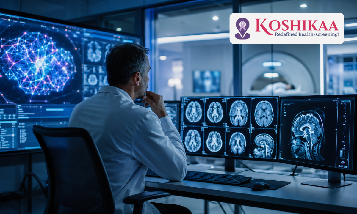
The integration of AI in diagnostic imaging represents a transformative paradigm shift in modern medicine, fundamentally elevating how clinicians detect, evaluate, and monitor complex health conditions. At Koshikaa, a leading proactive Health screening centre in Bangalore, we harness deep learning algorithms alongside high-resolution radiological technology to deliver unprecedented diagnostic accuracy, reduced reporting turnaround times, and earlier disease detection.
For decades, medical imaging relied entirely on manual visual inspection by human radiologists. While expert clinical judgment remains indispensable, the sheer volume and subtle complexity of modern high-slice CT scans, 3T MRIs, and digital mammograms present significant diagnostic challenges.
Subtle micro-calcifications, early-stage pulmonary nodules, or tiny vascular anomalies can easily escape detection during early disease phases.
Artificial intelligence functions as an intelligent, high-speed secondary reader. Powered by Convolutional Neural Networks (CNNs) trained on millions of anonymized radiological datasets, AI algorithms analyze pixel-level density shifts, spatial patterns, and tissue volume changes with remarkable mathematical precision.
Rather than replacing human expertise, these digital tools augment the specialist by flagging suspicious areas in real-time and ensuring no critical finding goes unnoticed. At Koshikaa, our mission focuses on bridging the gap between cutting-edge technology and accessible preventive healthcare.
Incorporating AI-driven image processing and automated protocol selection, we help patients across Bangalore gain actionable, reliable clarity regarding their internal organ health long before symptoms appear.
What You Will Learn in This Guide:
- How deep learning algorithms reduce human error and eliminate reader fatigue in complex scans.
- The role of AI triage in prioritizing urgent, life-threatening emergency findings.
- How neural network reconstruction lowers radiation dosage in modern CT imaging technology.
- Why combining expert radiological review with AI assistance creates a new gold standard for preventive health screenings.
By combining sophisticated computer-aided detection with expert radiologist oversight, modern diagnostic centers can dramatically lower false-positive rates, reduce unnecessary secondary biopsies, and provide patients with total peace of mind.
Medical Disclaimer
The clinical information, technological explanations, and diagnostic insights outlined in this article are provided solely for educational and public health awareness purposes. This content does not constitute formal medical advice, clinical diagnosis, or a personalized diagnostic recommendation. Individual imaging requirements and health screening protocols vary based on personal medical history and physician guidance. Always consult a qualified physician or certified radiologist to discuss your specific health concerns or diagnostic needs. Never delay seeking professional medical evaluation based on the general information provided in this publication.
Enhancing Accuracy & Reducing Human Error in Radiology
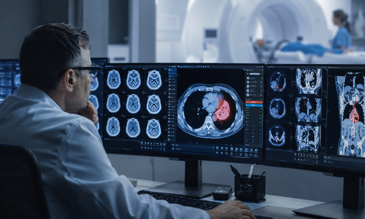
Diagnostic radiology involves interpreting intricate, high-dimensional visual data where even minute miscalculations can impact patient management. In busy clinical settings, radiologists often evaluate hundreds of complex scans daily, making cognitive fatigue, perceptual oversight, and inter-observer variability real challenges.
Modern AI radiology solutions address these challenges directly by functioning as an objective, highly sensitive analytical safety net across every phase of image interpretation.
Computer-Aided Detection (CAD) & Sub-Visual Pattern Recognition
Traditional visual evaluation relies entirely on the human eye to differentiate subtle variations in tissue density, grayscale contrast, and anatomical symmetry.
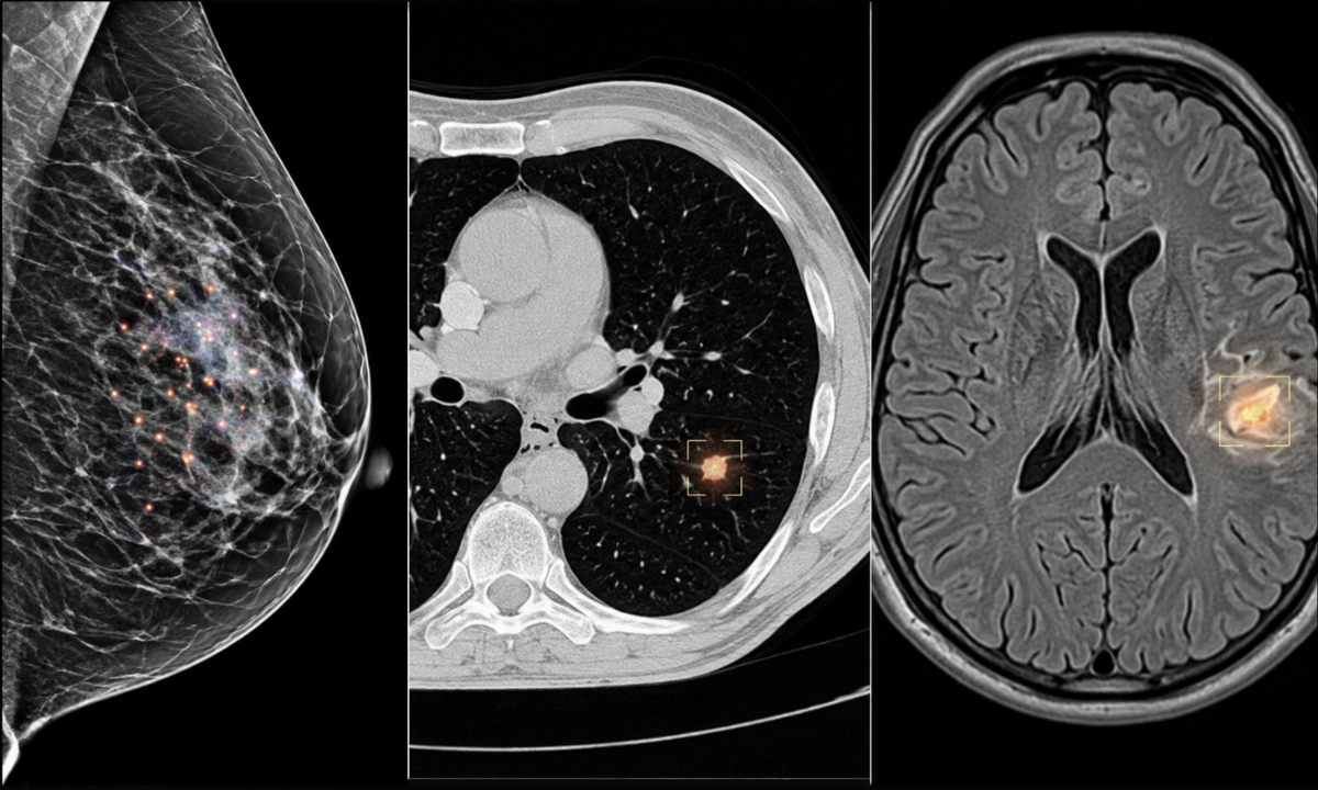
Deep learning models, however, evaluate pixel and voxel values at a granular mathematical level, detecting subtle structural deviations long before they manifest as distinct physical masses:
- Mammography & Breast Imaging: Deep neural networks highlight micro-calcification clusters and architectural distortions in dense breast tissue, reducing missed lesions and lowering unnecessary recall rates for benign findings.
- Chest & Pulmonary Scans: Computer vision tools systematically analyze multi-slice lung and chest scans to pinpoint sub-centimeter ground-glass opacities, solitary pulmonary nodules, and subtle pleural thickenings that might be overlooked during routine review.
- Musculoskeletal & Neurological Diagnostics: Automated detection systems identify subtle hairline stress fractures, early bone density loss, and micro-ischemic brain lesions that present with minimal contrast differences.
Mitigating Cognitive Fatigue & Reader Variability
Interpretation consistency is critical in preventive medicine and long-term health tracking.
Human visual perception naturally fluctuates due to high case volumes, long working hours, or environmental distractions. Algorithmic analysis remains completely unaffected by workload intensity, maintaining uniform analytical precision whether evaluating the first scan of the day or the fiftieth.
[Raw Imaging Dataset] ➔ [Parallel AI Algorithm Review] ➔ [Automated Anomaly Heatmap]
│
[Expert Radiologist Review] ◄─────────────────────────────────────┘
│
▼
[Consolidated High-Precision Final Report]
Providing concurrent feedback often rendering visual heatmaps or bounding boxes around suspicious tissue zones, these systems prompt radiologists to perform targeted second looks before finalizing diagnostic reports. Studies show that combining algorithmic screening with expert physician review achieves higher diagnostic sensitivity and specificity than either approach operating independently.
Precision Volumetric Quantification vs. Manual Measurement
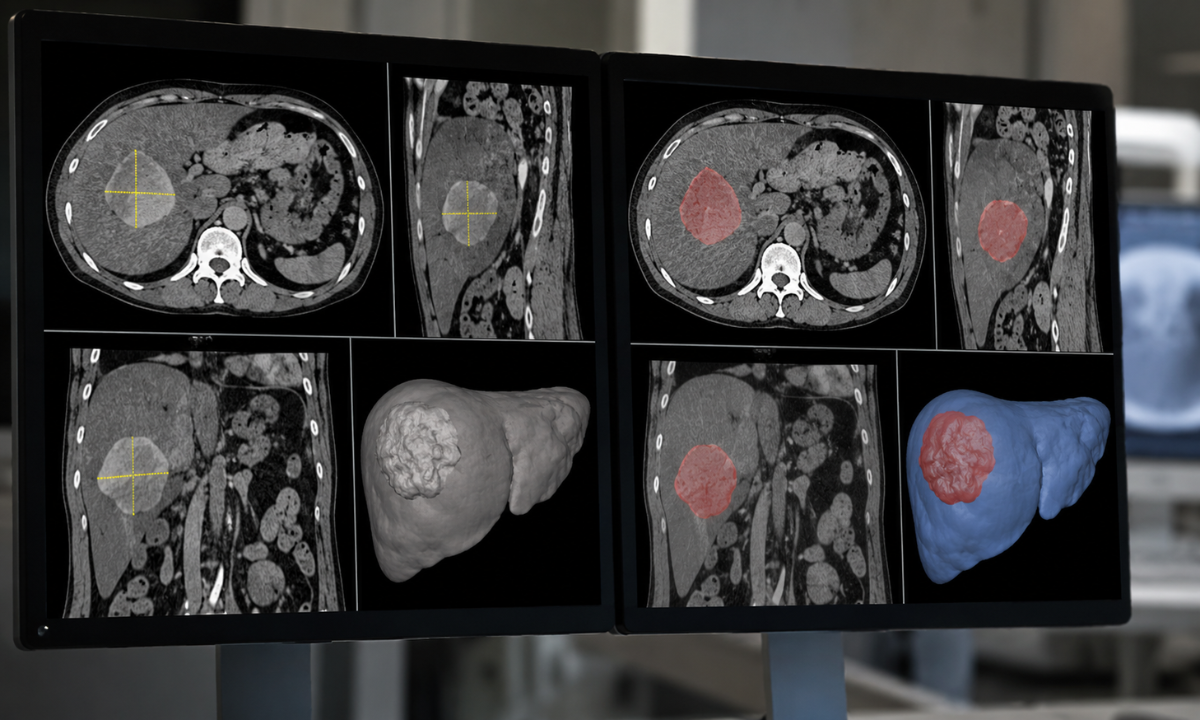
Beyond initial lesion detection, assessing whether a tumor, cyst, or inflammatory focus is responding to therapy requires exact measurement over time.
Volumetric Precision Advantage: Traditional manual measurements use two-dimensional linear calipers, which can vary depending on angle and slice selection. Deep learning algorithms perform automated 3D volumetric segmentation, calculating precise tissue volume and density metrics to accurately track disease progression or treatment response across sequential visits.
Speed, Triage, and Workflow Optimization in Emergency & Preventive Care
In modern medical imaging, diagnostic accuracy is deeply tied to processing speed. A delay in detecting a critical anomaly such as an acute intracranial hemorrhage, pulmonary embolism, or aortic dissection can significantly alter clinical outcomes.
The integration of modern digital diagnostics streamlines the clinical workflow, turning time-consuming image processing into a rapid, automated triage pipeline.
Automated Case Triage & Worklist Prioritization
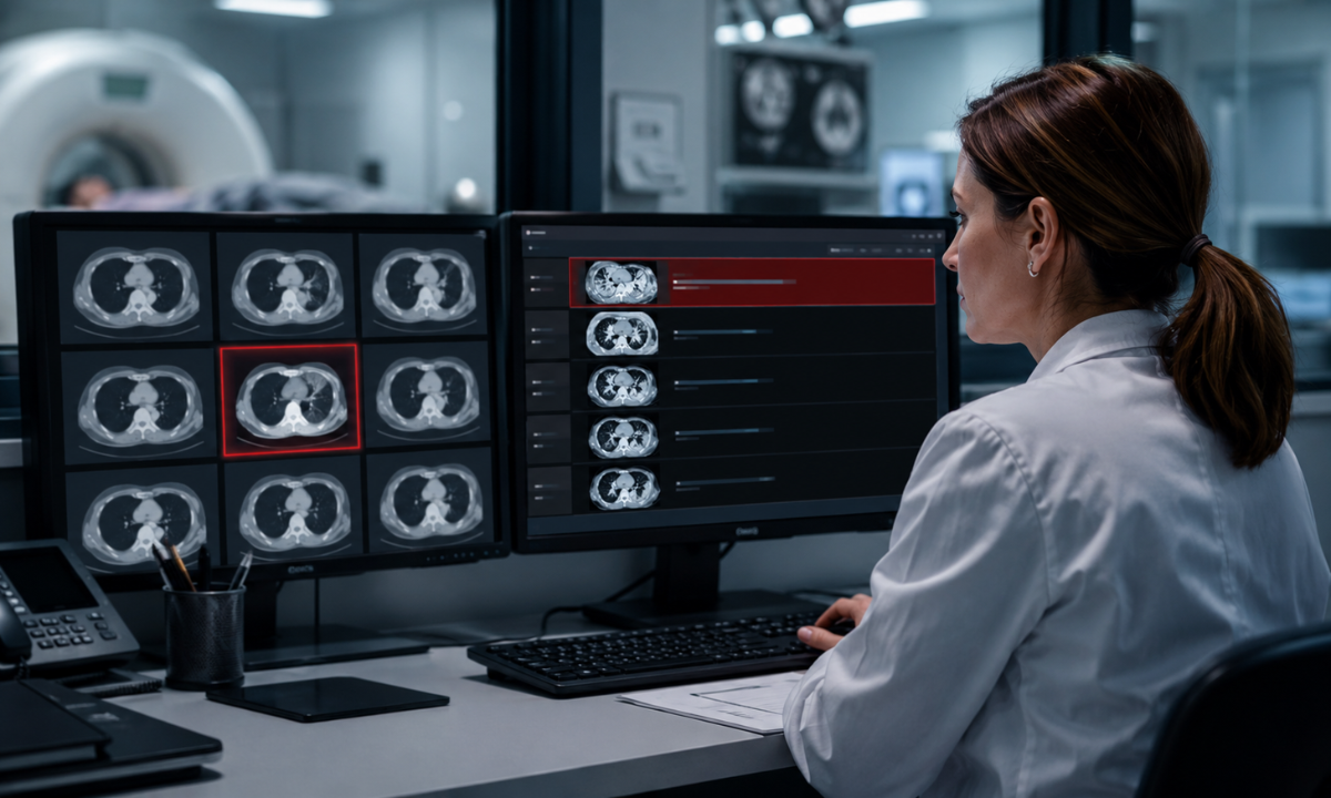
In traditional diagnostic workflows, radiological scans are typically reviewed in the chronological order they are acquired. While this works well for routine health checkups, it can introduce delays when an unscheduled, life-threatening finding occurs.
Deep learning triage algorithms scan raw image datasets immediately after acquisition, operating in the background before a physician opens the case file:
- Immediate Anomaly Flagging: As soon as a patient completes a scan, parallel processing algorithms analyze the cross-sectional image slices for critical emergency markers such as acute arterial blockages, free intra-abdominal air, or midline brain shifts.
- Dynamic Priority Re-Ordering: If a life-threatening abnormality is identified, the software instantly elevates the case to the top of the radiologist’s reading queue and issues an automated alert to the clinical team.
- Rapid Critical Intervention: By prioritizing urgent cases automatically, critical findings are escalated for immediate review, drastically reducing the time from scan completion to life-saving clinical action.
Streamlining Image Pre-Processing & Segmentation
Beyond emergency triage, advanced algorithm integration automates labor-intensive pre-processing tasks that previously consumed valuable physician time:
| Automated Workflow Task | Traditional Manual Process | AI-Assisted Automated Process | Clinical Impact & Patient Benefit |
|---|---|---|---|
| Organ & Lesion Segmentation | The radiologist manually traces organ boundaries and tumor margins slice-by-slice. | Neural networks automatically perform 3D volumetric segmentation in seconds. | Reduces processing time by up to 70%, allowing physicians to focus on diagnostic analysis. |
| Prior Study Registration | Manual side-by-side comparison and alignment of historical scans with current images. | Automated elastic registration aligns past and current cross-sectional slices. | Enables instant, precise tracking of subtle tumor growth or lesion regression. |
| Automated Draft Structuring | Manual typing and dictation of anatomical measurements and findings. | Software auto-populates quantitative measurements directly into structured draft templates. | Eliminates manual transcription errors and speeds up final report delivery. |
Faster Turnaround for Preventive Health Screenings: For patients undergoing routine annual health checkups, automated pre-processing means comprehensive diagnostic reports combining blood biomarkers and cross-sectional imaging are delivered significantly faster, eliminating unnecessary waiting anxiety and enabling quicker physician follow-ups.
Low-Dose CT Scanning & Deep Learning Image Reconstruction
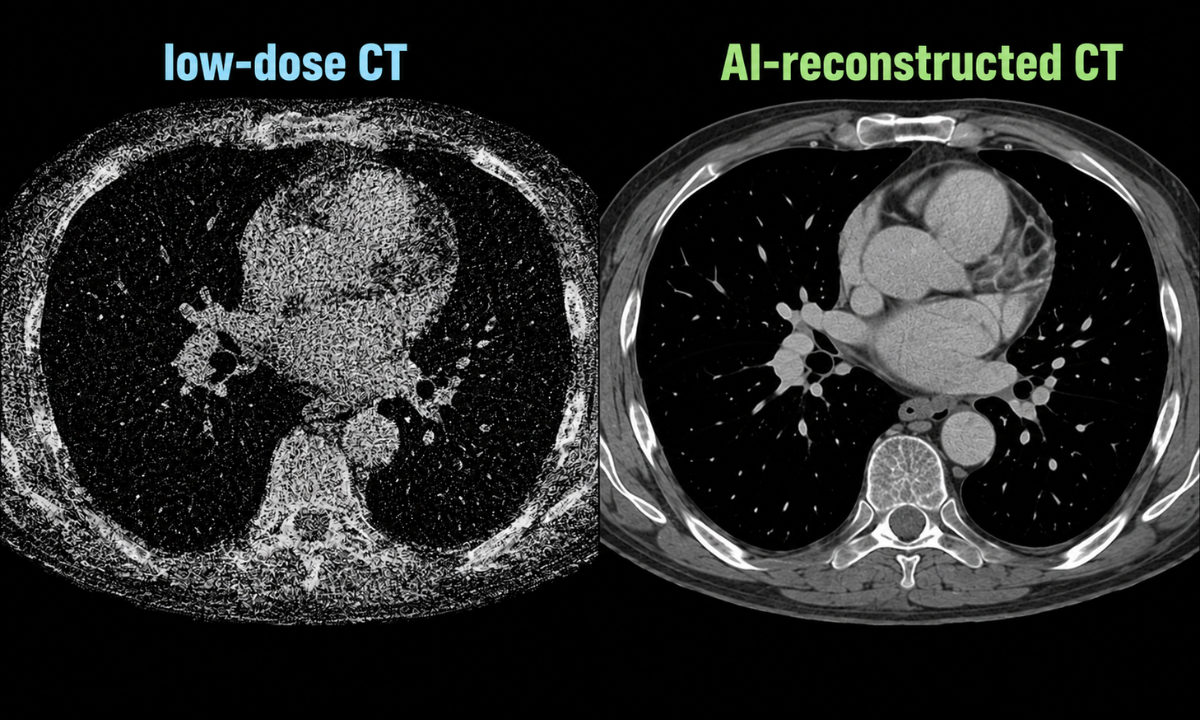
Computed Tomography (CT) is an essential diagnostic tool in modern medicine, offering detailed cross-sectional views of internal organs, vascular networks, and skeletal structures. However, a long-standing challenge in diagnostic radiology has been balancing image clarity against ionizing radiation exposure.
Modern advances in CT imaging technology, specifically Deep Learning Image Reconstruction (DLIR), have effectively resolved this dilemma by decoupling radiation dose from image resolution.
The Radiation vs. Resolution Challenge
Historically, CT image creation relied on Filtered Back Projection (FBP) or hybrid Iterative Reconstruction (IR) algorithms. Under low-dose radiation settings, these traditional mathematical models produce significant quantum noise, graininess, and streak artifacts particularly in dense anatomical regions like the shoulders, pelvis, or skull base. To obtain crystal-clear diagnostic images, clinicians often had to accept higher radiation doses, making frequent follow-up scans a safety concern.
How Deep Learning Image Reconstruction (DLIR) Works
Deep Learning Image Reconstruction replaces traditional mathematical approximations with deep convolutional neural networks trained on vast repositories of high-dose, artifact-free reference data. During scan acquisition, the neural network processes raw projection data in real time:
- Noise Discrimination: The algorithm intelligently distinguishes true biological tissue signals from random photon noise, selectively suppressing background grain without blunting sharp anatomical margins.
- Artifact Reduction: Deep learning models correct for beam-hardening and photon-starvation artifacts near heavy bone structures, producing uniform image clarity across the entire cross-section.
- Structural Preservation: Unlike older iterative algorithms that sometimes created a smoothed, “plastic-like” texture, deep learning maintains natural anatomical textures and subtle low-contrast boundaries.
Key Patient & Clinical Advantages
Standard-Dose Traditional CT ➜ Higher Radiation Exposure
⬇
Low-Dose AI-Reconstructed CT ➜ Up to 50–80% Dose Reduction + Superior Image Clarity
| Clinical Parameter | Traditional Reconstruction (FBP/IR) | Deep Learning Reconstruction (DLIR) | Patient Health Impact |
|---|---|---|---|
| Radiation Exposure | Standard diagnostic radiation dose required for clear resolution. | Reduced by 50% to 80% compared to standard protocols. | Dramatically lowers cumulative lifetime radiation exposure, particularly for pediatric patients and those needing repeat monitoring. |
| Image Noise & Grain | Higher noise levels when radiation tube current is lowered. | Significant noise suppression even at ultra-low radiation doses. | Ensures subtle soft-tissue lesions and micro-calcifications remain sharp and readable. |
| Contrast-to-Noise Ratio | Degrades rapidly under low-dose conditions. | Enhanced Contrast-to-Noise Ratio (CNR) across all tissue densities. | Improves diagnostic confidence when evaluating liver lesions, lung nodules, and vascular blockages. |
Safer Annual Health Screenings: The advent of low-dose, AI-reconstructed scanning makes preventive imaging such as low-dose chest screening for early lung lesions or coronary calcium scoring safer and more accessible for routine health tracking.
AI-Driven Advances in Magnetic Resonance Imaging
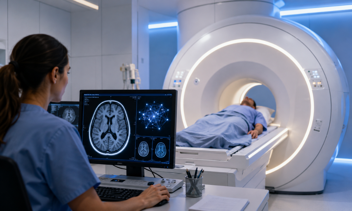
Magnetic Resonance Imaging (MRI) is the gold standard for soft-tissue diagnostics, offering unmatched clarity when evaluating the brain, spinal cord, joints, and abdominal organs.
However, traditional MRI technology protocols have historically presented operational challenges: long examination times (often lasting 30 to 45 minutes), sensitivity to patient movement, and discomfort for claustrophobic or pain-afflicted individuals.
The integration of deep neural networks into modern MRI technology has fundamentally reshaped soft-tissue imaging, solving these historic pain points by dramatically accelerating scan speeds while enhancing structural resolution.
Accelerating Image Acquisition with Deep Learning Reconstruction
Traditionally, achieving high signal-to-noise ratios (SNR) and sharp spatial resolution required acquiring dense raw data points, forcing patients to remain completely stationary for extended periods.
Deep Learning Reconstruction (DLR) algorithms change this dynamic by recovering high-fidelity images from significantly undersampled raw datasets:
- 30% to 50%+ Reduction in Scan Duration: Neural networks trained on pairs of rapid, low-resolution data and full-length, high-resolution reference scans learn to fill in missing data points in real time. This reduces acquisition times across routine neurological, spinal, and joint protocols by 30% to 50% or more without sacrificing diagnostic detail.
- Enhanced Scanner Throughput: Shorter examination windows allow diagnostic centers to serve more patients daily, reducing waiting times for critical diagnostic appointments.
- Improved Patient Comfort: Spending less time inside the narrow scanner bore significantly reduces patient anxiety and claustrophobia, making advanced scans far more accessible for elderly, pediatric, or acutely ill patients.
Eliminating Motion Artifacts in Soft-Tissue Scans
In soft-tissue diagnostics, subtle patient motion such as involuntary swallowing, breathing movements, or muscle twitching can introduce blurriness, ghosting artifacts, or streak distortions that render a scan unreadable.
[Involuntary Patient Motion / Respiration] ➔ [Raw K-Space Distortions]
⬇
[Deep Learning Motion Correction] ➔ [Restored Sharp Tissue Boundaries & Clear Detail]
Deep learning motion-correction models detect and compensate for patient movement mid-acquisition. After filtering out phase-shift errors caused by motion, the software restores sharp tissue boundaries and clear anatomical details. This minimizes the need for repeat scans, saving time and sparing patients additional stress.
High Contrast-to-Noise Ratios & Reduced Contrast Exposure
Deep learning algorithms selectively boost the Contrast-to-Noise Ratio (CNR) between healthy and diseased soft tissues. This enhanced clarity helps radiologists identify subtle micro-lesions, early cartilage degeneration, or small vascular malformations.
Safer Contrast Protocols: Advanced AI models can synthesize virtual high-contrast images from low-dose contrast agent data. This allows physicians to obtain clear, high-contrast diagnostic images while minimizing a patient’s overall exposure to gadolinium-based contrast agents.
Koshikaa’s AI-Augmented Care: Redefining Precision Diagnostics
We at Koshikaa believe that world-class healthcare begins with early, accurate, and accessible diagnosis. By integrating the latest medical imaging innovation with expert medical oversight, our diagnostic center brings high-grade radiological accuracy to every patient.
Whether you require a high-resolution MRI or a low-dose CT scan in Bangalore, our advanced facilities ensure your health screening is seamless, safe, and reliable.
Why Choose Koshikaa for Your Health Screening?
Selecting the right diagnostic partner directly impacts your treatment timeline and peace of mind. Here is why patients and physicians across Bangalore trust Koshikaa for their diagnostic needs:
- Dual-Layer Diagnostic Verification: Every scan undergoes parallel automated deep learning analysis paired with meticulous review by experienced radiologists for maximum diagnostic accuracy.
- Ultra-Low Radiation Protocols: Deep learning reconstruction delivers high-resolution cross-sectional clarity while reducing radiation exposure by up to 80%.
- All-in-One Testing Under One Roof: Access complete advanced radiology (CT, MRI, Ultrasound, Digital X-Ray, Mammography) alongside full-service pathology panels in a single visit.
- Rapid Digital Report Delivery: Automated case triage and pre-processing streamline workflows, ensuring fast report turnaround and eliminating unnecessary waiting anxiety.
- Patient-Centric Comfort: Our Banashankari facility features modern, wheelchair-accessible amenities with gentle, quick scanning workflows designed for elderly and anxious patients.
After harmonizing sophisticated algorithmic precision with human clinical empathy, Koshikaa empowers you and your healthcare providers to detect potential health risks early, when intervention is most effective.
Conclusion
Artificial intelligence is not replacing the human touch in modern medicine; it is elevating it. From detecting subtle micro-calcifications in early cancer screenings to lowering radiation doses in routine CT scans, AI-driven diagnostics are setting a new standard for patient safety and analytical precision. Taking a proactive approach to your health begins with choosing a diagnostic partner committed to precision technology, speed, and patient-first care.
Contact Koshikaa & Book Your Scan Today
Ready to schedule your proactive health checkup or advanced diagnostic scan? Connect with our specialist team at our Banashankari center:
- Address: 2nd Floor, 2227, 9th Main Road, Karesandra, Banashankari Stage II, Bengaluru, Karnataka – 560070
- Phone: +91 79966 66106
- Email: koshikaascreening@gmail.com
- Website: koshikaa.com