
Discovering structural changes in your organs can be deeply unsettling, yet understanding exactly what causes kidney scarring is the first step toward reclaiming complete control over your health.
When healthy renal tissue faces chronic irritation, it slowly replaces itself with firm, fibrous scar tissue that can alter how your body filters waste. Catching these changes in their infancy requires high-resolution visibility. By prioritizing an advanced CT scan in Bangalore, you can secure a clear, three-dimensional look at your internal anatomy.
This gives you the definitive data needed to stop cellular stress in its tracks, protecting your long-term wellness long before external symptoms ever manifest.
Rather than relying on how you feel, addressing structural health means looking directly at the root changes taking place beneath the surface. Kidney scarring isn’t an overnight event; it is a gradual process driven by manageable metabolic and physical stressors.
We bridge the gap between clinical complexity and proactive daily life, providing you with a clear roadmap to interpret your diagnostic data, stabilize your vibrant organ function, and build a lasting foundation for systemic vitality.
Medical Disclaimer
The clinical insights, pathological definitions, and preventative strategies detailed in this guide are intended strictly for educational and informational purposes. This content does not constitute professional medical advice, a formal clinical diagnosis, or a personalized treatment plan. Kidney scarring and disease progression are highly individualized processes heavily influenced by a patient’s unique genetic profile, history of systemic infections, and underlying conditions like chronic hypertension or diabetes. Always consult a qualified nephrologist or a board-certified healthcare provider to interpret your specific imaging and laboratory findings before initiating or altering any dietary, exercise, or medical regimen. Never delay seeking professional medical care based on the information read in this article.
Why Kidney Damage Happens and What Scarring Means?
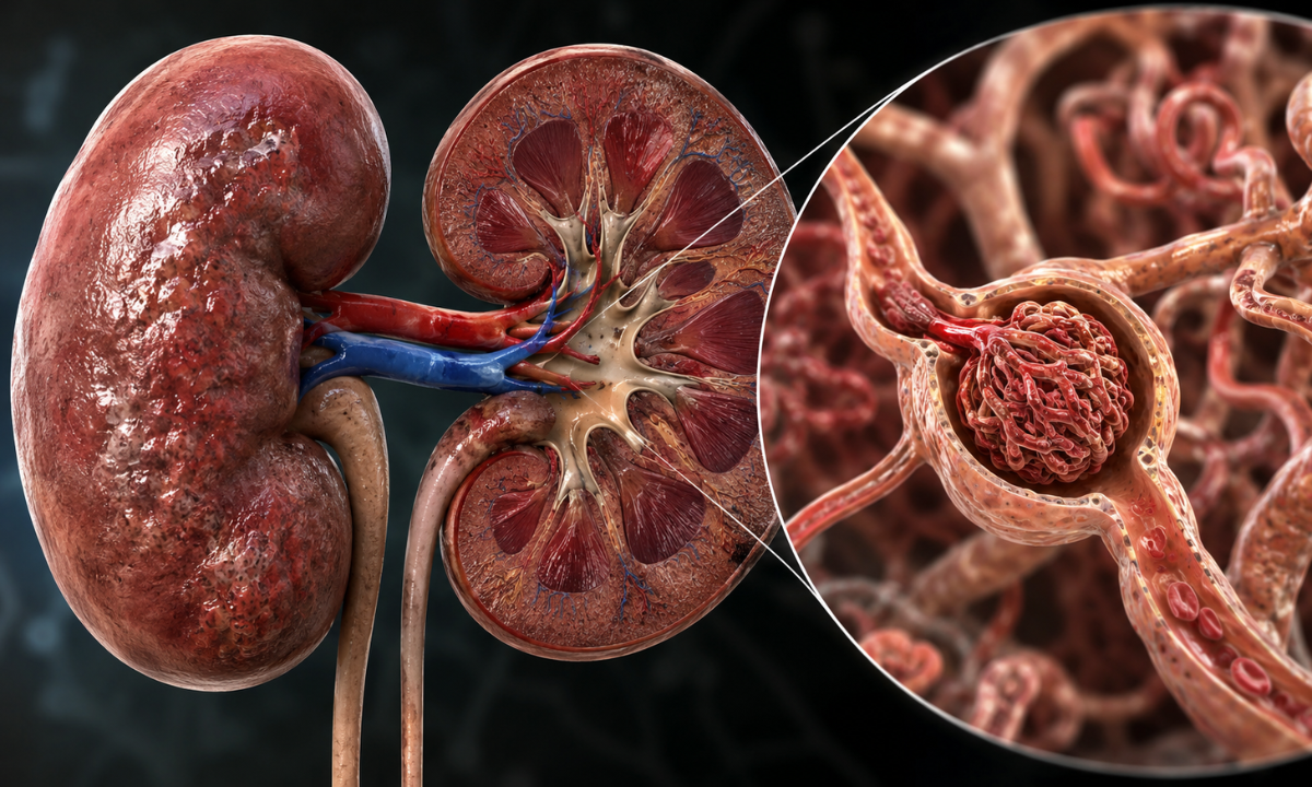
To understand the true nature of renal scarring, it helps to look closely at the microscopic architecture of your kidneys.
Your kidneys are not simple, solid blocks of tissue; they are highly intricate, living biological networks. Each kidney contains approximately one million microscopic filtering units called nephrons. Within every nephron lies a delicate cluster of loop-shaped blood vessels called a glomerulus, which acts as an ultra-fine, flexible colander designed to keep vital proteins in your blood while flushing waste out.
So, why kidney damage happens comes down to how these delicate colanders react to prolonged stress, inflammation, or physical injury. When these microscopic structures are exposed to continuous irritation, the body initiates a healing response.
However, much like a deep cut on your skin leaves behind a tough, inelastic mark, the delicate tissues inside your kidneys heal by building dense, fibrous connective tissue. This biological process is known as fibrosis or, more simply, kidney scarring.
As flexible, living tissue transforms into stiff, fibrous scar tissue, those specific nephrons lose their ability to filter blood. When a radiologist looks at a detailed imaging report, they will often evaluate the specific location of this structural change. One of the most common terms you might see flagged on a diagnostic report is kidney cortical scarring.
To understand what this means, we can look at the physical architecture of the kidney, which is divided into two distinct structural zones:
- The Renal Cortex (The Outer Filter): This is the outer crust or shell of the kidney. It contains the vast majority of your glomeruli and nephrons. As this is where the primary, high-volume blood filtration takes place, kidney cortical scarring means that the actual filtering units themselves have sustained damage.
- The Renal Medulla (The Inner Tubes): This is the deep, inner section of the kidney containing the specialized collecting ducts and tubules that transport the newly formed urine down into the bladder.
The Domino Effect of Untreated Scarring
When a localized area of the cortex develops scar tissue, those specific filters are permanently taken offline. As your body still has the same volume of blood to clean, the remaining healthy nephrons have to work significantly harder to compensate for the loss.
This creates a state of mechanical overwork known as hyperfiltration. If the underlying cause of the initial scar isn’t identified and corrected, this continuous, high-pressure overwork causes the remaining healthy filters to inflame, burn out, and develop scars of their own.
The Koshikaa Educational Perspective: A minor area of cortical scarring is not an immediate clinical failure.
Your kidneys possess an extraordinary functional reserve, meaning you can live a completely vibrant, healthy life with a small amount of scar tissue. The real purpose of early diagnostic tracking is to spot these focal scars while they are isolated, allowing you to change your lifestyle parameters and protect the surrounding healthy tissue from this progressive domino effect.
The Root Triggers and Common Causes of Renal Tissue Transformation
Kidney scarring does not happen at random. It is always the physical footprint of an underlying, unmanaged condition that has placed the renal tissue under prolonged stress. As the kidneys are highly vascular organs, meaning they are packed with miles of microscopic blood vessels, they are uniquely sensitive to shifts in your systemic blood pressure, blood sugar levels, and immune system health.
Identifying the specific root triggers that cause healthy nephrons to stiffen and form scar tissue, you can work with your medical team to neutralize the threat before it expands.
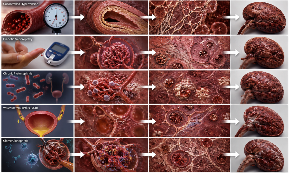
The primary medical conditions and structural anomalies that drive renal tissue transformation include:
| The Root Trigger | How It Causes Scar Tissue | The Long-Term Impact |
|---|---|---|
| Uncontrolled Hypertension | High blood pressure forces blood violently through the delicate glomerular capillaries, causing the walls of these tiny vessels to thicken, harden, and scar over time (a condition known as arteriolosclerosis). | Severely restricts blood flow to the nephrons, slowly starving them of oxygen and causing widespread cortical scarring. |
| Diabetic Nephropathy | Chronic, elevated blood glucose levels create harmful chemical byproducts that physically damage the delicate filtration mesh of the kidney, inducing chronic inflammation. | Causes the filters to leak vital protein and gradually replaces healthy structural tissue with dense, non-functional fibrous tissue. |
| Chronic Pyelonephritis | This is a severe, deep-tissue bacterial infection that travels up from the bladder into the actual functional tissue of the kidney itself. | The intense localized immune battle destroys the delicate renal architecture, leaving behind deep, permanent focal scars where the infection raged. |
| Vesicoureteral Reflux (VUR) | A structural, anatomical defect where urine abnormally flows backwards from the bladder up into the kidneys, creating high back-pressure and introducing bacteria. | Constant high-pressure stretching and recurrent infections combine to create severe, localized scarring, often beginning in childhood. |
| Glomerulonephritis | An autoimmune or inflammatory condition where your body’s own immune complexes mistakenly target and inflame the filtering units (glomeruli). | Rapidly compromises the structural integrity of the filters, leading to accelerated tissue scarring if not caught early. |
The Koshikaa Insight: Many of these conditions, especially early hypertension and diabetes, do not cause any physical pain or noticeable symptoms in their initial years. The scarring happens completely in the background.
By proactively checking your systemic numbers and monitoring your renal structure, you can catch these root triggers while they are still highly manageable, ensuring your internal filtration system remains uncompromised.
The Regeneration Myth: Will Kidney Tissue Grow Back?

When a patient reviews their diagnostic imaging report and sees words like “focal scar” or “cortical thinning,” the very first question they understandably ask their physician is: will the kidney grow back?
To answer this comprehensively, we have to look at the unique biological rules governing human organs. Different tissues in your body have completely different capacities for self-repair. For example, your liver is famous for its incredible regenerative capability; you can remove a significant portion of a healthy liver, and the remaining cells will rapidly multiply to restore the organ to its original size and shape.
Your skin operates similarly, constantly shedding old layers and weaving fresh cellular patches over cuts and scrapes.
The human kidney, unfortunately, follows a completely different biological blueprint.
The Cellular Reality of the Nephron
Your kidneys are born with a fixed endowment of filtering units. From the day you are born, you possess roughly one million nephrons in each kidney.
Unlike skin or liver cells, specialized kidney cells, specifically the podocytes (the highly advanced, star-shaped cells that form the ultimate microscopic mesh of the filtration barrier), cannot replicate or multiply once you reach adulthood.
- When a Nephron is stressed: If a kidney cell is merely irritated, inflamed, or sluggish due to poor hydration or temporary metabolic shifts, it can recover completely. With the right lifestyle interventions, cellular function can be fully restored.
- When a Nephron Scars: If the structural stress continues to the point where the nephron dies and is replaced by fibrous scar tissue, that specific filter is permanently offline. The body cannot grow new nephrons, nor can it dissolve dense, established collagen scars to regenerate pristine, functional kidney tissue.
Understanding Functional Reserve: The Silver Lining
While the fact that scar tissue is permanent might sound alarming at first, the human body has built an incredible safety net into your anatomy. Your renal system is engineered with a massive functional reserve.
In a healthy individual, both kidneys working together actually provide far more filtration power than your body strictly needs to survive. This is precisely why a living kidney donor can safely give away an entire kidney to someone in need. The single remaining kidney instantly adapts, growing slightly larger in a process called compensatory hypertrophy, and comfortably handles up to 70–80% of the filtration work that two kidneys used to share.
To understand how you can thrive even with localized scarring, think of your kidneys like a highly sophisticated business team of 100 people:
- Total Functional Capacity: 100% (2 Million Nephrons)
- Minor Scarring Occurs (e.g., 15% Lost): The remaining 85% of healthy filters step up to carry the workload.
- Outcome: Complete, vibrant filtration remains entirely stable.
If 10 to 15 workers face burnout and go permanently offline due to a focal scar from an old infection or a period of high blood pressure, the remaining 85 workers can easily step up, divide the daily task list among themselves, and keep the company running at peak efficiency.
Your overall kidney function (measured via your blood eGFR score) can remain completely normal and healthy even if a localized portion of the kidney contains permanent structural scars.
The True Goal of Proactive Renal Care
As kidney tissue does not grow back, the entire philosophy of renal medicine shifts from recreation to uncompromising preservation. You do not need to figure out how to regrow lost tissue; you simply need to protect the massive, healthy functional reserve you still have.
By proactively identifying minor areas of scarring through advanced screening, you gain the precise data needed to eliminate the stressors affecting your remaining healthy nephrons. Stopping the progression of scarring ensures that your remaining functional tissue stays completely healthy, vibrant, and capable of filtering your blood perfectly for the rest of your life.
Visualizing the Damage with Advanced Diagnostic Imaging

As your kidneys are tucked away deep inside your abdominal cavity, blood and urine tests can only provide a chemical report card of your renal health.
They can tell you if your overall filtration speed is slowing down or if your filters are actively leaking vital protein, but they cannot physically pinpoint where structural changes are taking place. To truly understand the physical shape, size, and layout of your renal tissue, clinicians rely on advanced diagnostic imaging.
Visualizing your internal anatomy allows doctors to differentiate between a temporary chemical imbalance and a permanent structural change, such as a localized scar. Depending on your medical history and initial lab work, evaluating your kidneys typically starts with non-invasive, radiation-free technology.
Opting for an advanced Ultrasound scan in Bangalore serves as the primary, high-impact tool for structural kidney evaluations. During this quick and completely painless procedure, a specialized clinical sonographer uses high-frequency sound waves to generate real-time, cross-sectional images of your renal system.
When analyzing an ultrasound or a high-resolution CT scan, radiologists look closely at several distinct physical markers to map out the presence of scar tissue:
- Organ Size and Contour: A healthy adult kidney is typically between 10 to 12 centimeters in length with a perfectly smooth, uniform outer border. If a kidney has significantly shrunk in size, or if its outer contour appears bumpy, irregular, or indented, it is a clear physical indication of long-term tissue scarring and chronic volume loss.
- Cortical Thickness: The renal cortex, the outer layer where your microscopic filtering units live, should possess a healthy, robust thickness. If an ultrasound reveals significant thinning of this outer shell, it flags a widespread loss of functioning nephrons due to advanced cortical scarring.
- Echogenicity (Tissue Density): Sound waves bounce off different types of bodily tissues in unique ways. Healthy, fluid-rich kidney tissue appears relatively dark on an ultrasound screen. Permanent scar tissue, however, is dense and fibrous, causing it to reflect sound waves intensely. If a report flags increased echogenicity or bright, echogenic foci, it means the scanner has physically detected areas of dense, fibrous scar tissue.
By pairing advanced structural imaging with your routine blood and urine panels, your medical team can accurately map out the exact geographical layout of your kidneys.
This unified diagnostic approach ensures that you aren’t just guessing about your internal health based on raw numbers, but actually looking at the physical structural integrity of your vital filtration system.
Treatment and Protection Strategies

Once a kidney-damaged patient discovers the presence of structural scar tissue, the clinical focus shifts entirely from anxiety to strategic action. While established scar tissue cannot be reversed, your remaining healthy renal tissue possesses an extraordinary capacity to maintain your body’s fluid and chemical balance.
Protecting this vital functional reserve requires a highly coordinated approach that targets the root mechanical and metabolic stressors running through your blood. Here is the definitive roadmap to stabilizing your kidney health and halting the progression of structural damage:
1. Tight Systemic Blood Pressure Management
High blood pressure is the leading driver of progressive cortical scarring. When blood forces its way too violently into the kidneys, it acts like a high-pressure power washer, damaging delicate internal mesh filters.
- The Target: For individuals with early-onset kidney changes or protein leakage, international clinical guidelines typically recommend keeping blood pressure strictly below 130/80 mmHg.
- The Clinical Toolkit: Physicians frequently utilize specific classes of blood pressure medications, such as ACE inhibitors or Angiotensin Receptor Blockers (ARBs). Beyond merely lowering pressure in your arm, these specialized medications actively dilate the exit pathways of the kidney’s filters, relieving internal mechanical stress and slowing down the scarring process.
2. Precise Glycemic and Metabolic Control
Elevated blood sugar creates inflammatory chemical byproducts that alter the cellular architecture of your nephrons. If you manage your metabolic health or diabetes, keeping your HbA1c levels within a strict, personalized target range prevents your blood vessels from hardening and turning into fibrous tissue.
3. Smart Dietary Engineering and Sodium Optimization
Your diet directly influences the workload placed on your remaining functional nephrons. Modifying what you consume can drastically reduce the metabolic stress your kidneys have to process:
- Sodium Reduction: Restricting your daily sodium intake to under 2,000 milligrams (about one teaspoon of salt) prevents fluid retention and shields your filtering units from volume overloads.
- Balanced Protein Intake: Consuming excessive amounts of dense protein forces your kidneys to generate high amounts of urea, increasing hyperfiltration stress. Shifting toward moderate, high-quality, or plant-based protein sources keeps the filtration workload highly manageable.
4. Continuous Diagnostic Auditing
Managing structural changes means tracking your progress through objective data. A proactive protection plan relies on routine, non-invasive lab evaluations to ensure your remaining tissue stays perfectly stable:
- Serum eGFR Blood Panel: Tracks overall filtration speed to ensure healthy tissue compensates perfectly (checked every 3 to 6 months).
- uACR Urine Tracking: Monitors filter integrity to ensure protein leakage is completely halted.
Aggressively managing these therapeutic targets, you change the environment inside your body. You step away from a state of progressive tissue decline and establish a stable, long-term equilibrium, ensuring your remaining healthy nephrons continue to filter your blood cleanly and efficiently for decades to come.
Why Choose Koshikaa for Your Preventative Renal Care?
When dealing with structural changes like renal scarring, catching cellular stress before it hardens into permanent fibrous tissue is the ultimate key to longevity. At Koshikaa, we have entirely reimagined the diagnostic experience.
We look past the rigid, clinical anxiety of traditional laboratories to provide an empathetic, patient-centric ecosystem focused squarely on proactive preservation.
As the best Health Screening Centre in Bangalore, we empower you to take complete command of your renal health journey through a highly sophisticated diagnostic approach:
- Integrated Structural and Metabolic Profiling: We understand that a single data point never tells the whole story. Our comprehensive screening packages seamlessly bundle high-precision metabolic blood panels, advanced urine protein metrics, and state-of-the-art imaging to evaluate both the function and physical structure of your kidneys simultaneously.
- Deep Lifestyle Contextualization: Before you ever undergo a scan, our digital health ecosystem maps your unique biological story including your daily hydration trends, nutritional habits, and genetic risk factors. This allows our clinical team to contextualize your results and look at the whole person, not just a row of laboratory metrics.
From the precision of our imaging systems to the exactness of our biochemical assays, Koshikaa delivers absolute clinical accuracy. This high-fidelity data gives you and your consulting physician the perfect window of opportunity to implement lifestyle changes while your functional reserve is at its strongest.
Conclusion
Discovering that your kidneys have developed localized scar tissue is not a signal of inevitable decline; it is a clear, definitive call to action. As kidney tissue does not grow back, your long-term vitality depends entirely on how aggressively you protect, optimize, and value the extraordinary functional reserve you still have.
Stepping away from guesswork and stepping into absolute diagnostic clarity, you gain the exact knowledge required to halt the progression of tissue damage. Managing your blood pressure, refining your metabolic health, and conducting regular checks on your internal filters are the ultimate steps toward a vibrant, uncompromised future.
Partner with Koshikaa today, establish your baseline data, and secure the strong structural foundation your body needs to thrive for decades to come.