
When it comes to proactive healthcare, many people assume that if they feel completely healthy on the outside, everything must be functioning perfectly on the inside.
However, your body often hides its most critical metabolic shifts deep within your bloodstream. A standard lipid panel blood test is far more than just a routine cholesterol check; it serves as a highly detailed health roadmap that evaluates your risk for cardiovascular disease, tracks how your body manages energy, and monitors your overall metabolic function. Understanding these hidden markers is the first step toward preventing long-term complications before they ever affect your quality of life.
Because your blood chemistry constantly changes based on your genetics, diet, and daily habits, getting an accurate assessment requires a precise and reliable diagnostic partner.
As a premier Health Screening centre in Bangalore, Koshikaa is dedicated to helping you decode these vital metrics with absolute clarity. By securing a comprehensive Blood test in Bangalore through our state-of-the-art facilities, you can transform complex laboratory numbers into actionable, personalized lifestyle insights.
Let us look past the clinical terms and explore exactly what this essential blood test reveals about your internal well-being and how it protects your future health.
Medical Disclaimer
The information provided in this article is strictly for educational purposes and does not replace professional medical advice, formal diagnosis, or specialized clinical treatment. Standard lipid ranges can vary based on individual health profiles. Always consult directly with a qualified physician regarding your specific laboratory results, dietary changes, and cardiovascular risk factors. Never ignore medical warnings or delay seeking professional care based on the contents of this guide.
Why Get Tested? Addressing the Silent Symptoms

One of the most dangerous misconceptions about high cholesterol and unbalanced triglycerides is that you will physically feel a warning sign when something is wrong. In reality, lipid imbalances are almost entirely asymptomatic.
Unlike a sudden fever or a sharp muscle ache, dangerous fat accumulation inside your arteries does not cause immediate physical distress. Waiting until you develop chest discomfort or breathing issues before getting checked means waiting until significant cardiovascular strain has already occurred.
When discussing the specific symptoms for a blood test, the symptoms are not physical pains; they are your unique lifestyle risk factors, your medical history, and your age. Your body relies on a lipid panel blood test to flag internal damage long before it manifests as a medical emergency.
If you experience or identify with any of the following underlying health markers, your body is already signaling the urgent need for a diagnostic evaluation:
- Unexplained Metabolic Fatigue: While high cholesterol itself doesn’t hurt, poor lipid metabolism frequently pairs with insulin resistance, sluggish blood circulation, and fluctuating energy levels that leave you feeling exhausted even after a full night’s rest.
- A Strong Family History: Your genetics heavily dictate how your liver processes dietary fats. If your parents or siblings have a history of high blood pressure, heart attacks, or early strokes, your internal baseline risk is naturally higher, making routine monitoring essential.
- Sedentary Routines and High-Stress Lifestyles: Long hours sitting at a desk combined with elevated daily stress trigger a surge in cortisol. This hormonal shift directly alters how your body stores fat, often driving up harmful lipid levels while depressing your protective cholesterol markers.
- Dietary Patterns Rich in Refined Carbs and Trans Fats: Consistently consuming processed foods, heavy oils, or excess sugars forces your liver to produce an abundance of baseline fats, which quietly circulate through your vascular system and slowly narrow your arterial walls.
Recognizing these structural risk factors as the true, silent indicators for a diagnostic check, you can take control of your health proactively.
Instead of waiting for a physical breakdown, scheduling a preventive evaluation allows you to see exactly what is happening beneath the surface, giving you the power to make simple, life-saving adjustments early.
What to Expect: The Procedure Demystified

Stepping into a clinic for a blood draw can occasionally bring up minor anxieties, especially if you are unsure of the exact requirements.
However, the actual procedure of lipid profile test screening is entirely straightforward, minimally invasive, and completed in a matter of minutes. Knowing how to prepare and understanding what happens during each phase ensures your results are perfectly accurate and reflective of your true baseline health.
To help eliminate any guesswork, let us walk through the simple timeline of your diagnostic journey, from your evening preparation to receiving your digital health insights.
The Diagnostic Journey Timeline
1. The Fasting Window (10 to 12 Hours Prior): As the food you consume heavily influences the immediate fats circulating in your bloodstream, a strict fasting period is necessary.
You should avoid all food, juices, tea, coffee, and alcohol for 10 to 12 hours before your appointment. Drinking plain water is highly encouraged, as staying well-hydrated makes your veins much easier to locate during the draw.
2. The Quick Sample Collection (At the Centre): Once you arrive at our facility, a highly trained phlebotomist will guide you to a comfortable seating area.
They will carefully cleanse a small area on your inner arm with an antiseptic wipe, apply a soft elastic band to locate a prominent vein, and use a sterile, ultra-fine needle to collect a small sample. Most patients report feeling nothing more than a minor, brief pinch.
3. Advanced Laboratory Processing: The moment your sample is secured, it is processed inside our advanced laboratory environment.
We utilize high-precision automated analyzers to ensure your lipid markers are evaluated down to the exact milligram, eliminating manual human error.
4. Rapid Digital Reporting (Within 24 Hours): You do not have to wait days in suspense to understand your health status.
Your comprehensive digital report is compiled, verified by our laboratory experts, and delivered straight to your smartphone within 24 hours, allowing you to instantly share the findings with your physician.
Procedure Protocol At a Glance
| Phase Requirements | What to Do | Why It Matters |
|---|---|---|
| Dietary Restriction | Fast completely for 10–12 hours. | Prevents recent dietary fats from artificially spiking your triglyceride readings. |
| Hydration Status | Drink plenty of plain water. | Plumps your veins for a smoother draw and prevents minor lightheadedness. |
| Activity Level | Avoid strenuous workouts right before. | Intense physical exertion immediately before a test can temporarily shift lipid baselines. |
Demystifying the process helps you approach your appointment with absolute peace of mind.
Spending just five minutes at a high-tech facility, you gain access to an incredibly detailed breakdown of your internal health. Now that you know how simple the testing process is, let us look directly at the five vital health revelations that will appear on your final report.
The 5 Key Things Your Lipid Panel Reveals
When you look at your final lab report, you will find a list of specific lipid markers. Rather than viewing these as isolated numbers, it helps to understand that each marker reveals a distinct story about your current arterial health, metabolic efficiency, and future cardiovascular wellness.
Here are the five critical health insights your lipid panel directly exposes:
1. Total Cholesterol Balance
This metric represents the total amount of cholesterol circulating in your bloodstream at the time of your test.
While it provides a helpful baseline view of your overall fat load, looking at total cholesterol alone is never enough. Your body uses this number as a starting point to see how heavily packed your vascular highways are before breaking them down into specific protective and high-risk components.
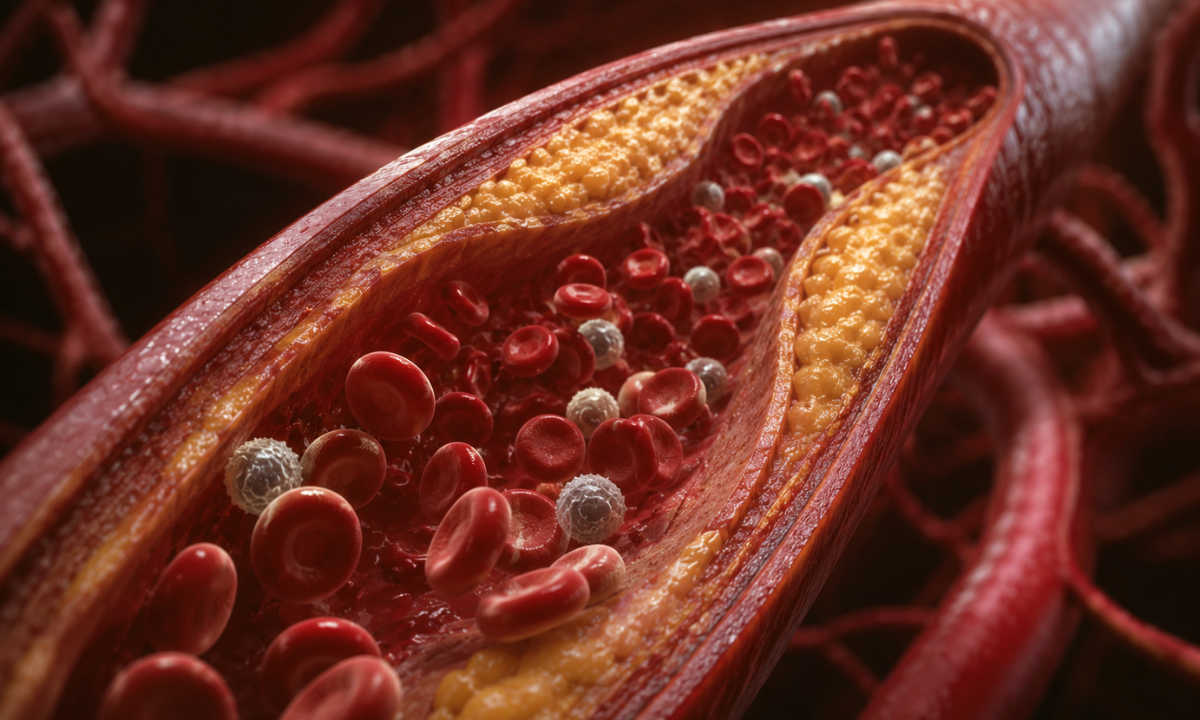
2. LDL Cholesterol (The Delivery Trucks)

Low-Density Lipoprotein, or LDL, is commonly referred to as “bad” cholesterol, but a more accurate way to visualize it is as a fleet of microscopic delivery trucks. LDL’s biological job is to carry vital fats out from your liver and deliver them to your cells to build protective membranes and hormones.
However, when you have an overabundance of these trucks circulating through your system, they drop their oily cargo along your arterial walls. Over time, this dropped fat oxidizes and transforms into hard plaque, a process called atherosclerosis that physically narrows your blood vessels and increases the risk of blockages.
3. HDL Cholesterol (The Clean-up Crew)
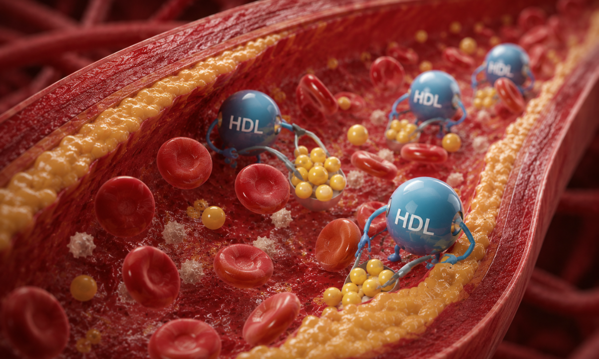
High-Density Lipoprotein, or HDL, is famously known as “good” cholesterol because it acts as your body’s natural internal recycling system. Think of HDL as a dedicated clean-up crew that constantly patrols your blood vessels.
It physically grabs the excess, dangerous cholesterol left behind by LDL trucks along your arterial walls, vacuums it up, and transports it safely back to your liver, where it is broken down and permanently excreted from your body. Maintaining a robust HDL level actively guards your heart against long-term structural damage.
4. Triglycerides (Stored Energy Reserves)

Unlike cholesterol, which is used strictly to build cellular walls and hormones, triglycerides are a pure chemical form of fat used entirely for energy storage.
When you consume excess calories, heavy carbohydrates, sugars, or alcohol that your body does not immediately burn for fuel, your liver instantly converts that surplus energy into triglycerides and stores them inside your fat cells. Elevated baseline triglycerides serve as a clear warning sign of a sluggish metabolism, often pointing toward insulin resistance, early fatty liver changes, or poor sugar processing.
5. The Total-to-HDL Ratio
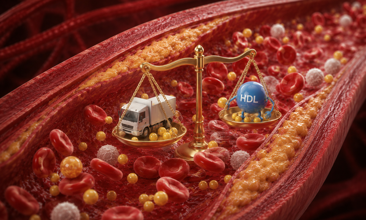
This final mathematical metric provides doctors with a highly accurate overview of your long-term cardiovascular health. It is calculated by dividing your total cholesterol number by your protective HDL number.
Instead of looking at your markers in isolation, this ratio reveals the exact balance of power between your “delivery trucks” and your “clean-up crew.” A lower ratio indicates that your body possesses an excellent internal defense system capable of keeping your vascular highways clean, balanced, and perfectly healthy.
Understanding Your Numbers: Normal Ranges At a Glance
When your secure digital report arrives, your specific lipid measurements will be expressed in milligrams per deciliter (mg/dL). While your doctor will always provide a highly personalized interpretation based on your family history and existing health conditions, knowing the standard normal values of lipid profile test results allows you to instantly gauge your baseline cardiovascular risk.
Use this simple reference guide to quickly decode your laboratory numbers.
| Lipid Marker | Optimal Target (Low Risk) | Borderline Risk (Monitor Closely) | High Risk (Requires Action) |
|---|---|---|---|
| Total Cholesterol | Less than 200 mg/dL | 200 – 239 mg/dL | 240 mg/dL and above |
| LDL (Bad Cholesterol) | Less than 100 mg/dL | 130 – 159 mg/dL | 160 mg/dL and above |
| HDL (Good Cholesterol) | 60 mg/dL and above | 41 – 59 mg/dL | Less than 40 mg/dL |
| Triglycerides | Less than 150 mg/dL | 150 – 199 mg/dL | 200 mg/dL and above |
If your numbers fall into the borderline or high-risk categories, there is no need to panic. For many patients, simple dietary adjustments, increased daily movement, and stress management can dramatically improve these numbers within a few short months. The true value of this test is simply knowing exactly where you stand so you can take control of your future.
Why Choose Koshikaa? The Koshikaa Advantage
At Koshikaa, we believe that world-class healthcare should be proactive, not reactive. As a leading diagnostic center in Banashankari, we do not just process lab samples; we provide a completely guided path to long-term wellness.
When you choose Koshikaa for your Blood test in Bangalore, you gain access to a truly personalized healthcare experience:
- Age-Specific Screening Packages: We recognize that a 30-year-old requires different health tracking than a 60-year-old. Our comprehensive health checkups are distinctly categorized by age and gender to ensure you only undergo the exact tests you actually need.
- Personalized Health Questionnaires: We are proud to be the first in the country to utilize an advanced personalized screening questionnaire. By analyzing your specific lifestyle habits and family history, we perfectly tailor your diagnostic experience.
- Complete Diagnostic Ecosystem: Should your blood work indicate the need for deeper investigation, our facility houses a complete suite of advanced imaging technology, including Low-Dose CT scans, MRI, and Ultrasound, allowing for immediate and seamless follow-up care.
Do not wait for invisible symptoms to become a physical emergency. By scheduling a comprehensive health assessment today, you take the ultimate step toward securing a healthier, brighter tomorrow.
Conclusion
A lipid panel is much more than a routine laboratory requirement; it is a highly accurate tool that gives you the power to protect your future. By understanding exactly what these five critical markers mean, you transition from worrying about silent symptoms to actively managing your cardiovascular well-being.
You do not have to wait for a medical emergency to start taking care of your body. With the quick, accurate, and highly personalized screening available at Koshikaa, securing your long-term metabolic health is easier, faster, and more accessible than ever before. Take the proactive step today, and give yourself the ultimate peace of mind.