
Early identification of structural abnormalities in the endocrine system is critical for preventing advanced metabolic complications. Clinicians routinely recommend a thyroid ultrasound to evaluate palpable neck masses or abnormal blood panels.
Securing a high-resolution Ultrasound Scan in Bangalore allows for the immediate, non-invasive visualization of the thyroid gland’s anatomy.
This diagnostic modality utilizes high-frequency sound waves to map soft tissues with exceptional clarity, completely eliminating the need for exposure to ionizing radiation. As a cornerstone of preventative medicine, regular screening tests help identify underlying nodules or tissue changes before external clinical symptoms manifest.
Undergoing the Best Health Screening Test in Bangalore ensures that subtle pathomorphological alterations are detected during their nascent stages. By evaluating the size, shape, and internal vascularity of the thyroid gland, this imaging protocol serves as a definitive tool for diagnosing asymptomatic thyroid disorders, thereby enabling timely clinical intervention and improved long-term patient outcomes.
Medical Disclaimer
The information provided in this clinical guide is strictly for educational purposes and does not constitute professional medical advice, diagnosis, or treatment. Always seek the direct guidance of a qualified endocrinologist, radiologist, or primary care physician regarding specific physical symptoms, diagnostic imaging requirements, and appropriate interventional protocols. Never disregard professional medical advice or delay seeking clinical evaluation based on the contents of this publication.
Key Clinical Highlights
- Radiation-Free Imaging: Utilizing acoustic wave propagation instead of ionizing radiation, making the procedure entirely safe for repeated diagnostic tracking.
- Soft Tissue Differentiation: Providing high-contrast resolution that allows radiologists to distinguish precisely between solid masses and fluid-filled cysts.
- Early Pathology Detection: Identifying microscopic nodules as small as two millimeters, long before they become palpable during a physical examination.
- Vascular Mapping: Integrating color Doppler technology to evaluate the blood flow dynamics within identified lesions or inflamed glandular tissue.
Identifying Thyroid Disorders
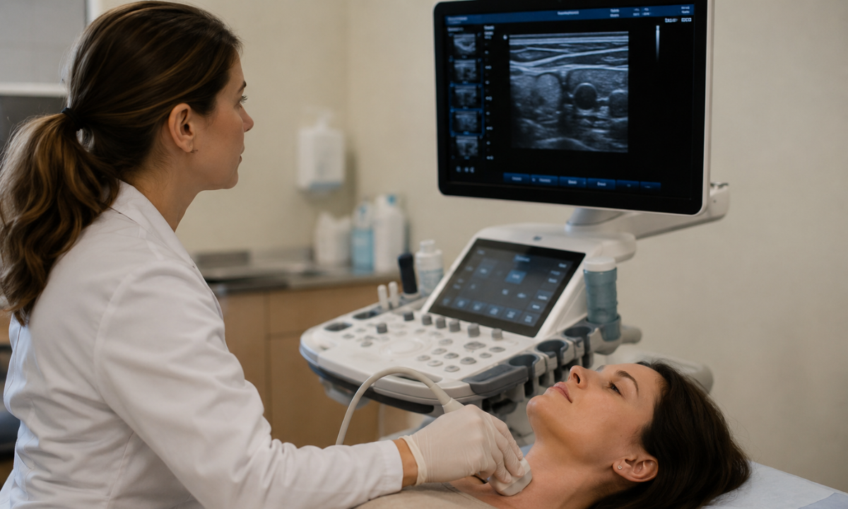
Diagnostic radiological imaging is rarely deployed without specific clinical justification. Physicians rely on the thyroid ultrasound as the primary investigative tool when physical examinations or serological (blood) tests indicate potential endocrine dysfunction. The primary objective is to transition from a generalized suspicion of illness to a precise, visually confirmed structural diagnosis.
Clinical specialists categorize the necessity for this specific ultrasound into three primary diagnostic pathways: physical anomalies, metabolic irregularities, and therapeutic monitoring.
Primary Diagnostic Pathways
The following table details the specific clinical presentations that immediately necessitate a high-resolution structural evaluation of the cervical region:
| Clinical Presentation | Physical or Physiological Manifestation | Diagnostic Objective of the Ultrasound |
|---|---|---|
| Palpable Cervical Nodules | A physician or the patient physically detects a localized, abnormal lump in the anterior neck region during a routine examination. | To determine the exact composition of the mass. The radiologist must differentiate whether the nodule is a harmless cystic (fluid-filled) structure or a complex solid mass requiring further oncological investigation. |
| Diffuse Thyromegaly (Goiter) | A generalized, visible swelling of the entire thyroid gland, often causing secondary symptoms such as dysphagia (difficulty swallowing) or a sensation of tracheal compression. | To accurately measure the total volumetric enlargement of the gland and identify if the swelling is uniform or caused by multiple hidden, internal nodules (multinodular goiter). |
| Abnormal Serological Panels | Routine blood work reveals severely elevated or depressed levels of Thyroid Stimulating Hormone (TSH), Free T3, or Free T4 (indicating Hyperthyroidism or Hypothyroidism). | To identify the structural root cause of the chemical imbalance. The scan can reveal patterns consistent with autoimmune conditions, such as Hashimoto’s thyroiditis, which alters the physical texture of the glandular tissue. |
| Cervical Lymphadenopathy | Abnormal, persistent swelling of the lymph nodes adjacent to the thyroid gland is often identified during an evaluation for chronic throat infections. | To map the precise location and vascularity of the enlarged nodes, determining if the swelling is a benign reactive response or indicative of a metastatic process. |
The Role of Ultrasound in Intervention Planning
Beyond initial diagnosis, the thyroid ultrasound serves a critical mechanical function for interventional procedures. If a solid nodule exhibits suspicious characteristics, the physician must perform a Fine Needle Aspiration (FNA) biopsy to extract cellular material for pathological review.
Performing a biopsy blindly based solely on physical palpation carries a high risk of missing the target lesion or puncturing adjacent vascular structures.
Therefore, clinicians utilize real-time ultrasound guidance. By continuously tracking the transducer on the monitor, the physician can visually navigate the biopsy needle directly into the exact center of the suspicious nodule with millimeter precision, guaranteeing an accurate cellular sample while maximizing patient safety.
The Thyroid Ultrasound Procedure
Demystifying the clinical environment is essential for reducing patient apprehension.
The thyroid ultrasound procedure is entirely non-invasive, executed on an outpatient basis, and typically concludes within twenty to thirty minutes. Because the protocol relies exclusively on acoustic wave technology, the patient experiences absolutely no exposure to ionizing radiation, and the process is entirely painless.
The radiological workflow follows a strict, standardized sequence to ensure complete anatomical capture and diagnostic accuracy.
The Sequential Scanning Protocol
Step 1: Patient Positioning and Anatomical Exposure
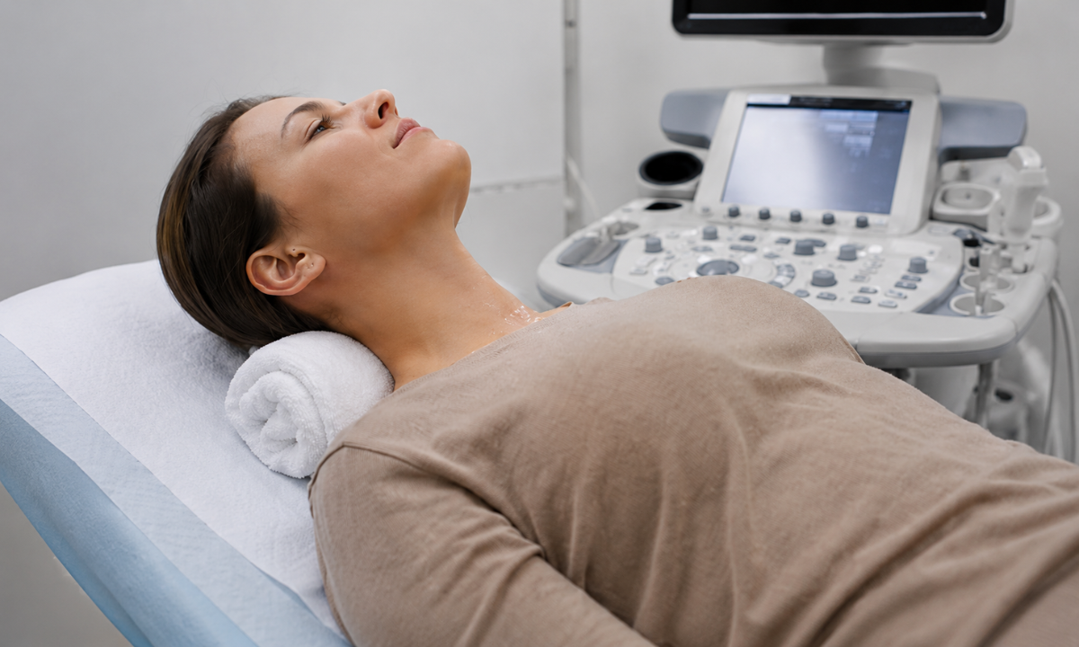
The patient is guided to lie in a supine position (flat on the back) on the examination table. To achieve optimal visualization of the anterior cervical region, the sonographer places a supportive clinical cushion directly beneath the patient’s shoulders. This specific positioning mildly hyperextends the neck.
Hyperextension brings the thyroid gland closer to the cutaneous surface (the skin) and simultaneously expands the physical scanning area, preventing the clavicles or the mandible from obscuring the acoustic window.
Step 2: Application of the Acoustic Coupling Medium
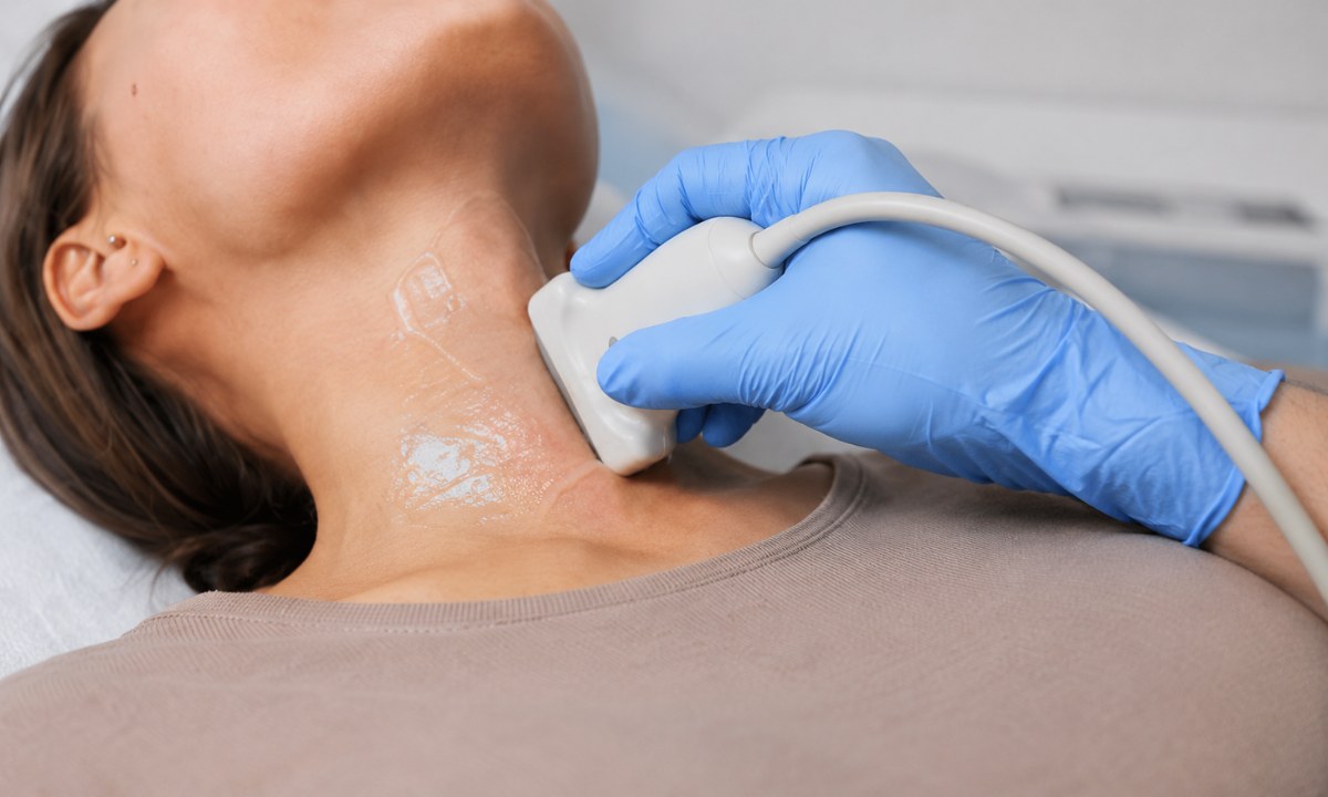
The sonographer applies a specialized, hypoallergenic, water-based conductive gel directly to the skin overlying the thyroid and trachea.
This gel serves a critical physical function. High-frequency sound waves cannot travel efficiently through air. The gel eliminates all microscopic air pockets between the medical instrument and the epidermis, providing a continuous fluid medium that ensures uninterrupted acoustic transmission into the deeper soft tissues.
Step 3: Transducer Manipulation and Image Acquisition
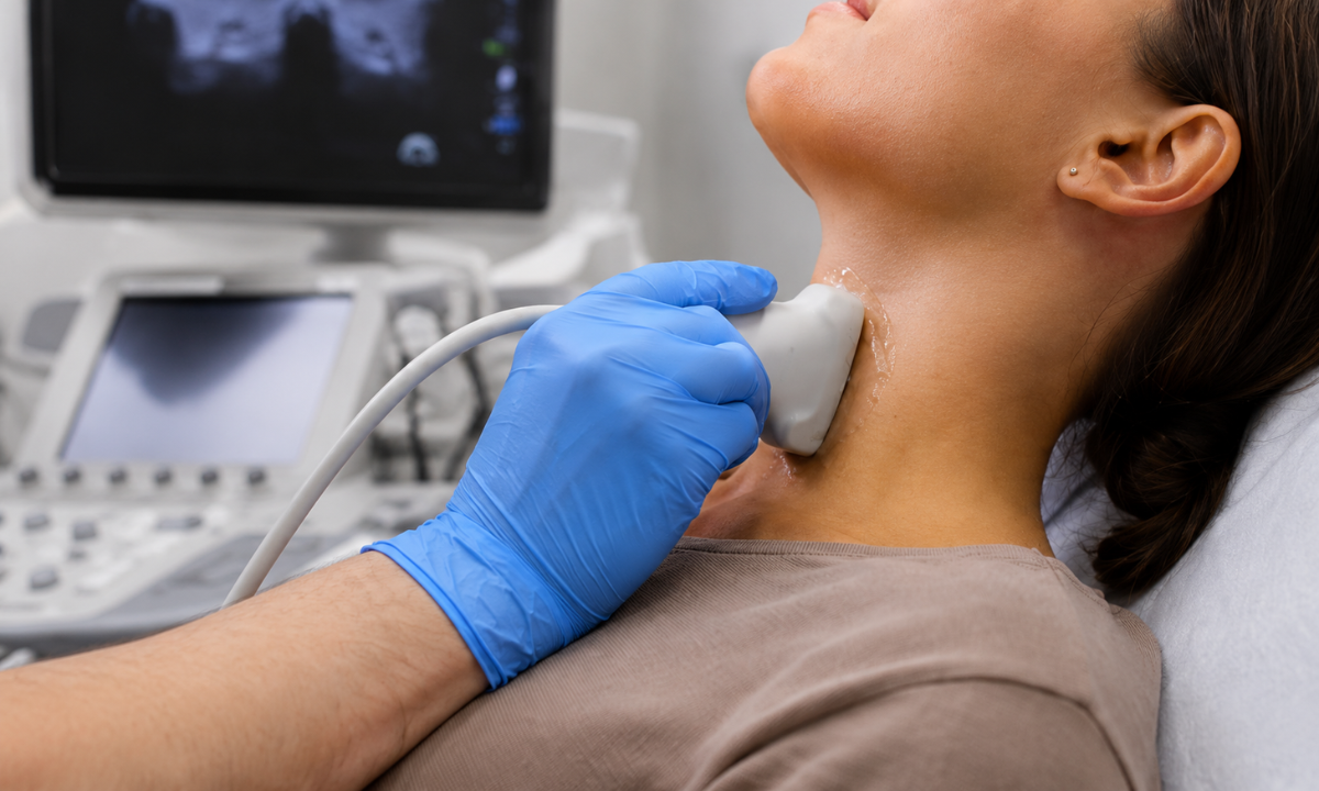
The clinician utilizes a specialized high-frequency linear array transducer. This handheld instrument is placed firmly against the gelled skin and continuously moved in deliberate, sweeping motions.
The sonographer must evaluate the gland from multiple spatial dimensions, scanning across both the transverse (horizontal) and longitudinal (vertical) anatomical planes. The transducer simultaneously emits the targeted sound waves and records the returning acoustic echoes as they bounce off the internal structures of the neck.
Step 4: Real-Time Data Conversion and Measurement
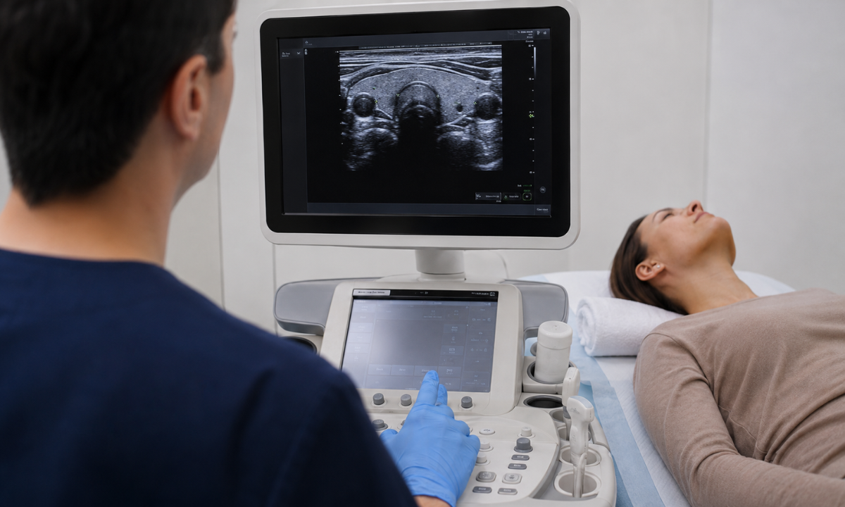
A highly advanced computer processor instantly translates the returning acoustic data into high-resolution grayscale images, displaying them in real-time on the clinical monitor. During the active sweep, the sonographer meticulously freezes the frame to capture static images and record exact diametric measurements.
Standard protocols require precise volumetric measurements of the right lobe, the left lobe, and the connecting central isthmus. If any nodules or cystic structures are identified, their exact spatial coordinates and physical dimensions are immediately documented for the attending radiologist.
Once the data collection is complete, the gel is simply wiped away, and the patient is immediately discharged without any post-procedural physical restrictions.
Benefits and Results Interpretation
Following the completion of image acquisition, the clinical focus shifts to precise radiological interpretation.
The diagnostic efficacy of the thyroid ultrasound lies in its ability to analyze the microscopic structural composition of glandular tissue and classify potential pathologies. Radiologists do not merely identify the presence of a mass; they evaluate specific sonographic features to determine its exact biological risk profile.
This detailed data synthesis directly dictates the subsequent clinical action plan.
Analyzing Structural Composition and Echogenicity
The primary diagnostic advantage of high-frequency ultrasound is its capacity to differentiate soft tissue density. The radiologist evaluates how the internal structures reflect acoustic waves, a property known as echogenicity.
The following clinical matrix details the specific sonographic features analyzed to differentiate between benign cysts and potentially malignant solid masses:
| Sonographic Feature | Clinical Observation | Diagnostic Implication |
|---|---|---|
| Echogenicity | Anechoic (Black): The structure completely transmits sound waves. Hypoechoic (Dark Gray): The mass is denser and absorbs more sound than the surrounding normal tissue. | Anechoic structures are strictly fluid-filled cysts and are overwhelmingly benign. Hypoechoic solid nodules require closer clinical scrutiny. |
| Internal Composition | Purely Cystic: Entirely fluid. Spongiform: Multiple tiny cystic spaces occupy the majority of the nodule volume. Solid: Composed entirely of dense cellular tissue. | Spongiform and purely cystic nodules represent the lowest risk of malignancy. Completely solid masses carry a statistically higher risk profile. |
| Margin Definition | Smooth/Well-Defined: The border is distinct and completely encapsulated. Irregular/Microlobulated: The edges are jagged or appear to infiltrate surrounding tissue. | Smooth margins strongly indicate benign growth. Irregular margins suggest aggressive cellular behavior and potential local tissue invasion. |
| Echogenic Foci (Calcifications) | Macrocalcifications: Large, dense calcium deposits producing an acoustic shadow. Microcalcifications: Tiny, bright punctate spots within the solid tissue. | Microcalcifications are highly correlated with specific malignant pathologies, such as papillary thyroid carcinoma, and mandate immediate biopsy evaluation. |
Vascular Mapping via Color Doppler Integration
Standard grayscale imaging evaluates static anatomical structure. By activating the Color Doppler function on the ultrasound machine, the radiologist can superimpose real-time physiological data over the structural map. This technology measures the speed and direction of blood cells within the localized vessels.
Evaluating vascularity is a critical diagnostic metric:
- Peripheral Vascularity: Blood flow restricted exclusively to the outer rim (the halo) of the nodule is typically a benign indicator.
- Central Vascularity: Rapidly dividing malignant cells require a robust nutrient supply, a process known as angiogenesis. The presence of significant internal blood flow penetrating the core of a solid nodule increases the clinical suspicion of malignancy.
The TI-RADS Classification and Biopsy Criteria
To eliminate subjective interpretation and standardize radiological reporting globally, clinicians utilize the Thyroid Imaging Reporting and Data System (TI-RADS). The radiologist assigns specific point values to the nodule based on its composition, echogenicity, shape, margins, and calcifications.
The cumulative TI-RADS score categorizes the nodule from TR1 (Benign) to TR5 (Highly Suspicious). This objective numerical framework, combined with the exact diametric measurements of the mass, provides the definitive criteria for determining if a patient requires a subsequent Fine Needle Aspiration biopsy to achieve a conclusive cellular diagnosis.
Financial Accessibility and Preparation Logistics
Preparing for a cervical sonogram is remarkably straightforward. Because the thyroid gland is located superficially in the anterior neck, the imaging process does not require the extensive physiological fasting mandated by abdominal or pelvic scans. Patients can schedule this diagnostic evaluation without disrupting their daily nutritional intake or occupational schedules.
Pre-Procedural Clinical Logistics
To ensure a seamless imaging experience and maximize the efficiency of the clinical workflow, diagnostic centers request that patients adhere to a few highly specific physical preparations:
- Nutritional and Hydration Status: Fasting is entirely unnecessary. Patients must consume their standard meals and maintain normal systemic hydration levels before the appointment.
- Pharmacological Regimens: Patients must continue taking all prescribed medications, including synthetic thyroid hormones (Levothyroxine), beta-blockers, or anti-thyroid agents, exactly as directed by their primary care physician. The scan strictly evaluates structural anatomy, which is not immediately altered by daily medication intake.
- Physical Access and Clothing: The sonographer requires unobstructed physical access to the entire anterior neck down to the clavicles. Patients are advised to wear loose-fitting, open-collared clothing. Furthermore, patients must temporarily remove all cervical jewelry, specifically metallic necklaces or heavy chains, as these objects physically block acoustic wave transmission and create severe diagnostic artifacts on the monitor.
Evaluating Diagnostic Affordability
A primary concern for patients navigating preventative healthcare involves financial predictability. The average cost of thyroid scan protocols in Bangalore varies based on the specific technological requirements dictated by the referring physician. Koshikaa maintains a strict institutional commitment to absolute financial transparency, ensuring patients understand the exact pricing structure before any diagnostic procedure commences.
To demystify the financial parameters, the following clinical matrix outlines the primary variables that influence the final cost of the ultrasound examination:
| Diagnostic Modality | Specific Clinical Application | Financial Impact Variable |
|---|---|---|
| Standard Grayscale Ultrasound | Basic structural evaluation to identify the presence, size, and precise anatomical location of nodules or generalized glandular enlargement (goiter). | Represents the accessible baseline standard cost for non-invasive cervical imaging. |
| Color Doppler Integration | Real-time mapping of vascularity and localized blood flow dynamics within identified solid nodules or inflamed glandular tissue. | Often requires a marginal increase over the baseline cost due to the deployment of advanced acoustic software analytics and prolonged scanning duration. |
| Ultrasound-Guided FNA Biopsy | Utilizing real-time imaging to physically direct a fine needle for cellular extraction from a highly suspicious lesion (TI-RADS 4 or 5). | Constitutes a specialized interventional procedure, significantly altering the cost structure due to the mandatory requirement for subsequent pathological laboratory analysis and specialized surgical instruments. |
Prioritizing transparent, highly competitive pricing models, Koshikaa ensures that essential structural diagnostics remain accessible to all demographics, actively preventing the financial deferment of critical endocrine evaluations.
Why Choose Koshikaa for Diagnostics
Executing precision diagnostics requires a clinical environment dedicated entirely to preventative screening and early pathology detection. Koshikaa operates as a premier diagnostic facility, providing the highly specific infrastructure necessary for accurate endocrine evaluations and comprehensive health monitoring in Bangalore.
The institution guarantees diagnostic excellence and patient-centric care through several specific operational advantages:
- Personalized Diagnostic Pathways: Koshikaa deploys a proprietary, highly detailed health screening questionnaire before any clinical evaluation. This algorithmic tool tailors the diagnostic assessment precisely to the patient’s biological age, genetic history, and specific physiological risk factors.
- Rapid Clinical Workflow: Medical uncertainty creates unnecessary physiological stress. The facility guarantees the delivery of highly accurate, radiologist-verified diagnostic reports within exactly twenty-four hours of the completed scan.
- Financial Accessibility: The institution operates on the principle that preventative healthcare must be accessible to all segments of society. Koshikaa structures its screening packages with highly competitive pricing and flexible payment parameters, ensuring superior diagnostics remain affordable.
The clinical imaging suites are equipped with state-of-the-art, high-frequency ultrasound systems. This specialized machinery ensures maximum acoustic resolution, allowing the sonography team to map microscopic soft-tissue anomalies with absolute precision.
Conclusion
Structural degradation of the thyroid gland frequently occurs without immediate systemic or visible physical symptoms. By systematically utilizing high-resolution ultrasound technology, clinicians can accurately identify benign nodules, cystic formations, and potentially malignant cellular changes at their earliest, most treatable stages.
Patients experiencing unexplained weight fluctuations, persistent cervical swelling, or abnormal serological reports must not delay structural evaluation. Schedule a comprehensive health assessment at Koshikaa to definitively map your endocrine anatomy, rule out complex pathologies, and secure your long-term metabolic stability.