What Is Blood Culture Test And Why Is It Done?

Table of Contents

“Why has my doctor asked for another blood test when I already feel so weak?”
If you have ever found yourself asking this during an illness, you are not alone. 

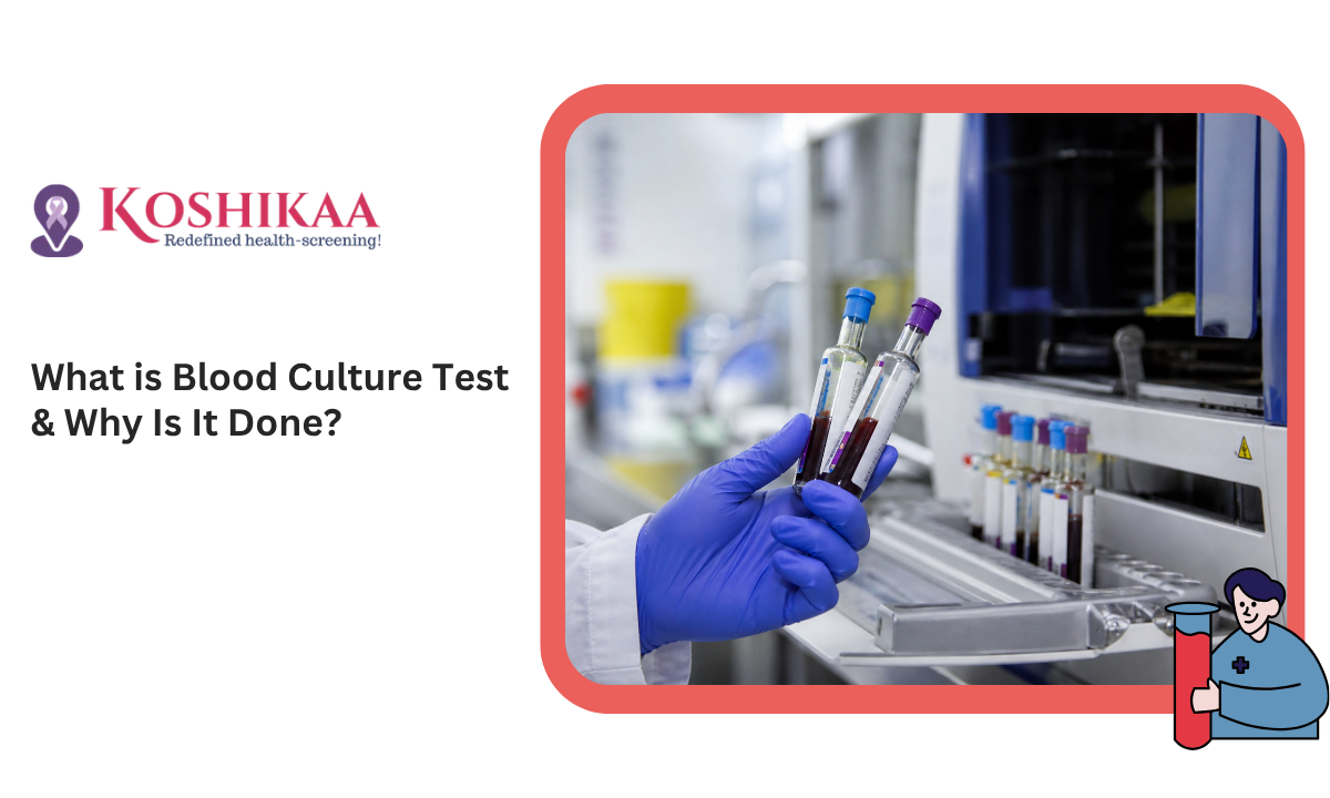
When infections become severe, unexplained, or don’t respond to regular treatment, doctors often recommend specialised diagnostics to identify the root cause. One such essential test is understanding what is blood culture test, especially when you are visiting a reliable blood test in Bangalore facility that prioritises accuracy and speed. 

What makes this test so powerful in detecting hidden infections before they turn dangerous? Let’s dive deeper.

Key Points at a Glance

  • A blood culture test detects harmful bacteria or fungi in the bloodstream
  • It plays a critical role in diagnosing life-threatening infections like sepsis
  • Helps doctors choose targeted antibiotics, improving recovery outcomes
  • Involves sterile sample collection and lab-based microbial analysis
  • Early detection significantly reduces complications and hospitalisation time
  • Widely recommended in emergency, ICU, and chronic infection cases

What Is Blood Culture Test?

Blood Culture Test Overview

A blood culture test is an advanced laboratory investigation designed to detect microorganisms such as bacteria, fungi, or yeast present in the blood. Under normal circumstances, blood is sterile, meaning it should not contain any infectious organisms.

However, when pathogens enter the bloodstream through wounds, infections, or medical procedures, they can spread rapidly across the body. This condition, known as ‘bacteremia’ or ‘fungemia’, can escalate into severe complications if not treated promptly.

This blood test doesn’t just confirm the presence of infection; it also helps pinpoint the exact organism responsible. That precision allows doctors to move from guesswork to targeted treatment, which is critical in urgent medical situations.

Why Blood Culture Test Is Done

Why Blood Culture Is Done

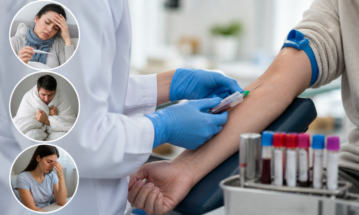
Understanding why blood culture test is done becomes crucial when symptoms are vague but potentially serious. Doctors typically recommend this test when there is a suspicion that an infection has entered the bloodstream and could affect multiple organs.

Here are some detailed scenarios where it is advised:

  • Unexplained high fever with chills or sweating that persists for days
  • Suspected sepsis, a medical emergency where infection spreads throughout the body
  • Symptoms like rapid heartbeat, confusion, or low blood pressure
  • Infections that are not improving despite antibiotic treatment
  • Patients with weakened immunity due to diabetes, cancer therapy, or organ transplants

Globally, studies show that sepsis contributes to nearly 1 in 5 deaths worldwide, highlighting the importance of early diagnosis through blood culture testing.

Blood Culture Test Purpose

Blood Culture Purpose

The blood culture test purpose is not limited to identifying infection; it is about enabling faster, smarter, and more effective medical decisions. In many cases, it can be the difference between timely recovery and prolonged illness.

Key Objectives Explained:

  • Detect infection early: Identifies pathogens before symptoms worsen
  • Guide antibiotic therapy: Helps doctors prescribe the most effective medication
  • Monitor ongoing infections: Tracks whether treatment is working
  • Prevent complications: Reduces the risk of organ damage or septic shock

For example, using broad-spectrum antibiotics without knowing the exact bacteria can lead to resistance. Blood culture tests eliminate this uncertainty and ensure precision-based care.

What Does Blood Culture Test Show

What Blood Culture test Shows

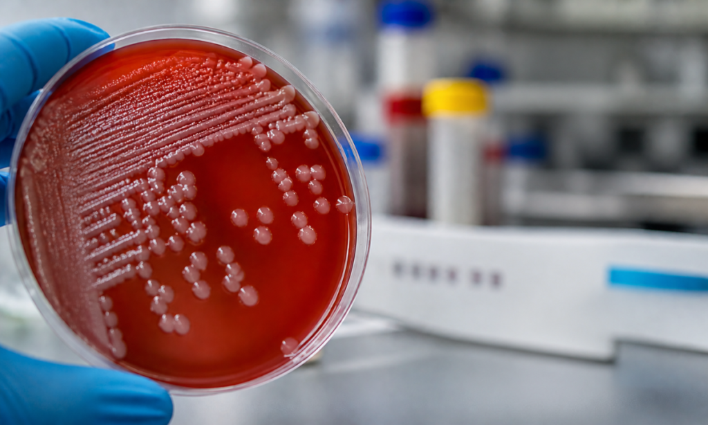
A frequently asked question is, ‘What does blood culture test show?’ The answer lies in both detection and detailed analysis.

The test results provide insights into whether microorganisms are present and, if so, their exact type and behaviour.

Result TypeMeaning
NegativeNo microbial growth detected; blood is likely infection-free
PositiveInfection confirmed; bacteria or fungi identified
ContaminatedExternal contamination during sample collection

In positive cases, the lab performs an additional antibiotic sensitivity test, which shows which medications will effectively eliminate the infection. This step is crucial in avoiding ineffective treatments and speeding up recovery.

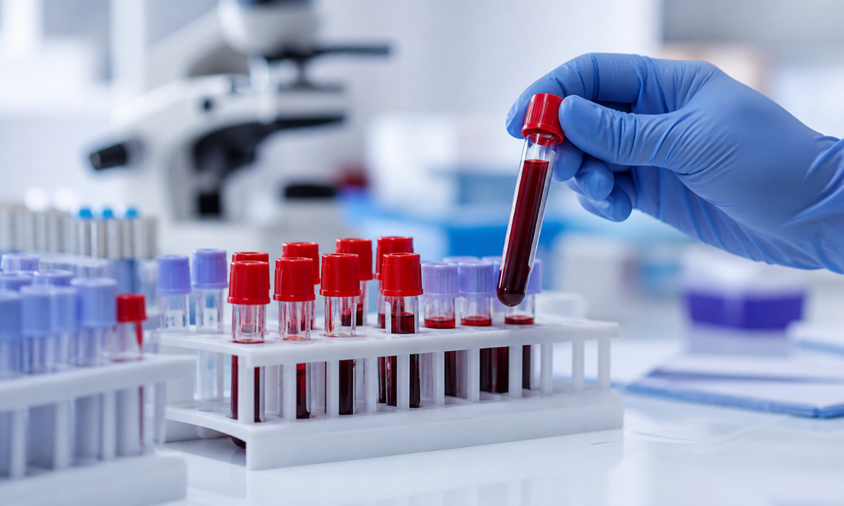
Blood Culture Test Procedure

Blood Culture Procedure

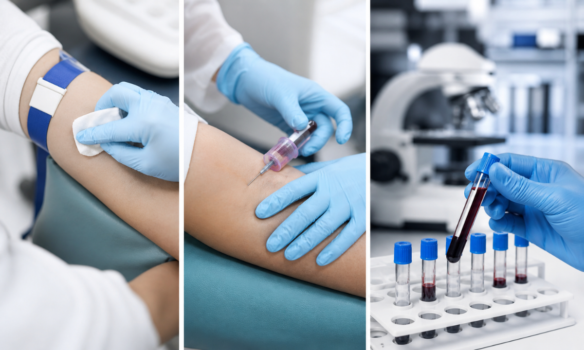
Knowing the blood culture test procedure can help reduce anxiety and prepare you for the process. While it may sound complex, it is actually straightforward and safe.

Step-by-Step Breakdown:

  1. Pre-Test Preparation
    You usually don’t need to fast, but informing your doctor about current medications is important. Antibiotics taken before the test can sometimes affect results.
  2. Sample Collection
    A trained technician cleans the skin thoroughly to prevent contamination. Blood is then drawn from a vein using sterile equipment, often from multiple sites to improve accuracy.
  3. Laboratory Processing
    The collected samples are placed in special culture bottles containing nutrients that encourage microbial growth. These are then monitored continuously.
  4. Observation Period
    The lab checks for signs of bacterial or fungal growth over 24–72 hours. Some infections may take longer to detect, depending on the organism.
  5. Final Analysis
    If growth is detected, further tests identify the organism and determine the best antibiotics for treatment.

This structured approach ensures high accuracy and reliable diagnosis.

When Should You Consider a Blood Culture Test?

When to Consider Blood Culture test?

Recognising the right time to take this test can significantly impact outcomes. It is often recommended when symptoms indicate a possible systemic infection.

You should consider it if you experience:

  • Persistent fever above 38°C lasting more than 2–3 days
  • Sudden chills, fatigue, or weakness
  • Rapid breathing or heart rate
  • Low blood pressure or dizziness
  • Confusion or altered mental state

These symptoms may seem general, but when combined, they can signal a serious underlying infection requiring immediate attention.

Benefits of Early Blood Culture Testing

Early diagnosis through blood culture testing offers several life-saving benefits. Acting quickly can prevent complications that may otherwise become difficult to manage.

Key Advantages:

  • Accurate diagnosis of bloodstream infections at an early stage
  • Targeted treatment, reducing unnecessary antibiotic use
  • Lower risk of severe complications like organ failure
  • Reduced hospital stays and medical costs
  • Improved survival rates in critical cases

Research indicates that early and appropriate antibiotic therapy, guided by blood culture results, can improve survival rates in sepsis patients by up to 30–40%.

Tips for Accurate Blood Culture Results

To ensure the most reliable results, a few precautions can make a significant difference.

  • Always choose a reputable diagnostic centre
  • Ensure strict sterile conditions during sample collection
  • Avoid taking antibiotics before the test unless prescribed
  • Inform your doctor about recent infections or treatments
  • Schedule the test during fever spikes for better detection

These small but important steps can greatly enhance test accuracy.

Common Myths About Blood Culture Tests

Despite its importance, there are several misconceptions surrounding this test.

Myth 1: It’s the same as a regular blood test
Reality: It is a specialised test specifically designed to detect infections in the bloodstream.

Myth 2: Results are immediate
Reality: Since it involves microbial growth, results typically take 1–3 days.

Myth 3: Only critically ill patients need it
Reality: It is recommended whenever a bloodstream infection is suspected, even in early stages.

Final Thoughts

Understanding what is blood culture test is essential in today’s healthcare landscape, where infections can escalate rapidly if left undiagnosed. This test plays a critical role in identifying hidden infections, guiding treatment, and preventing life-threatening complications.

If you’re looking for a dependable blood test in Bangalore, choosing a trusted health screening centre in Bangalore ensures accuracy, hygiene, and timely reporting. At Koshikaa, advanced diagnostic support combined with patient-focused care helps you stay informed, proactive, and protected against serious health risks.

FAQs

1. Is the blood culture test painful?

A blood culture test involves drawing a small amount of blood using a sterile needle, similar to any routine blood test. You may feel a quick pinch or slight discomfort at the puncture site, but it usually lasts only a few seconds. Most people tolerate it well, and any soreness afterwards is mild and temporary.

2. How long do results take?

Blood culture test results are not immediate because they require time for microorganisms to grow in a controlled lab environment. Preliminary results may be available within 24 hours, especially if bacteria are detected early. However, final results, including identification of the organism and antibiotic sensitivity, typically take 48 to 72 hours.

3. Can I eat before the test?

In most cases, you can eat and drink normally before a blood culture test, as fasting is not required. However, it is important to follow your doctor’s instructions, especially if other tests are scheduled alongside it. Also, inform your healthcare provider about any medications or antibiotics you are currently taking beforehand.

4. Is it safe for children and elderly patients?

Yes, the blood culture test is safe for both children and elderly patients when performed by trained healthcare professionals. Special care is taken during sample collection to ensure comfort and hygiene. Since these groups may have weaker immune systems, this test is especially important for detecting infections early and preventing serious complications.

MRI of the uterus is primarily used for diagnosing and evaluating conditions such as uterine fibroids, adenomyosis, and malignancies. It is particularly useful in the planning of surgical procedures and assessing the extent of gynecological diseases. This imaging test is valuable for its ability to deliver detailed cross-sectional images of the uterus and surrounding structures.
An MRI of the spleen can help diagnose various disorders affecting the spleen including splenomegaly (enlarged spleen), tumors, and other abnormalities. It provides clear, detailed images that are crucial for assessing the health of the spleen and planning potential surgical interventions.
Prostate MRI is a non-invasive imaging technique that provides detailed pictures of the prostate gland and surrounding tissues. It helps in the detection, staging, and monitoring of prostate cancer. Prostate MRIs are increasingly used to guide biopsy procedures and assist in the planning of treatment strategies for prostate health.
An MRI of the pancreas is used to identify issues such as pancreatitis, pancreatic cancer, and other pancreatic disorders. It offers high-resolution images that help in detecting small lesions and assessing the pancreatic ducts. This imaging technique is valuable for guiding biopsies and planning surgeries involving the pancreas.
MRI scans of the liver, kidney, and bile ducts provide detailed images that help diagnose various conditions, such as tumors, cysts, and blockages in the bile ducts. They are crucial for assessing organ damage, staging cancer, and planning liver or kidney-related surgeries. This type of MRI can also evaluate the health and function of these organs comprehensively.
Breast MRI is highly sensitive for detecting breast cancer, especially in individuals with dense breast tissue or a high risk of cancer. It is often used in conjunction with mammography to provide a more comprehensive evaluation. Breast MRIs can also monitor the response to breast cancer treatments and provide detailed pre-surgical mapping.
Brain and spine MRIs are critical tools for diagnosing and monitoring neurological conditions, including tumors, spinal cord injuries, and multiple sclerosis. These MRIs can reveal abnormalities in brain tissue and spinal cord structure that might not be visible with other imaging methods. They are essential for planning surgeries and assessing the effectiveness of treatments in neurology.
PET-CT scans of transplant organs like the liver, kidney, or pancreas are performed to assess metabolic activity, detect complications, and monitor post-transplant function. These scans play a vital role in the early detection of issues such as organ rejection, recurrence of disease, or infection, guiding timely interventions and optimizing transplant outcomes for patients.
PET-CT examinations of organs such as the liver, kidney, or prostate are crucial for assessing metabolic activity, detecting abnormalities, and monitoring disease progression. These scans help diagnose conditions such as liver metastases, renal cell carcinoma, or prostate cancer, guiding treatment decisions and monitoring response to therapy over time.
PET-CT scans of soft-tissue masses are utilized to assess metabolic activity and detect malignant tumors or other abnormalities. By combining PET imaging with CT imaging, these scans provide detailed information about tumor location, size, and metabolic characteristics, aiding in accurate diagnosis and treatment planning for soft-tissue cancers.
PET-CT scans are valuable for detecting cysts or fluid collections within the body, including cysts in the liver, kidneys, or other organs. These scans help characterize the nature and metabolic activity of cysts, guiding diagnosis and treatment planning for conditions such as cystic liver disease, renal cysts, or pancreatic cysts.
PET-CT scans of muscles are performed to assess metabolic activity and detect abnormalities such as inflammation, infection, or tumors. These scans provide valuable information about muscle function and pathology, aiding in the diagnosis and management of conditions like myositis, muscular dystrophy, or soft-tissue sarcomas.
PET-CT scans of the brain are utilized to evaluate brain metabolism and detect abnormalities such as tumors, epilepsy, or neurodegenerative diseases. By combining functional PET imaging with anatomical CT imaging, these scans offer comprehensive information about brain function and structure, guiding neurosurgical interventions and treatment decisions for neurological disorders
PET-CT scans of bones are commonly used to detect bone metastases in cancer patients, assess bone health in conditions like osteoporosis, and evaluate response to treatment. By combining PET and CT imaging, these scans provide detailed information about metabolic activity and anatomical changes in bone structures, aiding in accurate diagnosis and treatment planning for various bone-related conditions.
CT scans of the spine are conducted to assess the vertebrae, spinal cord, and surrounding soft tissues for conditions like herniated discs, spinal fractures, or spinal stenosis, aiding in the diagnosis and management of spinal disorders with detailed imaging essential for surgical planning and intervention.
CT scans of the pelvis are utilized to visualize the pelvic bones, reproductive organs, bladder, and surrounding structures for conditions such as fractures, tumors, or pelvic inflammatory disease, guiding treatment plans and surgical interventions with precise anatomical information.
CT scans of the neck are performed to evaluate structures such as the throat, thyroid gland, lymph nodes, and blood vessels for abnormalities like tumors, infections, or injuries, assisting in diagnosis and management of head and neck disorders with detailed imaging.
CT scans of the extremities are employed to assess bones, joints, and soft tissues in the arms or legs, aiding in the diagnosis of fractures, arthritis, or other musculoskeletal conditions, guiding orthopedic treatment and rehabilitation plans for optimal recovery.
CT scans of the chest are utilized to examine the lungs, heart, blood vessels, and surrounding structures for conditions such as lung nodules, pneumonia, or pulmonary embolism, facilitating accurate diagnosis and management of respiratory and cardiovascular diseases.
CT scans of the brain are conducted to assess brain structures, identify abnormalities like tumors, hemorrhages, or strokes, and aid in diagnosing neurological conditions, guiding neurosurgical interventions and treatment strategies with precise imaging of the brain’s anatomy.
CT scans of the abdomen are performed to visualize and evaluate the internal organs such as the liver, kidneys, pancreas, and intestines for conditions like tumors, infections, or injuries, providing detailed images essential for diagnosis and treatment planning.
Transplant organ ultrasound scans, including those of the liver, kidney, or pancreas, are performed to assess organ viability, detect complications, and monitor post-transplant function. These scans play a vital role in the early detection of issues such as organ rejection, vascular complications, or fluid collections, guiding timely interventions and optimizing transplant outcomes for patients.
Ultrasound examinations of organs such as the liver, kidney, or prostate are crucial for assessing organ structure, detecting abnormalities, and monitoring disease progression. These scans help diagnose conditions such as liver cirrhosis, kidney stones, or prostate enlargement, guiding treatment decisions and monitoring response to therapy over time.
Soft-tissue masses can be evaluated using ultrasound to determine their size, location, and characteristics. Ultrasound imaging helps differentiate between benign and malignant masses, guiding further diagnostic evaluation and treatment planning. By providing real-time imaging, ultrasound assists in the precise localization of soft-tissue masses for biopsy or surgical removal if needed.
Ultrasound scans are valuable for detecting cysts or fluid collections within the body, including cysts in the liver, kidneys, or other organs. These scans help characterize the nature and size of cysts, aiding in diagnosis and treatment planning for conditions such as cystic liver disease or renal cysts, and guiding interventions such as cyst drainage or sclerotherapy.
Musculoskeletal ultrasound scans are used to assess the health and integrity of muscles, identifying issues such as strains, tears, or muscle inflammation. These scans provide detailed images of muscle structures, allowing healthcare providers to diagnose muscle injuries accurately and develop personalized rehabilitation plans to promote recovery and restore function.
Ultrasound examinations of joints are essential for evaluating joint health, detecting abnormalities such as inflammation, fluid accumulation, or cartilage damage. By visualizing the joint structures in real-time, ultrasound helps diagnose conditions like arthritis, bursitis, or ligament injuries, guiding treatment decisions and monitoring disease progression over time.
Ultrasound scans of tendons are performed to assess for injuries, inflammation, or tears, aiding in the diagnosis and management of conditions such as tendonitis or tendon tears. These scans provide detailed images of tendon structures, helping healthcare providers determine the extent of damage and plan appropriate treatment strategies, including physical therapy or surgical intervention if necessary.
Pelvis X-rays play a crucial role in diagnosing injuries, diseases, and developmental conditions affecting the pelvic region. They provide detailed images of the hip bones, sacrum, and coccyx, aiding in the assessment of fractures, dislocations, or signs of arthritis. Pelvic X-rays are instrumental in guiding surgical interventions, monitoring healing progress, and evaluating conditions such as osteoporosis or tumors. By providing precise anatomical information, these imaging studies enable healthcare professionals to deliver personalized care and improve patient outcomes.
Teeth X-rays, known as dental radiographs, are indispensable tools for dentists to assess oral health. They aid in diagnosing dental decay, gum disease, and abnormalities in tooth structure or alignment. These images provide valuable insights into hidden dental issues, allowing for early intervention and prevention of complications. Additionally, dental X-rays are essential for planning treatments such as orthodontics, root canals, or dental implants, ensuring optimal outcomes and patient satisfaction.
Spine X-rays are pivotal in diagnosing spinal fractures, degenerative conditions, and spinal misalignments, providing critical information for treatment planning and monitoring spinal health. They offer detailed images of the vertebrae and surrounding structures, enabling healthcare professionals to identify issues such as scoliosis, disc herniation, or spinal stenosis
Skull X-rays are valuable for detecting fractures, assessing head trauma, and evaluating the alignment of facial bones, crucial in diagnosing conditions like fractures, tumors, or developmental abnormalities in the skull. They provide detailed views of the cranial structure, aiding clinicians in determining appropriate treatment strategies and ensuring patient safety.
For the heart, X-ray solutions can aid in the detection of various cardiac problems, ranging from mild to severe, including – Heart failure, Cardiac congestion and other heart-related conditions. In addition to cardiac assessments, X-ray solutions are also used for breast examinations to identify tumors or signs of breast cancer.

Bone X-rays are essential for diagnosing fractures and assessing bone alignment, aiding doctors in developing treatment plans and monitoring healing progress. They provide detailed images of the skeletal system, helping identify abnormalities or injuries that may not be visible through other imaging techniques

Abdominal X-rays are performed when patients experience symptoms such as significant nausea, ongoing vomiting, abdominal pain, flank or back pain, or abdominal swelling. These X-rays can help diagnose various conditions.

Frequently Booked Tests

Frequently Booked Checkups