
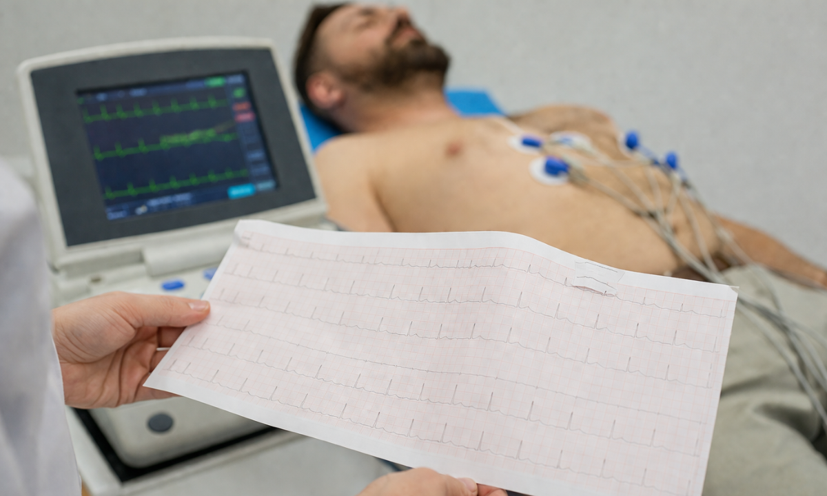
Evaluating sudden cardiovascular distress strictly requires precise electrical monitoring. Patients requiring an immediate ECG test in Bangalore must secure rapid clinical evaluation to prevent catastrophic myocardial infarction. As a specialized Health screening centre in Bangalore, Koshikaa provides unparalleled diagnostic accuracy for acute coronary events.
Utilizing advanced ECG services in Bangalore allows our dedicated cardiology team to instantly map internal myocardial conduction. The primary clinical objective during this initial assessment involves definitively establishing the ECG report normal values for the patient.
To secure absolute biological stability during a suspected cardiac event, the multidisciplinary cardiology board strictly enforces the following diagnostic sequence.
- Rapid Electrical Acquisition: Instantly capturing the microscopic electrical potential traveling through the cardiac muscle utilizing specialized dermal electrodes
- Baseline Mathematical Comparison: Precisely evaluating the visual waveforms against standardized clinical parameters to identify severe conduction delays
- Immediate Ischemic Identification: Detecting specific repolarization abnormalities strictly indicating active biological tissue death within the ventricular myocardium
This comprehensive diagnostic guide will precisely detail the exact physiological mechanisms dictating the cardiac cycle and outline the specific electrical metrics required to identify critical structural failures.
The Anatomical Mechanism Understanding the Cardiac Cycle
Comprehending an electrocardiogram strictly requires understanding the internal electrical conduction system governing the human heart.
The cardiac cycle does not rely on external neurological stimulation to initiate physical muscular contraction. Instead, the myocardium utilizes a highly specialized internal network of autorhythmic cellular structures to generate and propagate specific electrical impulses.
This precise electrical cascade directly dictates the mechanical pumping action required to sustain systemic physiological perfusion.
To accurately interpret diagnostic waveforms, medical professionals must strictly track the internal path of this electrical action potential.
- The Sinoatrial Node: Which is located within the superior wall of the right atrium, this specific cellular cluster functions as the primary biological pacemaker.
It spontaneously generates an electrical action potential, causing immediate atrial depolarization and subsequent mechanical contraction.
- The Atrioventricular Node: As the electrical impulse travels inferiorly, it encounters this secondary cellular junction located between the atria and the ventricles.
This specific node purposefully delays the electrical transmission by a fraction of a second. This critical chronological pause ensures the atria empty their blood volume into the ventricles before massive ventricular contraction occurs.
- The Bundle of His and Purkinje Fibers: Following the critical delay, the electrical signal rapidly accelerates down the ventricular septum through specialized conduction pathways.
The impulse finally disperses throughout the Purkinje network, penetrating the deep ventricular myocardium. This final rapid propagation triggers simultaneous and massive ventricular depolarization forcing oxygenated blood into the systemic arterial network.
Surface electrodes capture this exact internal electrical sequence, transforming microscopic biological voltage into the visible structural waveforms analyzed during clinical evaluation.
Clinical Methodology ECG Report How to Read
Understanding how to read an ECG report strictly requires analyzing the standardized electrocardiographic grid paper. Medical professionals utilize highly calibrated thermal paper advancing at exactly twenty-five millimeters per second.
This precise mechanical standardization allows cardiologists to translate visual spatial distances directly into exact physiological timeframes and exact electrical voltage amplitudes.
Attempting to interpret structural waveforms without establishing this mathematical baseline guarantees severe diagnostic failure. The standardized electrocardiographic grid features strict geometrical parameters defining both chronological duration and electrical intensity.
- Chronological Measurement: The horizontal axis strictly measures elapsed physiological time. One single millimeter square exactly represents zero point zero four seconds. One large five millimeter square exactly represents zero point two zero seconds.
- Electrical Amplitude: The vertical axis strictly measures the internal myocardial voltage generation. One single millimeter vertical square exactly represents zero point one millivolts of electrical potential.
Once the medical professional establishes this absolute structural baseline, they initiate a highly rigorous systematic evaluation protocol. Clinical cardiologists never simply glance at the total diagnostic waveform. They strictly deconstruct the electrical data utilizing a precise sequential methodology.
The Standardized Electrocardiographic Evaluation Protocol
| Diagnostic Phase | Clinical Action | Physiological Objective |
|---|---|---|
| Rate Calculation | Measuring the exact chronological distance between continuous R waves | Establishing the primary mechanical pumping frequency to identify severe bradycardia or fatal tachycardia |
| Rhythm Verification | Analyzing the continuous presence and precise placement of the P wave | Confirming that the electrical impulse strictly originates from the standardized sinoatrial node pacemaker |
| Axis Determination | Evaluating the primary direction of the total ventricular depolarization vector | Identifying severe anatomical structural displacement or massive internal conduction blockages |
| Morphological Analysis | Scrutinizing every individual electrical complex for exact structural abnormalities | Detecting severe localized cellular necrosis or massive anatomical ventricular hypertrophy |
Strictly adhering to this rigorous mathematical and structural protocol medical professionals eliminate diagnostic subjectivity.
This standardized methodology directly transforms abstract electrical data into a highly precise biological blueprint detailing the exact functional status of the human heart.
Baseline Metrics: Establishing ECG Report Normal Values
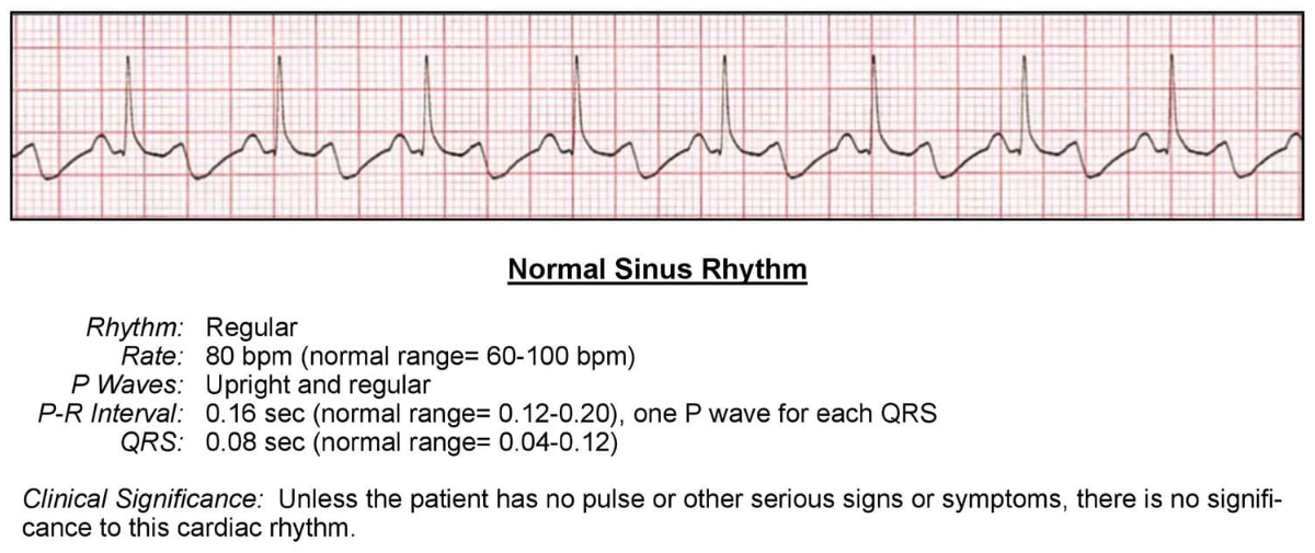
Establishing absolute clinical accuracy during an electrocardiographic evaluation requires strict adherence to standardized mathematical parameters. Medical professionals strictly evaluate every individual waveform against universally accepted physiological baselines.
Deviating from these specific mathematical ranges strictly indicates underlying pathological cellular behavior. To accurately interpret the diagnostic data, cardiologists must definitively establish the ECG report normal values for each specific structural component of the cardiac cycle.
The standardized electrocardiographic waveform consists of distinct electrical events dictating precise mechanical actions.
Standardized Electrocardiographic Waveform Parameters
| Waveform Component | Physiological Event | Standard Chronological Duration | Standard Voltage Amplitude |
|---|---|---|---|
| The P Wave | Primary atrial depolarization initiating superior mechanical contraction | Less than zero point one two seconds | Less than zero point two five millivolts |
| The PR Interval | The critical electrical delay within the atrioventricular node | Between zero point one two and zero point two zero seconds | Isoelectric baseline maintenance |
| The QRS Complex | Massive bilateral ventricular depolarization triggers primary mechanical pumping | Between zero point zero eight and zero point one two seconds | Highly variable, strictly depending on specific anatomical leads |
| The T Wave | Cellular repolarization resetting the ventricular myocardium | Variable duration strictly aligning with the overall cardiac rate | Less than zero point five millivolts in standard limb leads |
Systematically measuring these exact structural components against the established mathematical baselines, cardiologists instantly identify microscopic conduction delays.
Any chronological extension beyond these strict numerical boundaries mathematically confirms severe internal physiological blockages or localized anatomical tissue necrosis.
Pathological Indicators: ECG Report Normal and Abnormal
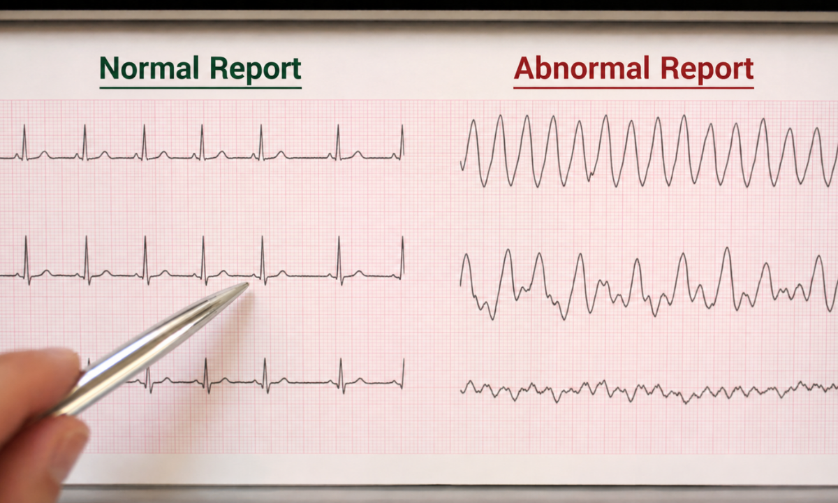
Differentiating an ECG report as normal or abnormal strictly requires identifying precise mathematical deviations from the established physiological baseline.
When cardiac cellular structures suffer severe oxygen deprivation, they immediately alter their electrical transmission capabilities. These localized cellular mutations physically manifest as severe structural distortions on the standardized diagnostic grid.
Clinical cardiologists categorize these specific waveform alterations into distinct pathological classifications, strictly dictating the necessary medical intervention.
The absolute most critical diagnostic evaluation involves analyzing the specific ST segment. This precise electrical interval connects massive ventricular depolarization directly to cellular repolarization.
Under standard physiological conditions, this segment must remain perfectly flat along the standardized isoelectric baseline. When a patient experiences a massive coronary artery occlusion, the affected myocardial tissue rapidly undergoes severe ischemic necrosis.
This immediate cellular death forces a drastic upward shift in the diagnostic electrical waveform, strictly defining an ECG report of a heart attack. Medical professionals classify this exact structural deviation as an ST elevation myocardial infarction, mandating immediate emergency surgical intervention.
Beyond localized structural tissue death, the electrocardiogram precisely identifies fatal global electrical conduction failures. Severe ventricular arrhythmias destroy the coordinated mechanical pumping action of the human heart, rapidly inducing systemic biological failure.
- Ventricular Tachycardia: The diagnostic waveform displays a rapid, continuous series of massive, widened QRS complexes, strictly indicating the ventricles are firing independently at an extreme frequency without adequate physical diastolic blood filling time
- Ventricular Fibrillation: The standardized structural waveform completely degenerates into chaotic, highly erratic electrical oscillations, strictly confirming absolute cardiac arrest and immediate biological death without instant external electrical defibrillation
This rapid structural interpretation remains the absolute determining factor regarding long-term patient survival.
Why Choose Koshikaa? Specialized Cardiac Diagnostics at Koshikaa
At Koshikaa, we recognize that acute cardiac symptoms strictly require immediate diagnostic precision. Evaluating potential myocardial infarction necessitates unparalleled electrocardiographic expertise combined with advanced infrastructure. By choosing our specialized center, patients secure direct access to critical medical advantages.
- Rapid Diagnostic Protocols Our facility utilizes highly sensitive electrocardiography equipment strictly operated by specialized cardiological technicians. This ensures every patient undergoes complex electrical mapping immediately following the initial onset of sudden chest pain.
- Immediate Clinical Reporting During a potential cardiac event, we completely prioritize immediate diagnostic interpretation. Our streamlined reporting processes ensure rapid transition of critical pathological data directly to waiting emergency physicians or cardiologists.
- Integrated Preventive Strategy Koshikaa functions as a dedicated Health screening centre in Bangalore. We strongly encourage continuous preventive cardiovascular evaluation. Our proactive ECG services in Bangalore allow patients to identify subtle cardiac conduction abnormalities before they escalate into severe acute coronary syndromes.
Choosing Koshikaa for your next ECG test in Bangalore guarantees access to an elite medical environment where diagnostic speed and biological accuracy strictly dictate emergency cardiac management.
Conclusion
Navigating a complex cardiovascular event strictly requires an immediate evidence-based medical response. Delaying diagnostic clarification during acute chest pain guarantees massive irreversible myocardial tissue death. Rapid electrocardiographic monitoring utilizes advanced structural imaging to provide the absolute foundation of early medical intervention and long term cardiac survival.
If you observe sudden, severe cardiac symptoms, seek emergency medical evaluation immediately. Secure your comprehensive ECG services in Bangalore at Koshikaa to obtain the precise diagnostic data strictly required to optimize your long term functional recovery.