Warning Signs That Indicate You Need A Heart Health Checkup

Table of Contents

Have you ever brushed off chest discomfort or unusual fatigue, telling yourself it’s just stress or a busy day? Many of us ignore early signs until they become serious. Still, a timely heart health checkup can make all the difference, especially when access to advanced diagnostics, such as a CT scan in Bangalore, is readily available. 

Understanding what your body is trying to tell you could be the key to preventing life-threatening conditions, so what are the subtle signs you should never ignore?

Key Points at a Glance

  • Early detection through a heart health checkup can reduce cardiovascular risks by up to 80%.
  • Common signs of heart disease often go unnoticed or are mistaken for minor issues.
  • Symptoms may differ between men and women; awareness is crucial.
  • A complete heart health checkup includes blood tests, ECG, imaging, and lifestyle assessment.
  • Advanced tests, such as a CT scan of the heart, help detect blockages early.
  • Regular screening at a trusted health screening centre in Bangalore ensures long-term heart wellness.

What is a Heart Health Check?

Heart Health Check Explained

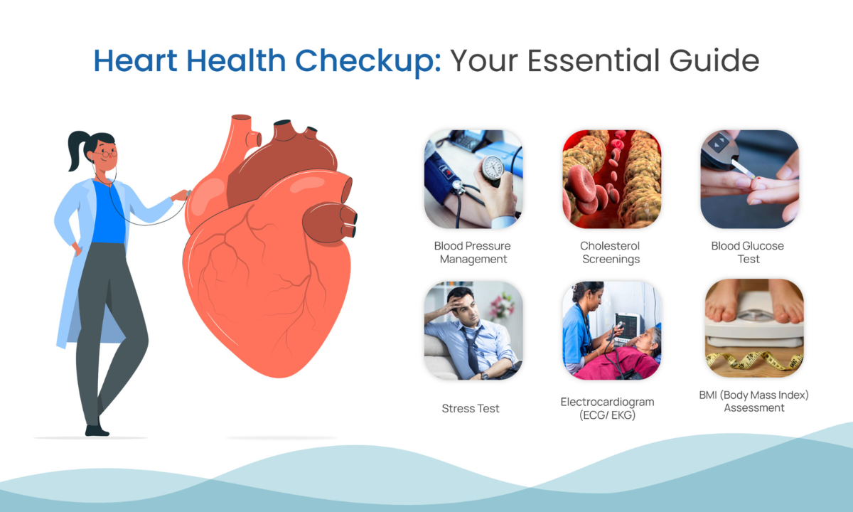
A common question people ask is, ‘What is a heart health check?’ Simply put, it is a comprehensive evaluation of your cardiovascular system to assess risks, detect early disease, and prevent complications.

A complete heart health checkup usually includes:

Test TypePurpose
Blood TestsCheck cholesterol, sugar, and triglycerides
ECG (Electrocardiogram)Measures heart rhythm and electrical activity
EchocardiogramAssesses heart structure and function
Stress TestEvaluates heart performance under exertion
CT Scan for HeartDetects blockages or plaque buildup

These tests together provide a detailed picture of your heart’s condition and future risk factors.

10 Warning Signs You Should Never Ignore

Your body often sends signals before a serious cardiac event. Recognizing these signs of heart disease early can save your life and prevent complications.

1. Chest Pain or Discomfort

This is the most well-known symptom, yet it’s often ignored or misinterpreted as acidity or muscle strain. It may feel like pressure, tightness, squeezing, or even a burning sensation in the chest.

  • Can last a few minutes or come and go
  • May worsen with physical activity or emotional stress
  • Sometimes accompanied by sweating or nausea

2. Shortness of Breath

Breathlessness

Struggling to breathe after minimal exertion, or even while resting, is a major red flag. It may indicate that your heart is not pumping blood efficiently.

  • Can occur while lying down or during sleep
  • Often linked with fluid buildup in the lungs
  • May appear before chest pain in some cases

3. Unusual Fatigue

Fatigue

Feeling excessively tired even after proper rest is one of the more subtle symptoms of heart problems, especially in women. This kind of fatigue can interfere with daily activities.

  • You may feel drained after simple tasks like walking or climbing stairs
  • Can persist for days or weeks before a major cardiac event
  • Often mistaken for stress or lack of sleep

4. Irregular Heartbeat

Abnormal heartbeat

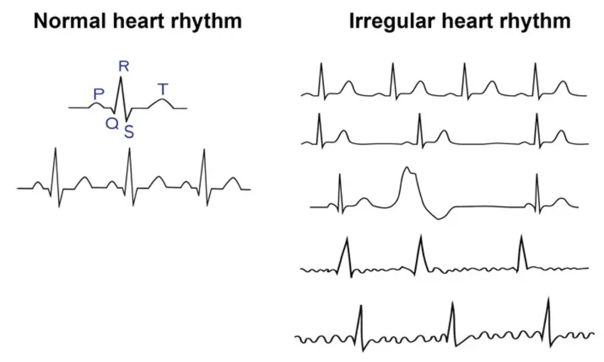
A racing, fluttering, or skipping heartbeat (palpitations) should not be ignored. It could signal arrhythmia or other underlying heart conditions.

  • May feel like your heart is “pounding” or “flipping”
  • Can occur with dizziness or fainting
  • Sometimes triggered by caffeine, stress, or underlying disease

5. Pain Radiating to Arm, Neck, or Jaw

Radiation pain in left arm

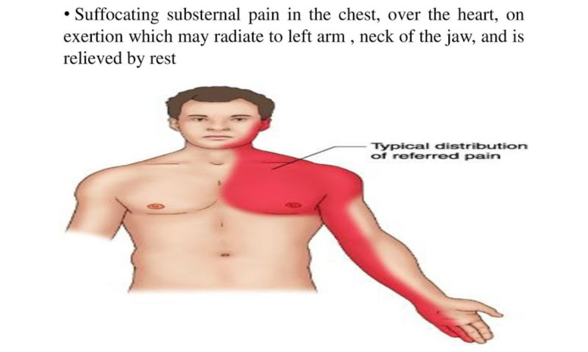
Pain that starts in the chest and spreads to other areas is a classic warning sign of a heart attack. However, in some cases, the pain may begin in these areas first.

  • Commonly affects the left arm but can occur on both sides
  • Jaw or neck pain without chest discomfort can also be significant
  • Often accompanied by a feeling of heaviness or pressure

6. Swelling in Legs, Ankles, or Feet

Swelling in Whole Foot

Fluid retention (oedema) can indicate heart failure, where the heart cannot pump blood effectively, causing fluid to accumulate in the body.

  • Shoes or socks may feel tighter than usual
  • Swelling may worsen by the end of the day
  • It can also be accompanied by weight gain due to fluid buildup

7. Dizziness or Lightheadedness

Dizziness

Frequent dizziness, feeling faint, or sudden loss of balance may indicate poor blood flow to the brain due to heart issues.

  • It can occur suddenly while standing or walking
  • May be linked to low blood pressure or arrhythmia
  • In severe cases, it can lead to fainting (syncope)

8. Persistent Cough

Chronic cough

A chronic cough that doesn’t go away, especially when producing white or pink mucus, may be related to heart failure rather than a respiratory issue.

  • Often worse when lying down
  • May be accompanied by shortness of breath
  • Indicates fluid accumulation in the lungs

9. Nausea or Loss of Appetite

Nausea or Loss of Appetite

Digestive discomfort is often overlooked but can be an early warning sign of heart trouble, particularly in women.

  • Feeling full quickly or loss of appetite
  • Nausea may occur without any clear cause
  • Sometimes confused with gastric issues or food poisoning

10. Cold Sweats

Cold Sweats

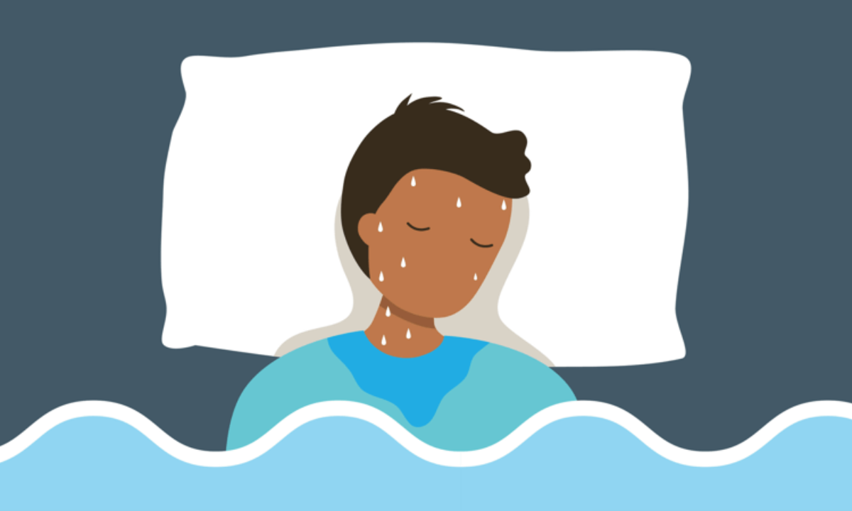
Sudden, unexplained sweating, especially cold, clammy skin, can be a serious warning sign of a heart attack.

  • Occurs without physical exertion or heat
  • Often accompanied by chest discomfort or dizziness
  • Requires immediate medical attention

Recognising these symptoms early and acting promptly can make a life-saving difference. If you notice even a few of these warning signs, don’t delay seeking medical advice.

Who Should Get a Heart Health Checkup?

While everyone benefits from screening, certain groups should prioritize it. Heart disease often develops silently over time, showing symptoms only at advanced stages. That’s why identifying your risk factors early and opting for preventive screening can significantly improve long-term heart health and reduce complications.

If you fall into any of the following categories, scheduling a heart health checkup should be a priority:

  • Individuals above 30 years
  • People with diabetes or high blood pressure
  • Smokers or those with sedentary lifestyles
  • Family history of heart disease
  • High-stress professionals

Regular checkups at a reliable health screening centre in Bangalore can significantly reduce risks and help you stay one step ahead of potential heart conditions.

Why Early Detection Matters

According to global health studies, cardiovascular diseases account for nearly 32% of all deaths worldwide. However, early detection can prevent most complications and significantly improve survival rates. The biggest challenge is that many heart conditions develop silently, without obvious symptoms, making regular screening crucial for timely intervention.

A proactive approach not only protects your heart but also helps you take control of your overall well-being before issues become severe or life-threatening.

Here’s why a heart health checkup is essential:

  • Identifies hidden risk factors
  • Prevents sudden cardiac events
  • Enables timely lifestyle changes
  • Reduces long-term treatment costs

Role of Advanced Imaging in Heart Diagnosis

Modern diagnostics have transformed cardiac care, making it easier to detect problems at a very early stage. A CT scan for the heart is one of the most effective tools available today, offering detailed images of the heart and blood vessels that traditional tests may not capture. It plays a crucial role in identifying silent conditions, especially in individuals who may not yet show obvious symptoms.

By providing precise and quick results, this advanced imaging technique allows doctors to make accurate diagnoses and recommend timely treatment plans, reducing the risk of major cardiac events.

Benefits of a CT Scan:

  • Detects plaque buildup in arteries
  • Identifies blockages before symptoms worsen
  • Non-invasive and highly accurate
  • Helps in early intervention planning

This makes it a critical part of a complete heart health checkup.

Heart Health Checkup vs Full Body Checkup

Many people wonder whether they need a heart-specific test or a general screening like a full body checkup in Bangalore.

Here’s a quick comparison:

FeatureHeart Health CheckupFull Body Checkup
FocusCardiovascular systemOverall health
TestsECG, Echo, CT scanBlood tests, organ screening
PurposeDetect heart diseaseGeneral wellness check
FrequencyBased on risk factorsAnnually

Both a heart health checkup and a full body checkup are important, but a targeted heart evaluation is essential if you notice symptoms.

Tips to Protect Your Heart

Prevention is always better than a cure. Here are actionable tips to maintain heart health:

  • Exercise for at least 30 minutes daily
  • Eat a balanced, low-fat diet
  • Manage stress through meditation or yoga
  • Avoid smoking and limit alcohol
  • Monitor blood pressure and cholesterol regularly

Small lifestyle changes can significantly reduce your risk.

When Should You Act Immediately?

Do not delay seeking help if you experience:

  • Severe chest pain lasting more than 5 minutes
  • Sudden breathlessness
  • Fainting or loss of consciousness
  • Pain radiating to the arm or jaw

These symptoms require emergency medical attention.

Final Thoughts

Your heart works tirelessly for you every second. Don’t wait for a crisis to start caring for it. Recognising the early signs of heart disease and scheduling a timely heart health checkup can save your life, especially with access to advanced diagnostics like a CT scan in Bangalore

At Koshikaa, prioritising preventive care through comprehensive screening ensures you stay ahead of potential risks and lead a healthier, longer life.

Frequently Asked Questions 

1. How often should I get a heart health checkup?

If you are above 30 or have existing risk factors like diabetes, high blood pressure, or a family history of heart disease, getting a heart health checkup once a year is highly recommended. Regular screening helps detect hidden issues early, monitor changes over time, and take preventive action before conditions become serious or life-threatening.

2. Are heart problems always accompanied by chest pain?

No, heart problems are not always accompanied by chest pain. Many symptoms of heart problems can be subtle or atypical, especially in women. These may include fatigue, shortness of breath, nausea, or dizziness. Because symptoms can vary widely, it’s important to stay alert and not ignore unusual changes in your body.

3. Is a CT scan safe for heart screening?

Yes, a CT scan for the heart is generally safe, non-invasive, and highly effective when recommended by a doctor. It provides detailed images of the heart and blood vessels, helping detect blockages or plaque buildup early. Modern technology ensures minimal radiation exposure, making it a reliable option for accurate diagnosis and preventive screening.

4. Can young people have heart disease?

Yes, heart disease is no longer limited to older adults. Due to factors like stress, unhealthy eating habits, lack of exercise, smoking, and genetic predisposition, even younger individuals are at risk today. Early screening and lifestyle modifications are crucial to prevent long-term complications and ensure better heart health from a younger age.

MRI of the uterus is primarily used for diagnosing and evaluating conditions such as uterine fibroids, adenomyosis, and malignancies. It is particularly useful in the planning of surgical procedures and assessing the extent of gynecological diseases. This imaging test is valuable for its ability to deliver detailed cross-sectional images of the uterus and surrounding structures.
An MRI of the spleen can help diagnose various disorders affecting the spleen including splenomegaly (enlarged spleen), tumors, and other abnormalities. It provides clear, detailed images that are crucial for assessing the health of the spleen and planning potential surgical interventions.
Prostate MRI is a non-invasive imaging technique that provides detailed pictures of the prostate gland and surrounding tissues. It helps in the detection, staging, and monitoring of prostate cancer. Prostate MRIs are increasingly used to guide biopsy procedures and assist in the planning of treatment strategies for prostate health.
An MRI of the pancreas is used to identify issues such as pancreatitis, pancreatic cancer, and other pancreatic disorders. It offers high-resolution images that help in detecting small lesions and assessing the pancreatic ducts. This imaging technique is valuable for guiding biopsies and planning surgeries involving the pancreas.
MRI scans of the liver, kidney, and bile ducts provide detailed images that help diagnose various conditions, such as tumors, cysts, and blockages in the bile ducts. They are crucial for assessing organ damage, staging cancer, and planning liver or kidney-related surgeries. This type of MRI can also evaluate the health and function of these organs comprehensively.
Breast MRI is highly sensitive for detecting breast cancer, especially in individuals with dense breast tissue or a high risk of cancer. It is often used in conjunction with mammography to provide a more comprehensive evaluation. Breast MRIs can also monitor the response to breast cancer treatments and provide detailed pre-surgical mapping.
Brain and spine MRIs are critical tools for diagnosing and monitoring neurological conditions, including tumors, spinal cord injuries, and multiple sclerosis. These MRIs can reveal abnormalities in brain tissue and spinal cord structure that might not be visible with other imaging methods. They are essential for planning surgeries and assessing the effectiveness of treatments in neurology.
PET-CT scans of transplant organs like the liver, kidney, or pancreas are performed to assess metabolic activity, detect complications, and monitor post-transplant function. These scans play a vital role in the early detection of issues such as organ rejection, recurrence of disease, or infection, guiding timely interventions and optimizing transplant outcomes for patients.
PET-CT examinations of organs such as the liver, kidney, or prostate are crucial for assessing metabolic activity, detecting abnormalities, and monitoring disease progression. These scans help diagnose conditions such as liver metastases, renal cell carcinoma, or prostate cancer, guiding treatment decisions and monitoring response to therapy over time.
PET-CT scans of soft-tissue masses are utilized to assess metabolic activity and detect malignant tumors or other abnormalities. By combining PET imaging with CT imaging, these scans provide detailed information about tumor location, size, and metabolic characteristics, aiding in accurate diagnosis and treatment planning for soft-tissue cancers.
PET-CT scans are valuable for detecting cysts or fluid collections within the body, including cysts in the liver, kidneys, or other organs. These scans help characterize the nature and metabolic activity of cysts, guiding diagnosis and treatment planning for conditions such as cystic liver disease, renal cysts, or pancreatic cysts.
PET-CT scans of muscles are performed to assess metabolic activity and detect abnormalities such as inflammation, infection, or tumors. These scans provide valuable information about muscle function and pathology, aiding in the diagnosis and management of conditions like myositis, muscular dystrophy, or soft-tissue sarcomas.
PET-CT scans of the brain are utilized to evaluate brain metabolism and detect abnormalities such as tumors, epilepsy, or neurodegenerative diseases. By combining functional PET imaging with anatomical CT imaging, these scans offer comprehensive information about brain function and structure, guiding neurosurgical interventions and treatment decisions for neurological disorders
PET-CT scans of bones are commonly used to detect bone metastases in cancer patients, assess bone health in conditions like osteoporosis, and evaluate response to treatment. By combining PET and CT imaging, these scans provide detailed information about metabolic activity and anatomical changes in bone structures, aiding in accurate diagnosis and treatment planning for various bone-related conditions.
CT scans of the spine are conducted to assess the vertebrae, spinal cord, and surrounding soft tissues for conditions like herniated discs, spinal fractures, or spinal stenosis, aiding in the diagnosis and management of spinal disorders with detailed imaging essential for surgical planning and intervention.
CT scans of the pelvis are utilized to visualize the pelvic bones, reproductive organs, bladder, and surrounding structures for conditions such as fractures, tumors, or pelvic inflammatory disease, guiding treatment plans and surgical interventions with precise anatomical information.
CT scans of the neck are performed to evaluate structures such as the throat, thyroid gland, lymph nodes, and blood vessels for abnormalities like tumors, infections, or injuries, assisting in diagnosis and management of head and neck disorders with detailed imaging.
CT scans of the extremities are employed to assess bones, joints, and soft tissues in the arms or legs, aiding in the diagnosis of fractures, arthritis, or other musculoskeletal conditions, guiding orthopedic treatment and rehabilitation plans for optimal recovery.
CT scans of the chest are utilized to examine the lungs, heart, blood vessels, and surrounding structures for conditions such as lung nodules, pneumonia, or pulmonary embolism, facilitating accurate diagnosis and management of respiratory and cardiovascular diseases.
CT scans of the brain are conducted to assess brain structures, identify abnormalities like tumors, hemorrhages, or strokes, and aid in diagnosing neurological conditions, guiding neurosurgical interventions and treatment strategies with precise imaging of the brain’s anatomy.
CT scans of the abdomen are performed to visualize and evaluate the internal organs such as the liver, kidneys, pancreas, and intestines for conditions like tumors, infections, or injuries, providing detailed images essential for diagnosis and treatment planning.
Transplant organ ultrasound scans, including those of the liver, kidney, or pancreas, are performed to assess organ viability, detect complications, and monitor post-transplant function. These scans play a vital role in the early detection of issues such as organ rejection, vascular complications, or fluid collections, guiding timely interventions and optimizing transplant outcomes for patients.
Ultrasound examinations of organs such as the liver, kidney, or prostate are crucial for assessing organ structure, detecting abnormalities, and monitoring disease progression. These scans help diagnose conditions such as liver cirrhosis, kidney stones, or prostate enlargement, guiding treatment decisions and monitoring response to therapy over time.
Soft-tissue masses can be evaluated using ultrasound to determine their size, location, and characteristics. Ultrasound imaging helps differentiate between benign and malignant masses, guiding further diagnostic evaluation and treatment planning. By providing real-time imaging, ultrasound assists in the precise localization of soft-tissue masses for biopsy or surgical removal if needed.
Ultrasound scans are valuable for detecting cysts or fluid collections within the body, including cysts in the liver, kidneys, or other organs. These scans help characterize the nature and size of cysts, aiding in diagnosis and treatment planning for conditions such as cystic liver disease or renal cysts, and guiding interventions such as cyst drainage or sclerotherapy.
Musculoskeletal ultrasound scans are used to assess the health and integrity of muscles, identifying issues such as strains, tears, or muscle inflammation. These scans provide detailed images of muscle structures, allowing healthcare providers to diagnose muscle injuries accurately and develop personalized rehabilitation plans to promote recovery and restore function.
Ultrasound examinations of joints are essential for evaluating joint health, detecting abnormalities such as inflammation, fluid accumulation, or cartilage damage. By visualizing the joint structures in real-time, ultrasound helps diagnose conditions like arthritis, bursitis, or ligament injuries, guiding treatment decisions and monitoring disease progression over time.
Ultrasound scans of tendons are performed to assess for injuries, inflammation, or tears, aiding in the diagnosis and management of conditions such as tendonitis or tendon tears. These scans provide detailed images of tendon structures, helping healthcare providers determine the extent of damage and plan appropriate treatment strategies, including physical therapy or surgical intervention if necessary.
Pelvis X-rays play a crucial role in diagnosing injuries, diseases, and developmental conditions affecting the pelvic region. They provide detailed images of the hip bones, sacrum, and coccyx, aiding in the assessment of fractures, dislocations, or signs of arthritis. Pelvic X-rays are instrumental in guiding surgical interventions, monitoring healing progress, and evaluating conditions such as osteoporosis or tumors. By providing precise anatomical information, these imaging studies enable healthcare professionals to deliver personalized care and improve patient outcomes.
Teeth X-rays, known as dental radiographs, are indispensable tools for dentists to assess oral health. They aid in diagnosing dental decay, gum disease, and abnormalities in tooth structure or alignment. These images provide valuable insights into hidden dental issues, allowing for early intervention and prevention of complications. Additionally, dental X-rays are essential for planning treatments such as orthodontics, root canals, or dental implants, ensuring optimal outcomes and patient satisfaction.
Spine X-rays are pivotal in diagnosing spinal fractures, degenerative conditions, and spinal misalignments, providing critical information for treatment planning and monitoring spinal health. They offer detailed images of the vertebrae and surrounding structures, enabling healthcare professionals to identify issues such as scoliosis, disc herniation, or spinal stenosis
Skull X-rays are valuable for detecting fractures, assessing head trauma, and evaluating the alignment of facial bones, crucial in diagnosing conditions like fractures, tumors, or developmental abnormalities in the skull. They provide detailed views of the cranial structure, aiding clinicians in determining appropriate treatment strategies and ensuring patient safety.
For the heart, X-ray solutions can aid in the detection of various cardiac problems, ranging from mild to severe, including – Heart failure, Cardiac congestion and other heart-related conditions. In addition to cardiac assessments, X-ray solutions are also used for breast examinations to identify tumors or signs of breast cancer.

Bone X-rays are essential for diagnosing fractures and assessing bone alignment, aiding doctors in developing treatment plans and monitoring healing progress. They provide detailed images of the skeletal system, helping identify abnormalities or injuries that may not be visible through other imaging techniques

Abdominal X-rays are performed when patients experience symptoms such as significant nausea, ongoing vomiting, abdominal pain, flank or back pain, or abdominal swelling. These X-rays can help diagnose various conditions.

Frequently Booked Tests

Frequently Booked Checkups