
Have you ever wondered how doctors ensure your baby is receiving enough oxygen and nutrients inside the womb? During my pregnancy journey, one test that often comes up, especially in critical situations, is the Doppler scan during pregnancy. If you are planning an ultrasound scan in Bangalore, understanding this advanced scan can make you feel more confident and prepared.
Unlike routine ultrasounds, this scan goes deeper, giving real-time insights into your baby’s blood flow and overall well-being. Curious to know when, why, and how this scan can safeguard your baby’s health? Let’s explore everything step by step.
Medical Disclaimer:
This blog is intended for informational purposes only and should not be considered medical advice. Always consult a qualified healthcare professional or your doctor for diagnosis, recommendations, and treatment related to your pregnancy.
Key Points at a Glance
- A Doppler scan pregnancy test measures blood flow in the fetus, placenta, and umbilical cord
- It is non-invasive, safe, and radiation-free
- Commonly recommended in high-risk pregnancies
- Usually performed between 24 and 40 weeks, often around 32 weeks
- Helps detect growth issues, placental problems, and oxygen supply concerns
- Results are quick and help doctors take timely action
What is a Doppler Scan in Pregnancy?
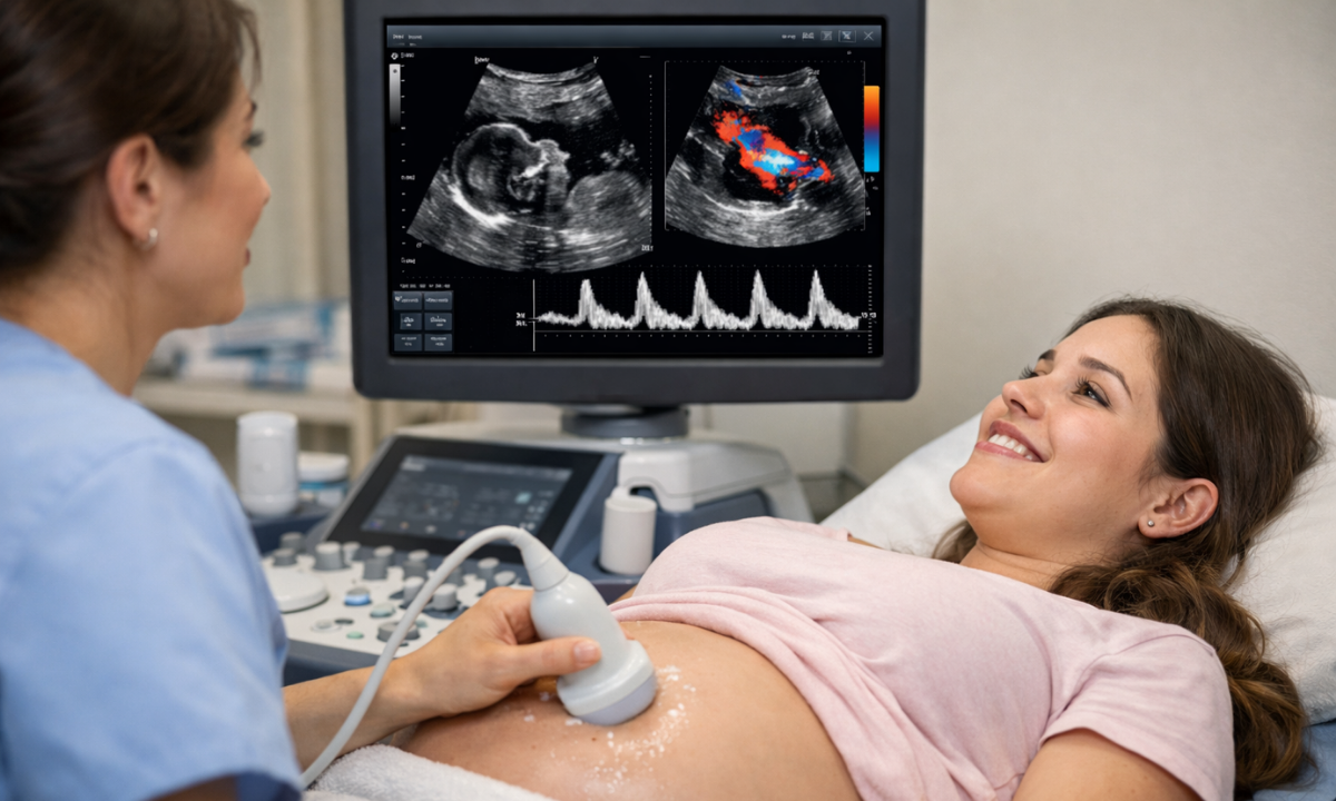
If you are asking, ‘What is a Doppler scan in pregnancy?’ let me simplify it for you. It’s an advanced ultrasound technique that uses sound waves to study the speed and direction of blood flow in your baby’s body.
Unlike regular scans that show images, this one shows how well blood circulates through critical areas like the placenta and umbilical cord. This makes it incredibly valuable in assessing whether your baby is developing safely.
Why the Doppler Test Is Done in Pregnancy
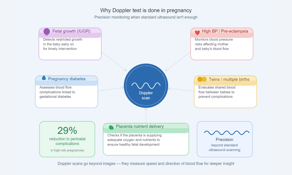
Now you might be wondering, why is a Doppler test done in pregnancy when regular ultrasounds already exist? The answer lies in precision and prevention.
Doctors recommend this scan when they need a deeper understanding of your baby’s condition. It helps in:
- Detecting restricted fetal growth (IUGR)
- Monitoring high blood pressure or pre-eclampsia
- Assessing pregnancies with diabetes
- Evaluating twins or multiple pregnancies
- Checking if the placenta is delivering enough nutrients
In fact, studies suggest that Doppler scans can reduce perinatal complications by up to 29% in high-risk pregnancies, making them a crucial diagnostic tool.
When a Doppler Scan Is Done in Pregnancy
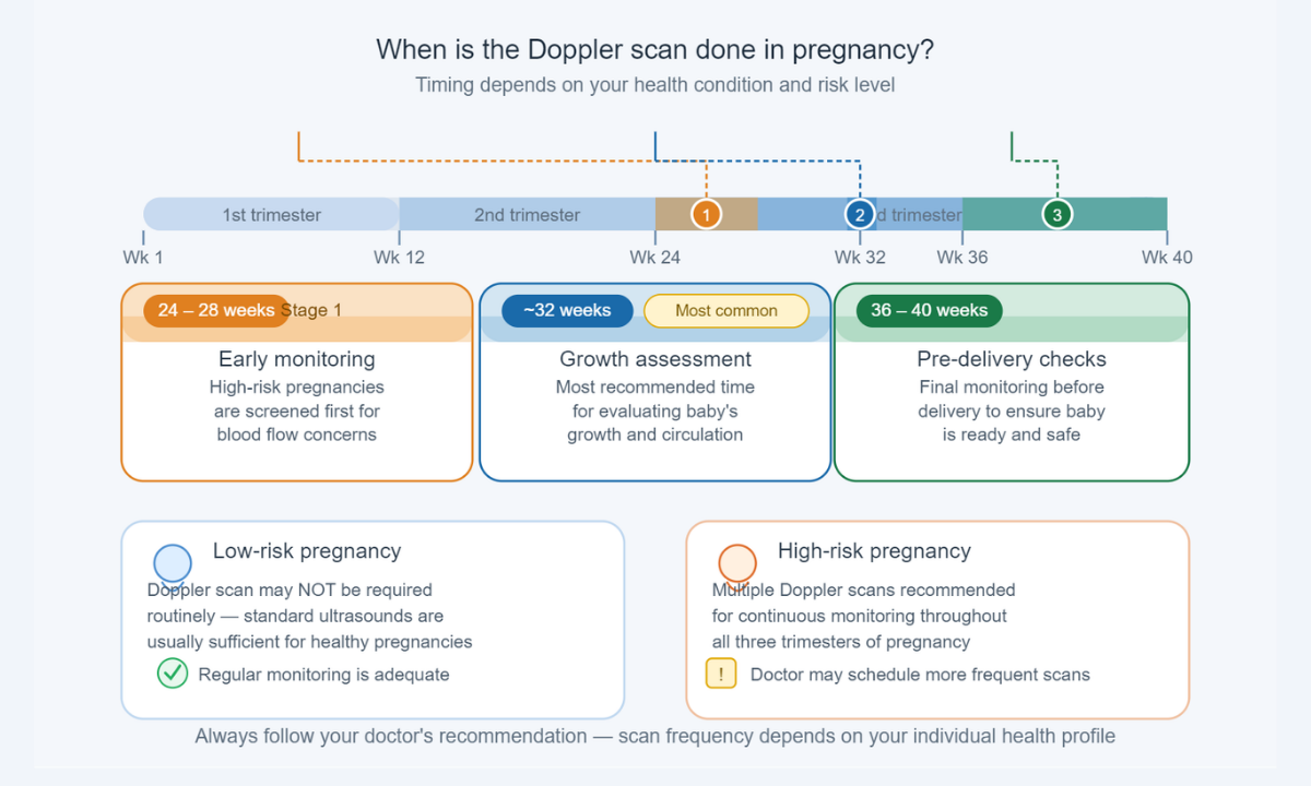
Timing plays a key role. So, when a Doppler scan is done in pregnancy, it depends on your health condition.
Here’s a quick breakdown:
| Stage of Pregnancy | Purpose |
|---|---|
| 24–28 weeks | Early monitoring in high-risk cases |
| Around 32 weeks | The most common time for growth assessment |
| 36–40 weeks | Final checks before delivery |
For low-risk pregnancies, it may not be required routinely. However, in high-risk cases, your doctor may recommend multiple scans for continuous monitoring.
What Does the Doppler Scan Measure?

This scan focuses on three vital blood flow areas that directly impact your baby’s health and overall development:
- Umbilical Artery – Ensures nutrients and oxygen reach the baby. It also helps detect if the placenta is functioning properly or if there is any resistance in blood flow, which could indicate growth concerns.
- Uterine Artery – Checks blood flow from the mother to the placenta. Abnormal flow here may signal conditions like pre-eclampsia or reduced placental efficiency.
- Middle Cerebral Artery (MCA) – Evaluates the blood supply to the baby’s brain. Increased blood flow in this artery can sometimes indicate that the baby is compensating for low oxygen levels.
Each of these measurements provides crucial insights, helping doctors identify potential complications early and take timely steps to ensure a safe and healthy pregnancy.
Types of Doppler Scans
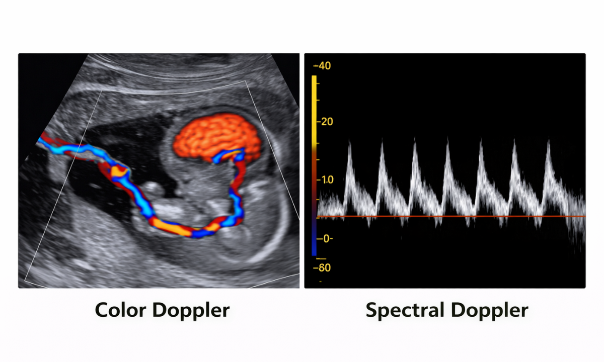
There are mainly two types used during pregnancy, and each serves a slightly different purpose in assessing your baby’s well-being:
Color Doppler
- Shows blood flow using different colours (usually red and blue)
- Helps visualise circulation patterns clearly in real time
- Makes it easier for doctors to identify blockages, reduced flow, or abnormal circulation in the placenta and umbilical cord
Spectral Doppler
- Displays blood flow as waveforms on a graph
- Helps measure the speed, direction, and resistance of blood flow
- Provides detailed numerical values that doctors use to assess whether the blood supply is normal or needs closer monitoring
Both are equally safe and non-invasive, and doctors often use them together for a more comprehensive evaluation, depending on what needs to be assessed in your pregnancy.
What Happens During the Procedure?
If you are feeling anxious, let me reassure you; the procedure is simple, safe, and completely painless. It is very similar to a regular ultrasound, with a slightly more detailed focus on blood flow.
- A clear gel is applied to your abdomen to help transmit sound waves effectively
- A handheld device (transducer) is gently moved across the skin
- Sound waves create detailed images along with colour or waveform patterns showing blood flow
- You may hear a soft “whooshing” sound, which represents the baby’s blood circulation
Duration: 20–60 minutes, depending on how detailed the assessment needs to be
Results: Usually available within 15–60 minutes, and your doctor will explain them to you
In most cases, no special preparation is needed, and you can eat and drink normally before the scan. You can walk in and out without any discomfort, which makes it a stress-free and reassuring experience for expectant mothers.
Understanding Doppler Scan Pregnancy Results
The next big question is about Doppler scan pregnancy results: what do they really indicate, and how should you interpret them?
Doctors carefully analyse blood flow patterns and specific indices (like resistance and pulsatility) to understand how well oxygen and nutrients are reaching your baby. Based on this, results are typically interpreted as follows:
- Normal blood flow: Indicates healthy fetal development and efficient placental function. This means your baby is receiving adequate oxygen and nutrients, and routine monitoring will usually continue.
- Reduced flow: May signal placental insufficiency, where the placenta is not delivering enough nutrients. In such cases, doctors may recommend more frequent scans, rest, or medication to support better outcomes.
- Absent or reversed flow: This is a serious finding that suggests significant compromise in blood circulation. It requires immediate medical attention, close monitoring, and sometimes early delivery to protect the baby’s health.
Doctors may also compare these results with your baby’s growth, heartbeat, and other ultrasound findings to get a complete picture. These insights help them decide whether to continue monitoring, adjust treatment, or take timely action, ensuring the safest possible outcome for both mother and baby.
Is There Any Doppler Scan Pregnancy Risk?
One of the most common concerns I hear is about the Doppler scan pregnancy risk.
Here’s the truth:
- It uses sound waves, not radiation
- It is completely non-invasive
- It is considered safe for both mother and baby
However, it is only recommended when medically necessary, ensuring there’s no overuse.
Who Should Definitely Get a Doppler Scan?
While not mandatory for everyone, some pregnancies benefit greatly from this scan:
- Mothers with high blood pressure or diabetes
- Cases of slow fetal growth
- Multiple pregnancies (twins/triplets)
- Previous history of pregnancy complications
If you fall into any of these categories, your doctor may strongly recommend it.
Tips to Prepare for Your Doppler Scan
To make the most of your ultrasound Doppler scan, here are a few simple tips I always suggest:
- Wear comfortable, loose clothing
- Stay hydrated before the test
- Carry your previous reports
- Ask questions and clarify doubts immediately
Being prepared helps you feel more relaxed and informed.
Final Thoughts
A Doppler scan during pregnancy is more than just a test; it’s a powerful tool that ensures your baby’s safety by monitoring vital blood flow. If you are planning an ultrasound scan in Bangalore, choosing a trusted health screening centre in Bangalore can make all the difference in accuracy and care.
At Koshikaa, the focus is always on combining advanced technology with compassionate care, ensuring every mother feels informed, reassured, and supported throughout her pregnancy journey.
FAQs
1. Is a Doppler scan painful?
No, a Doppler scan is completely painless and non-invasive. It feels very similar to a regular ultrasound, where a gel is applied, and a device is moved over your abdomen. There are no needles, injections, or discomfort involved, making it a safe and reassuring experience for expectant mothers.
2. How long does the scan take?
A Doppler scan usually takes between 20 and 60 minutes, depending on the level of detail required. In some cases, it may take slightly longer if the baby’s position makes imaging difficult. However, the procedure is generally quick, smooth, and completed within a single outpatient visit.
3. Can it harm my baby?
No, a Doppler scan is considered completely safe for your baby when performed under medical supervision. It uses sound waves, not radiation, to assess blood flow. Doctors recommend it only when necessary, ensuring that both mother and baby benefit from accurate monitoring without any known risks.
4. Do all pregnant women need it?
Not all pregnant women require a Doppler scan. It is usually recommended for high-risk pregnancies, such as those involving high blood pressure, diabetes, or concerns about the baby’s growth. Your doctor will decide if it is necessary based on your medical condition and overall pregnancy progress.
5. How soon will I get the results?
In most cases, Doppler scan results are available within 30 to 60 minutes after the procedure. A radiologist analyses the blood flow patterns and shares the findings with your doctor. Your doctor will then explain the results and guide you on any next steps if required.
Reference
1. From Google