
Have you ever felt anxious about a strange pain or symptom and wished there was a safe, simple way to see what’s happening inside your body? That’s exactly where medical ultrasound becomes a powerful and reassuring tool in modern healthcare.
Today, many people choose an ultrasound scan in Bangalore as a quick, safe, and reliable way to understand their health without fear of radiation or complex procedures. Whether it is pregnancy, abdominal pain, thyroid swelling, or kidney stones, an ultrasound gives us real-time answers in a painless way.
In this blog, we will clearly understand how diagnostic medical ultrasound works, the different types of ultrasound, and why it is one of the safest imaging tests available today, so keep reading to discover how this simple scan can protect your health.
Key Points at a Glance
- Diagnostic medical ultrasound is a radiation-free imaging test.
- It uses high-frequency sound waves to create real-time images.
- It is safe for pregnant women and children who require regular monitoring.
- There are many types of ultrasound, including abdominal, thyroid, pregnancy, Doppler, and more.
- It helps detect kidney stones, liver issues, thyroid nodules, and fetal growth.
- It is affordable compared to CT or MRI.
- Minimal preparation is required for most scans.
What is Diagnostic Medical Ultrasound?
Diagnostic medical ultrasound, also called sonography, is a non-invasive imaging technique that uses high-frequency sound waves to produce images of organs, tissues, and blood vessels inside the body.
Unlike X-rays or CT scans, ultrasound does not use radiation. This makes it extremely safe for repeated use and for sensitive groups like pregnant women and children.
We often use ultrasound to detect, monitor, or guide treatment for various health conditions. It is widely used in cardiology, obstetrics, gastroenterology, urology, and general medicine.
How does Medical Ultrasound Work?
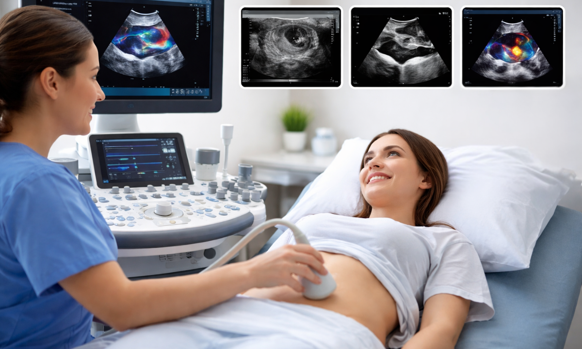
Ultrasound works in a way similar to sonar technology used by ships or bats. It sends sound waves into the body and listens to the echoes that bounce back.
Let’s understand the process step by step:
| Step | What Happens | Why It Matters |
|---|---|---|
| 1. Gel Application | A water-based gel is applied to the skin | Removes air pockets and improves sound transmission |
| 2. Transducer Use | A hand-held probe sends sound waves into the body | Sound waves are inaudible to humans |
| 3. Echo Reflection | Sound waves bounce off organs and tissues | Different tissues reflect waves differently |
| 4. Image Formation | A computer converts echoes into images | Creates real-time 2D or 3D images |
The image produced is called a sonogram. We can see movements in real time, such as blood flow or a baby’s heartbeat.
Common Types of Ultrasound
There are several diagnostic medical ultrasound types used, depending on the organ or condition being examined. Each type focuses on specific organs and helps doctors identify problems quickly and accurately.
Understanding which scan is recommended for you can reduce anxiety and help you prepare better for the procedure. Let us look at the most commonly performed ultrasound scans and what they evaluate.
1. Abdominal Ultrasound

An abdominal ultrasound is one of the most frequently recommended scans when someone experiences stomach pain, bloating, or digestive discomfort. It provides a clear view of major abdominal organs and helps detect infections, stones, inflammation, or tumors at an early stage.
An abdominal scan checks organs such as:
- Liver
- Gallbladder
- Spleen
- Pancreas
- Kidneys
It is commonly used for:
- Ultrasound scan for liver conditions like fatty liver or tumors
- Detecting gallstones
- Identifying infections
- Evaluating abdominal pain
2. USG Abdomen and Pelvis

A USG abdomen and pelvis is often recommended when symptoms involve both the lower abdomen and pelvic region. It provides a more comprehensive view, helping doctors assess urinary, reproductive, and digestive organs together in a single scan.
This scan is especially useful for identifying causes of persistent pelvic pain, urinary discomfort, irregular menstrual cycles, or prostate-related concerns. Examining multiple organs at once, it allows for quicker diagnosis and better treatment planning.
It helps examine:
- Uterus and ovaries (in women)
- Prostate (in men)
- Urinary bladder
- Kidneys
Doctors often recommend it for pelvic pain, fibroids, cysts, urinary infections, or unexplained abdominal discomfort.
3. Ultrasound Scan for Kidney Stones
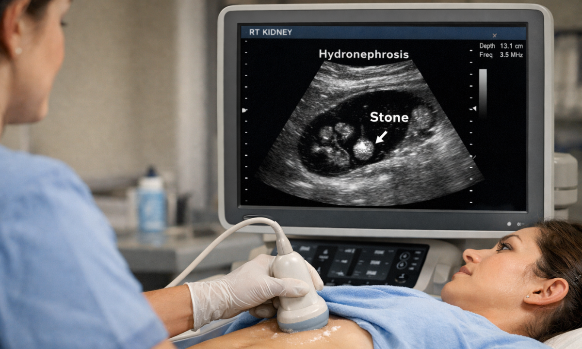
Kidney stones can cause sudden, sharp pain in the lower back or abdomen, often making daily activities difficult. An ultrasound scan for kidney stones is commonly recommended to quickly identify the cause of this discomfort in a safe and non-invasive way.
This scan helps doctors understand whether a stone is blocking urine flow or causing swelling in the kidney. Early detection allows timely treatment and prevents complications.
An ultrasound scan for kidney stones helps detect:
- Stone size
- Stone location
- Kidney swelling
- Urinary obstruction
It is a safe first-line imaging test, especially for pregnant women or patients who should avoid radiation.
4. Thyroid Ultrasound
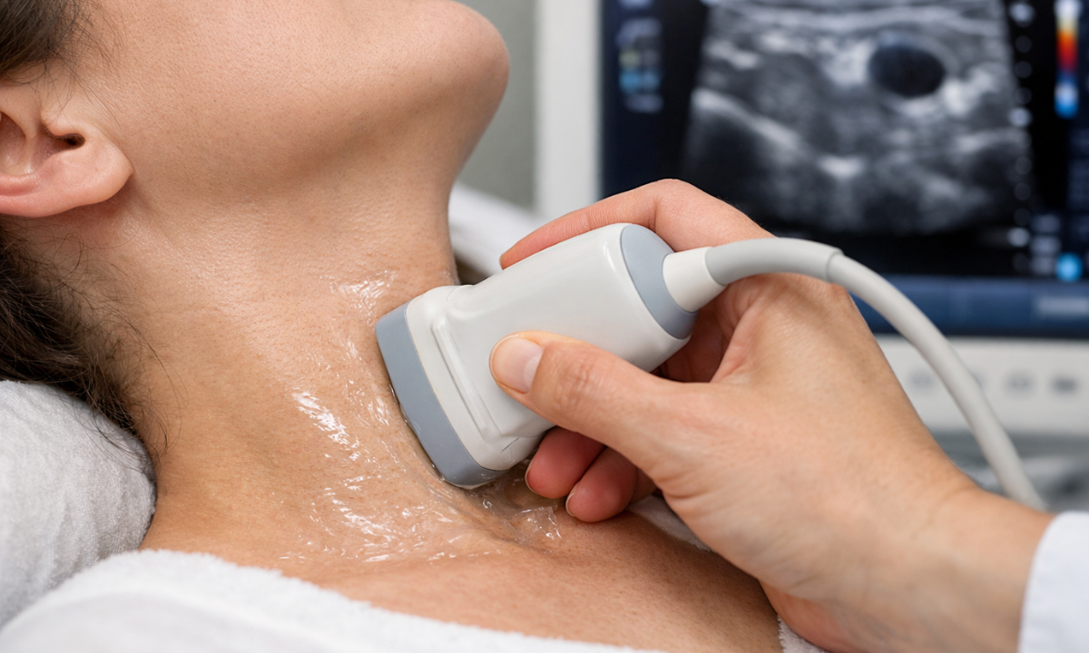
A thyroid ultrasound evaluates the thyroid gland located at the front of the neck. It is usually recommended when we notice swelling in the neck, abnormal thyroid blood reports, difficulty swallowing, or a visible lump.
A Thyroid ultrasound scan provides detailed images of the size, shape, and texture of the thyroid gland. It helps doctors differentiate between solid nodules and fluid-filled cysts, which is important for deciding the next steps in treatment.
It helps detect:
- Thyroid nodules
- Cysts
- Enlargement
- Suspicious lumps
Also helps guide fine-needle aspiration biopsy if needed, ensuring accurate sampling of thyroid nodules for further evaluation.
5. Ultrasound Scan for Pregnancy

One of the most common uses of diagnostic imaging is the ultrasound scan for pregnancy. It plays a vital role in confirming pregnancy, estimating the due date, and ensuring both mother and baby are healthy throughout the journey.
Regular pregnancy ultrasounds provide reassurance and help doctors detect any concerns early. They also allow parents to see their baby’s development, which can be a deeply emotional and comforting experience.
It helps monitor:
- Fetal growth
- Baby’s heartbeat
- Placenta position
- Amniotic fluid levels
During pregnancy, a growth scan is performed in the later months to check whether the baby is developing normally, gaining appropriate weight, and receiving enough nutrients and oxygen through the placenta.
6. Doppler Ultrasound
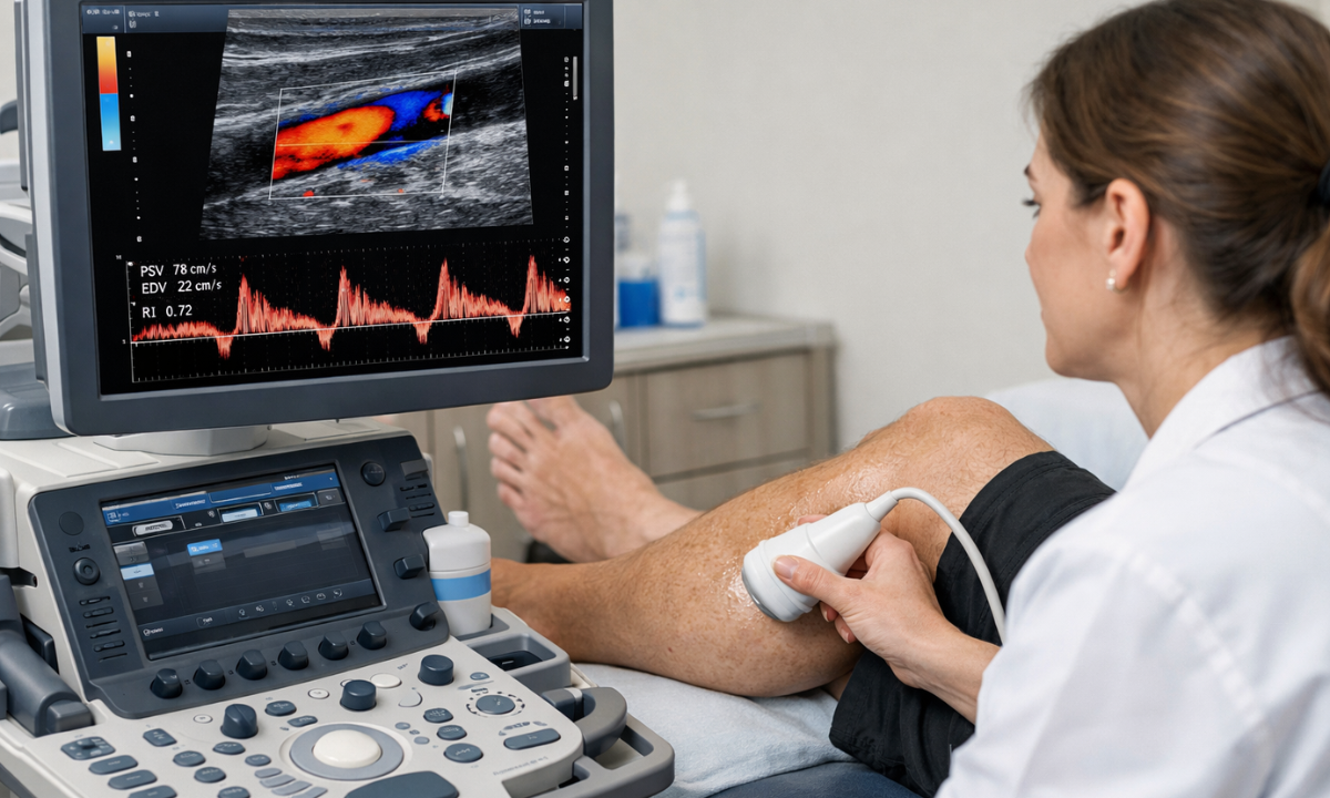
Doppler ultrasound checks blood flow in arteries and veins using sound waves to measure the movement of blood cells. It helps us understand whether blood is flowing normally or if there is any blockage or narrowing in the vessels.
This type of scan is especially useful for patients experiencing leg swelling, chest pain, stroke risk, or circulation issues. It provides real-time information about blood speed and direction, which helps doctors plan the right treatment quickly.
It helps identify:
- Blood clots
- Blocked arteries
- Varicose veins
- Circulation problems
7. Echocardiogram
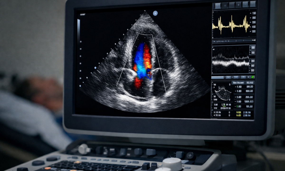
An echocardiogram is a special ultrasound of the heart that provides moving images of the heart in action. It allows doctors to see how well the heart muscles and valves are working without any invasive procedure.
This scan is often recommended if we experience symptoms like chest pain, shortness of breath, irregular heartbeat, or high blood pressure. It helps detect heart conditions early and guides proper treatment planning.
It shows:
- Heart chambers
- Valve function
- Pumping ability
- Structural abnormalities
An echocardiogram is completely safe and painless, and it plays a crucial role in diagnosing and monitoring various heart-related conditions.
Benefits of Medical Ultrasound
Let us understand why medical ultrasound is so widely recommended in modern healthcare. It combines safety, accuracy, comfort, and affordability, making it one of the most trusted diagnostic tools available today.
1. Safe – No Radiation
Ultrasound does not use ionising radiation, unlike X-rays or CT scans. Instead, it relies on harmless sound waves, which makes it suitable even for repeated monitoring.
This makes it especially safe for:
- Pregnant women
- Children
- Regular follow-ups
- Patients who require frequent imaging
Because of this safety profile, doctors confidently recommend ultrasound for both routine and critical evaluations.
2. Non-Invasive and Painless
There are no cuts, stitches, or injections involved in most ultrasound procedures. The scan is performed externally using a handheld probe.
We simply lie down comfortably while the probe moves gently over the skin with a layer of gel. Most people experience little to no discomfort, and the test is usually completed within 15–30 minutes.
3. Real-Time Imaging
One of the biggest advantages of ultrasound is that it provides real-time imaging. This means doctors can see movement instantly as it happens inside the body.
For example, they can observe:
- Blood flows through the vessels
- A baby moving in the womb
- The heart beating and pumping blood
This real-time view helps doctors make faster and more accurate decisions, especially in emergencies or during guided medical procedures.
4. Affordable
Compared to MRI or CT scans, the cost for ultrasound scan is generally lower.
Here’s a simple ultrasound scan cost breakdown at Koshikaa:
| Scan | Price |
|---|---|
| Abdominal ultrasound | ₹1250/- |
| USG Abdomen and Pelvis | ₹1000/- |
| Kidney ultrasound | ₹850/- |
| Breast ultrasound | ₹1000/- |
| Pelvis ultrasound | ₹850/- |
| Transvaginal ultrasound | ₹1000/- |
| Thyroid ultrasound | ₹1000/- |
5. Minimal Preparation
Most ultrasound scans require very little preparation, making the process simple and convenient. The instructions depend on the type of scan, but they are usually easy to follow.
Examples:
- Fasting for an abdominal scan (6–8 hours)
- Drinking water for a pelvic scan (full bladder improves clarity)
- No preparation for a thyroid scan
Because the preparation is minimal, we can usually complete the scan without disrupting our daily routine.
6. Procedure Guidance
Ultrasound also acts as a real-time guide during certain medical procedures. Doctors can clearly see internal structures while performing the procedure, which improves precision.
It is commonly used during:
- Biopsies
- Fluid drainage
- Needle placement
This live guidance improves safety, accuracy, and overall treatment outcomes.
How to Prepare for an Ultrasound
Preparation depends on the type of scan.
| Type of Scan | Preparation Needed |
|---|---|
| Abdominal | 6–8 hours fasting |
| Pelvic | Drink 3–4 glasses of water |
| Thyroid | No preparation |
| Pregnancy | Usually, no fasting is required |
| Kidney Scan | May require a full bladder |
Always follow the instructions given by your diagnostic centre.
When Should We Consider an Ultrasound?
You may need an ultrasound if you experience:
- Persistent abdominal pain – To check for issues like gallstones, liver problems, infections, or inflammation in abdominal organs.
- Thyroid swelling – To evaluate thyroid enlargement, nodules, cysts, or other abnormalities in the neck.
- Irregular menstrual cycles – To examine the uterus and ovaries for conditions such as PCOS, fibroids, or cysts.
- Kidney stone symptoms – To detect stones, blockage, or swelling in the kidneys and urinary tract.
- Pregnancy confirmation – To confirm pregnancy, estimate due date, and assess early fetal development.
- Liver abnormalities – To identify fatty liver, infections, tumours, or other structural changes in the liver.
- Unexplained pelvic pain – To examine pelvic organs and identify possible causes like cysts, infections, or reproductive concerns.
A reliable health screening centre in Bangalore often includes an ultrasound as part of preventive health packages.
Limitations of Ultrasound
While highly effective, ultrasound has some limitations.
- Sound waves do not travel well through bone or air.
- Deep structures may be difficult to visualize.
- Image quality depends on the technician’s skill.
Despite these limitations, it remains one of the most trusted diagnostic tools.
Final Thoughts
Diagnostic ultrasound has transformed modern healthcare by offering a safe, affordable, and reliable way to look inside the body without surgery or radiation. From a simple ultrasound scan for liver issues to a detailed growth scan during pregnancy, it helps us detect problems early and take timely action.
If you are considering an ultrasound scan in Bangalore, choosing a trusted diagnostic centre matters for accuracy and comfort. At Koshikaa, we focus on patient safety, skilled professionals, and high-quality imaging to give you clarity and confidence about your health.
When we understand what is happening inside our body, we make better decisions, and sometimes, that simple scan can bring the peace of mind we truly need.
Frequently Asked Questions
1. Is ultrasound safe during pregnancy?
Yes, ultrasound is considered very safe during pregnancy because it uses high-frequency sound waves, not radiation. It has been used for decades to monitor fetal growth and development without harming the mother or baby.
Doctors may recommend multiple scans throughout pregnancy to ensure everything is progressing normally and safely.
2. How long does an ultrasound take?
An ultrasound scan usually takes about 15 to 30 minutes, depending on the type of examination.
A simple thyroid or abdominal scan may be quicker, while a detailed pregnancy or Doppler scan may take slightly longer. The procedure is done on an outpatient basis, and you can typically resume normal activities immediately afterwards.
3. Is the ultrasound procedure painful?
No, an ultrasound is generally painless and non-invasive. You may feel slight pressure from the probe as it moves over the skin, but it does not cause discomfort in most cases. The gel applied during the scan may feel cool, but the entire process is usually comfortable and stress-free.
4. What is the average cost for an ultrasound scan?
The cost for an ultrasound scan varies depending on the type of scan, the diagnostic centre, and the city. However, it is generally more affordable than CT scans or MRI.
Basic scans like abdominal or pelvic ultrasound are usually reasonably priced, making them accessible for routine diagnosis and preventive health checkups.
5. Can ultrasound detect all diseases?
While ultrasound is highly effective for examining soft tissues and organs, it cannot detect every condition. Sound waves do not pass well through bone or air-filled structures, so certain lung or bone issues may require a CT or an MRI.
Doctors choose the most appropriate imaging test based on your symptoms and medical needs.
Reference:
1. From Google
2. https://my.clevelandclinic.org/health/diagnostics/4995-ultrasound