Guide on Essential Vitamin and Mineral Deficiency Blood Tests

Table of Contents

You slept eight hours last night. You drank your morning coffee. and even had a healthy lunch. So why are you struggling to keep your eyes open at 3 PM?

We often blame stress, traffic, or “burnout” for our low energy. But very often, the culprit isn’t your lifestyle—it’s your biology.

You might be suffering from what nutritionists call “Hidden Hunger” (Micronutrient Deficiency). 

This happens when you look healthy on the outside, but on the inside, your cells are starving for the specific fuel they need to function.

We believe that “feeling okay” isn’t enough.

You deserve to feel vital. Whether you are dealing with unexplained hair fall, constant fatigue, or brittle nails, a simple check-up can reveal the invisible gaps in your nutrition. 

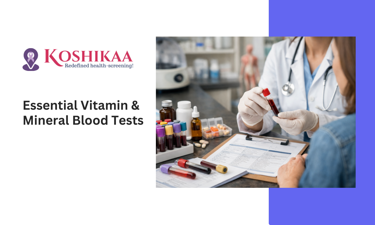
In this guide, we will break down the essential vitamin and mineral deficiency blood test options, decipher the signs of vitamin D deficiency in blood test results, and explain why getting tested might be the most productive thing you do this year.

The “Big Two”: Vitamins You Cannot Ignore

The invisible crash of Energy and Mood.

When we talk about a vitamin and mineral deficiency blood test, we aren’t usually looking for rare, exotic diseases.

Especially in urban hubs like Bangalore, 80% of deficiencies come down to just two culprits: Vitamin D and Vitamin B12.

A. Vitamin D: The “Indoor” Epidemic

Vitamin D

It is ironic, isn’t it? We live in a tropical country with abundant sunshine, yet nearly 70% of Indians are Vitamin D deficient.

Why? Because the modern lifestyle keeps us indoors—in air-conditioned offices, cars, and metro stations—during the prime sunlight hours (10 AM to 3 PM).

  • Why you need it: It acts more like a hormone than a vitamin. It controls calcium absorption, immune function, and mood regulation.
  • The Warning Signs:
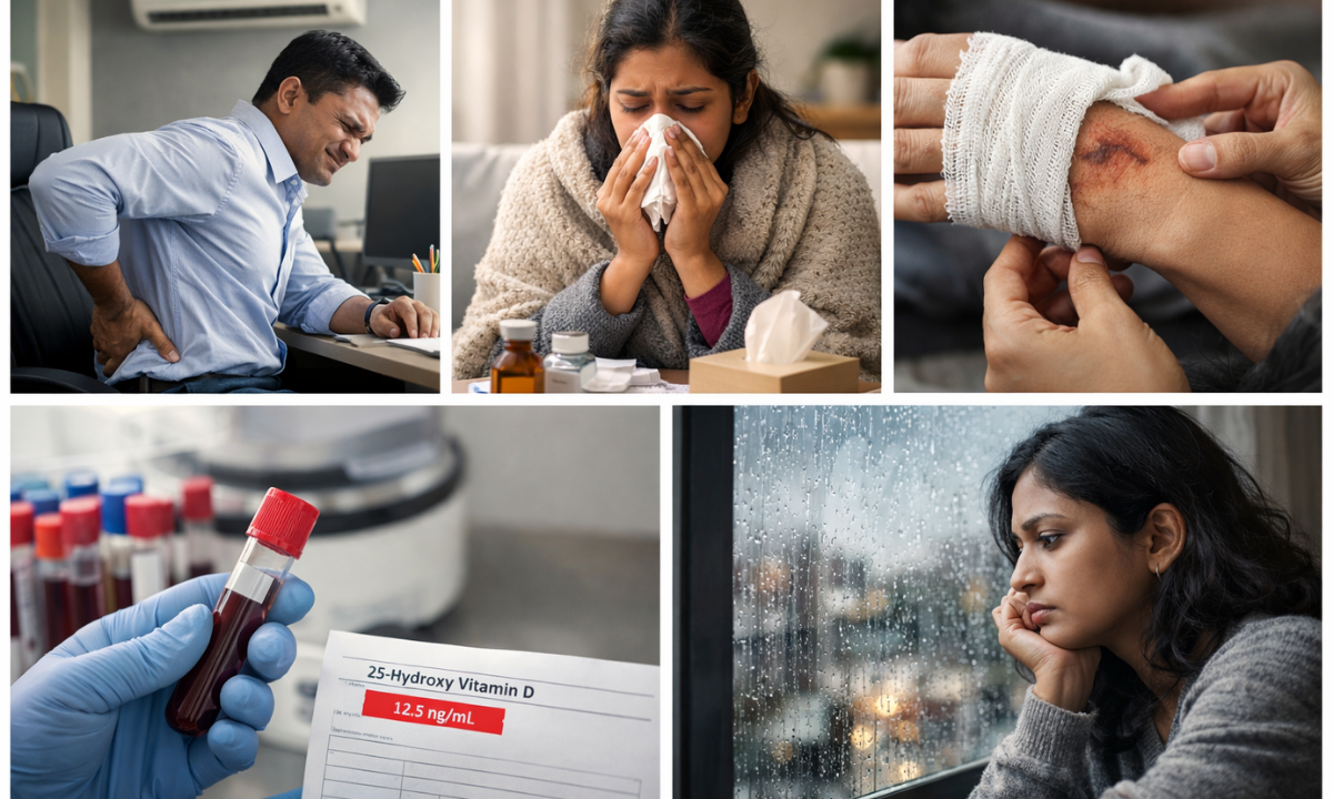
    • Unexplained bone or lower back pain.
    • Frequent infections (falling sick often).
    • Slow healing of wounds.
    • “The Blues”: Mood swings or feelings of depression, especially during monsoons.
  • The Test: A Vitamin D deficiency blood test measures “25-Hydroxy Vitamin D.” If your level is below 20 ng/mL, your “battery” is critically low.

B. Vitamin B12: The “Vegetarian’s Challenge”

Vitamin B12

If Vitamin D is the battery, Vitamin B12 is the wiring.

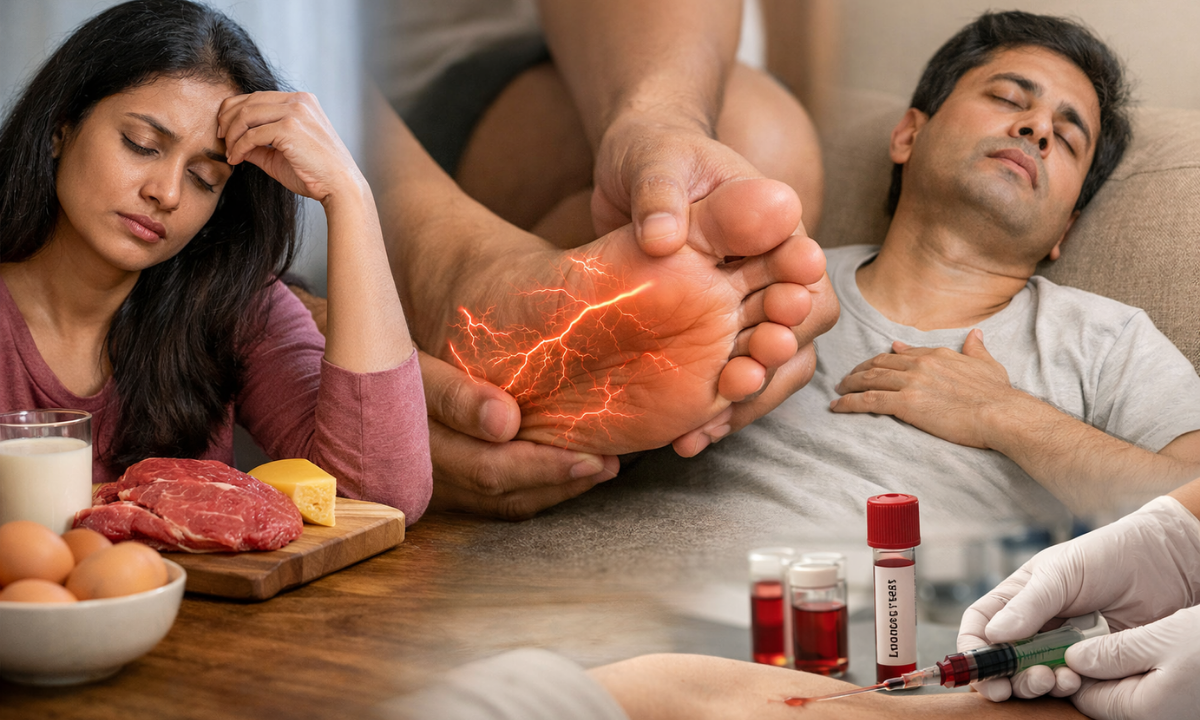
It protects your nerves and helps create red blood cells. Since B12 is found almost exclusively in animal products (meat, eggs, dairy), vegetarians and vegans are at extremely high risk.

Even non-vegetarians in India often have low levels because we don’t consume meat daily.

  • Why you need it: It keeps your brain sharp and your nerves firing correctly.
  • The Warning Signs:
    • Brain Fog: Forgetting names or feeling confused.
    • Pins and Needles: Tingling sensations in hands or feet.
    • Pale Skin: Due to a specific type of anemia.
    • Extreme lethargy (feeling “heavy”).
  • The Test: A vitamin B12 deficiency blood test is often paired with a “Homocysteine” test for a complete picture of your nerve health.

Many patients treat fatigue with more coffee. But caffeine cannot fix a biological deficit. If you are drinking 4 cups a day just to function, checking your B12 and D3 is not optional—it is essential tests.

The Supporting Cast: Essential Minerals

Iron, Calcium, and the “Relaxation Mineral.

Vitamins are organic (made by plants/animals), while minerals are inorganic (from the soil/water).

Your body needs them for strong bones, transmitting nerve signals, and keeping your heart beating. A deficiency here doesn’t just make you tired; it can physically weaken you.

A. Iron (Ferritin): The Oxygen Carrier

Iron

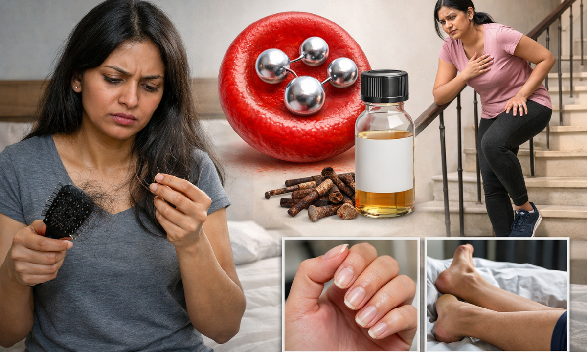
Iron deficiency is the most common nutritional disorder in the world. In India, it affects nearly 50% of women.

Iron helps create hemoglobin, which carries oxygen from your lungs to the rest of your body.

  • The “Ferritin” Trap: Many people test their Hemoglobin and think they are fine. But you must test Serum Ferritin.

This measures your stored iron. You can have normal hemoglobin but “empty” iron stores, which still causes symptoms.

  • The Warning Signs:
    • Hair Fall: This is often the first sign.
    • Breathlessness when climbing stairs.
    • Pale skin and brittle nails.
    • Restless Leg Syndrome (uncomfortable urge to move legs at night).

B. Calcium: Not Just for Bones

Calcium

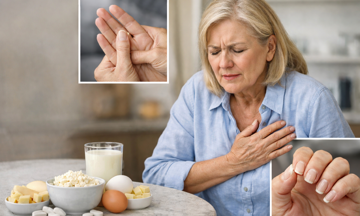
We all know calcium builds strong bones. But did you know your heart needs calcium to beat?

If your blood calcium drops too low, your body literally “steals” calcium from your bones to keep your heart working. Over time, this makes bones weak (Osteoporosis).

  • The Warning Signs:
    • Muscle cramps (especially in the calves/toes).
    • Numbness in fingers.
    • Weak, brittle nails.

C. Magnesium: The “Ignored” Mineral

Magnesium

Magnesium is involved in over 300 chemical reactions in the body, yet it is rarely talked about.

It is called the “Relaxation Mineral.” It helps your muscles relax after contracting and helps your brain shut down for sleep.

  • The Warning Signs:
    • Insomnia: Waking often at night.
    • Eye Twitching: That annoying flutter in your eyelid is often a magnesium alert.
    • High stress and anxiety.
    • Frequent headaches or migraines.

Calcium and Iron compete for absorption. If you are taking supplements, never take them together. Take Iron with Vitamin C (lemon juice) and Calcium with Vitamin D for the best results.

Signs You Need This Test (The Checklist)

Many of us ignore subtle signs, thinking they are just part of “aging” or “stress.”

However, your body has very specific ways of signaling a shortage. Use this checklist to see if your symptoms match a potential deficiency.

What’s Missing? Symptom Table

If you are experiencing…It could be a lack of…Why? (The Science)
Extreme Fatigue / WeaknessVitamin D, B12, IronYour cells aren’t getting enough oxygen (Iron) or your metabolism is stalling (B12).
Hair Fall / ThinningIron (Ferritin), Biotin, ZincHair follicles are “non-essential” tissue; the body cuts their supply first when nutrients are low.
Mouth Ulcers / CracksVitamin B12, B-ComplexB-vitamins maintain the moist lining (mucosa) of your mouth and tongue.
Muscle Cramps / TwitchingMagnesium, Calcium, Vitamin DYour muscles need these minerals to “relax” after contracting. Without them, they seize up.
Bone or Lower Back PainVitamin D, CalciumYour bones are softening (Osteomalacia) due to a lack of mineralization.
Numbness / Tingling (Hands)Vitamin B12The protective sheath (myelin) around your nerves is wearing thin.
Brittle or Spoon-shaped NailsIronLack of oxygenated blood reaching the nail bed changes its shape and texture.

Self-Check: If you ticked more than two boxes in this table, it is highly recommended to book a blood test for deficiency of vitamin profiles.

Guessing with supplements can be dangerous (some vitamins are toxic in high doses), so testing is the only safe first step.

The Logistics: How to Prepare for the Test

Do I need to fast? Does it hurt?

Getting a blood test in Bengaluru used to mean driving through traffic to a lab and waiting in a queue.

Today, with the Health Screening Centre in Bangalore, services like Koshikaa, the lab comes to your doorstep. But before you book, here is what you need to know about preparation.

To Fast or Not to Fast?

This depends on which vitamins you are testing, but since most people opt for a complete profile (checking everything at once), fasting is highly recommended.

  • Strict Fasting (10–12 Hours): Required if your package includes Iron Studies, Calcium, or Blood Sugar. Food can temporarily spike these levels, giving you a false result.
  • No Fasting Needed: Purely testing Vitamin D or Vitamin B12 does not technically require fasting. However, doctors prefer fasting samples to keep the blood serum clear (non-lipemic) for more accurate analysis.

The Golden Rule: If you are booking a “Full Body Vitamin Profile,” schedule your home collection for 8:00 AM. Have your dinner by 9:00 PM the previous night, and drink only water in the morning.

The Procedure: A Simple Prick

  1. Home Collection: A trained phlebotomist visits your home.
  2. The Draw: They draw a small amount of blood (usually 3–5 ml) from a vein in your arm.
  3. The Processing: The sample is transported in a cold-chain box (crucial for sensitive enzymes) to our NABL-accredited lab.

The Report: You typically receive a detailed digital report within 24 hours.

Cost & Packages: Why “Bundling” Saves You Money

Don’t pay double by testing single.

One of the biggest hesitations people have is the cost.

  • “Is it expensive?”
  • “Should I just buy the pills instead?”

Here is the financial reality: Guessing is expensive.

Buying supplements you don’t need (or taking the wrong dose) wastes money and can be dangerous.

However, booking individual tests can also add up quickly. That is why most Health Screening Centres in Bangalore offer “Profiles” or “Packages.”

Market Price Comparison in Bangalore (2026 Estimates):

Test NameAverage Cost (Single Test)Smart Choice (Profile)
Vitamin D Total₹1,200 – ₹1,800Most Vitamin Deficiency Packages bundle these together for approx. ₹1,999 – ₹2,999.(You save nearly 50% compared to an individual booking)
Vitamin B12₹800 – ₹1,300
Iron Profile (Ferritin)₹650 – ₹900
Calcium & Magnesium₹400 – ₹600
Total Value~₹3,000+

Pro Tip: Look for a “Full Body Vitamin Profile.” It usually includes Vitamin D, B12, Calcium, Iron, and sometimes Electrolytes for a single discounted price.

Why Choose Koshikaa?

We don’t just give you a number; we give you a plan.

Most labs hand you a report full of confusing numbers and leave you to figure it out. Koshikaa is different. We are not just a testing lab; we are a Preventive Health Partner.

  1. Contextual Reports: We explain what your low Vitamin D actually means for your daily energy.
  2. Actionable Advice: We don’t just say “Low.” We tell you which foods (available in Bangalore markets) can help boost your levels naturally.
  3. Convenience: Our phlebotomists are trained for painless home collection across Bangalore—from Whitefield to Malleshwaram.

Conclusion

Fatigue is not a personality trait. Brain fog is not “just stress.” Hair fall is not “just the water.”

Very often, these are your body’s cries for help. They are biological signals that your fuel tank is running empty.

A simple vitamin and mineral deficiency blood test can turn the lights back on. Imagine waking up without needing three coffees. Imagine finishing your workday with energy left for the gym or your family.

That version of you is waiting. Don’t let Hidden Hunger steal another day of your potential.

MRI of the uterus is primarily used for diagnosing and evaluating conditions such as uterine fibroids, adenomyosis, and malignancies. It is particularly useful in the planning of surgical procedures and assessing the extent of gynecological diseases. This imaging test is valuable for its ability to deliver detailed cross-sectional images of the uterus and surrounding structures.
An MRI of the spleen can help diagnose various disorders affecting the spleen including splenomegaly (enlarged spleen), tumors, and other abnormalities. It provides clear, detailed images that are crucial for assessing the health of the spleen and planning potential surgical interventions.
Prostate MRI is a non-invasive imaging technique that provides detailed pictures of the prostate gland and surrounding tissues. It helps in the detection, staging, and monitoring of prostate cancer. Prostate MRIs are increasingly used to guide biopsy procedures and assist in the planning of treatment strategies for prostate health.
An MRI of the pancreas is used to identify issues such as pancreatitis, pancreatic cancer, and other pancreatic disorders. It offers high-resolution images that help in detecting small lesions and assessing the pancreatic ducts. This imaging technique is valuable for guiding biopsies and planning surgeries involving the pancreas.
MRI scans of the liver, kidney, and bile ducts provide detailed images that help diagnose various conditions, such as tumors, cysts, and blockages in the bile ducts. They are crucial for assessing organ damage, staging cancer, and planning liver or kidney-related surgeries. This type of MRI can also evaluate the health and function of these organs comprehensively.
Breast MRI is highly sensitive for detecting breast cancer, especially in individuals with dense breast tissue or a high risk of cancer. It is often used in conjunction with mammography to provide a more comprehensive evaluation. Breast MRIs can also monitor the response to breast cancer treatments and provide detailed pre-surgical mapping.
Brain and spine MRIs are critical tools for diagnosing and monitoring neurological conditions, including tumors, spinal cord injuries, and multiple sclerosis. These MRIs can reveal abnormalities in brain tissue and spinal cord structure that might not be visible with other imaging methods. They are essential for planning surgeries and assessing the effectiveness of treatments in neurology.
PET-CT scans of transplant organs like the liver, kidney, or pancreas are performed to assess metabolic activity, detect complications, and monitor post-transplant function. These scans play a vital role in the early detection of issues such as organ rejection, recurrence of disease, or infection, guiding timely interventions and optimizing transplant outcomes for patients.
PET-CT examinations of organs such as the liver, kidney, or prostate are crucial for assessing metabolic activity, detecting abnormalities, and monitoring disease progression. These scans help diagnose conditions such as liver metastases, renal cell carcinoma, or prostate cancer, guiding treatment decisions and monitoring response to therapy over time.
PET-CT scans of soft-tissue masses are utilized to assess metabolic activity and detect malignant tumors or other abnormalities. By combining PET imaging with CT imaging, these scans provide detailed information about tumor location, size, and metabolic characteristics, aiding in accurate diagnosis and treatment planning for soft-tissue cancers.
PET-CT scans are valuable for detecting cysts or fluid collections within the body, including cysts in the liver, kidneys, or other organs. These scans help characterize the nature and metabolic activity of cysts, guiding diagnosis and treatment planning for conditions such as cystic liver disease, renal cysts, or pancreatic cysts.
PET-CT scans of muscles are performed to assess metabolic activity and detect abnormalities such as inflammation, infection, or tumors. These scans provide valuable information about muscle function and pathology, aiding in the diagnosis and management of conditions like myositis, muscular dystrophy, or soft-tissue sarcomas.
PET-CT scans of the brain are utilized to evaluate brain metabolism and detect abnormalities such as tumors, epilepsy, or neurodegenerative diseases. By combining functional PET imaging with anatomical CT imaging, these scans offer comprehensive information about brain function and structure, guiding neurosurgical interventions and treatment decisions for neurological disorders
PET-CT scans of bones are commonly used to detect bone metastases in cancer patients, assess bone health in conditions like osteoporosis, and evaluate response to treatment. By combining PET and CT imaging, these scans provide detailed information about metabolic activity and anatomical changes in bone structures, aiding in accurate diagnosis and treatment planning for various bone-related conditions.
CT scans of the spine are conducted to assess the vertebrae, spinal cord, and surrounding soft tissues for conditions like herniated discs, spinal fractures, or spinal stenosis, aiding in the diagnosis and management of spinal disorders with detailed imaging essential for surgical planning and intervention.
CT scans of the pelvis are utilized to visualize the pelvic bones, reproductive organs, bladder, and surrounding structures for conditions such as fractures, tumors, or pelvic inflammatory disease, guiding treatment plans and surgical interventions with precise anatomical information.
CT scans of the neck are performed to evaluate structures such as the throat, thyroid gland, lymph nodes, and blood vessels for abnormalities like tumors, infections, or injuries, assisting in diagnosis and management of head and neck disorders with detailed imaging.
CT scans of the extremities are employed to assess bones, joints, and soft tissues in the arms or legs, aiding in the diagnosis of fractures, arthritis, or other musculoskeletal conditions, guiding orthopedic treatment and rehabilitation plans for optimal recovery.
CT scans of the chest are utilized to examine the lungs, heart, blood vessels, and surrounding structures for conditions such as lung nodules, pneumonia, or pulmonary embolism, facilitating accurate diagnosis and management of respiratory and cardiovascular diseases.
CT scans of the brain are conducted to assess brain structures, identify abnormalities like tumors, hemorrhages, or strokes, and aid in diagnosing neurological conditions, guiding neurosurgical interventions and treatment strategies with precise imaging of the brain’s anatomy.
CT scans of the abdomen are performed to visualize and evaluate the internal organs such as the liver, kidneys, pancreas, and intestines for conditions like tumors, infections, or injuries, providing detailed images essential for diagnosis and treatment planning.
Transplant organ ultrasound scans, including those of the liver, kidney, or pancreas, are performed to assess organ viability, detect complications, and monitor post-transplant function. These scans play a vital role in the early detection of issues such as organ rejection, vascular complications, or fluid collections, guiding timely interventions and optimizing transplant outcomes for patients.
Ultrasound examinations of organs such as the liver, kidney, or prostate are crucial for assessing organ structure, detecting abnormalities, and monitoring disease progression. These scans help diagnose conditions such as liver cirrhosis, kidney stones, or prostate enlargement, guiding treatment decisions and monitoring response to therapy over time.
Soft-tissue masses can be evaluated using ultrasound to determine their size, location, and characteristics. Ultrasound imaging helps differentiate between benign and malignant masses, guiding further diagnostic evaluation and treatment planning. By providing real-time imaging, ultrasound assists in the precise localization of soft-tissue masses for biopsy or surgical removal if needed.
Ultrasound scans are valuable for detecting cysts or fluid collections within the body, including cysts in the liver, kidneys, or other organs. These scans help characterize the nature and size of cysts, aiding in diagnosis and treatment planning for conditions such as cystic liver disease or renal cysts, and guiding interventions such as cyst drainage or sclerotherapy.
Musculoskeletal ultrasound scans are used to assess the health and integrity of muscles, identifying issues such as strains, tears, or muscle inflammation. These scans provide detailed images of muscle structures, allowing healthcare providers to diagnose muscle injuries accurately and develop personalized rehabilitation plans to promote recovery and restore function.
Ultrasound examinations of joints are essential for evaluating joint health, detecting abnormalities such as inflammation, fluid accumulation, or cartilage damage. By visualizing the joint structures in real-time, ultrasound helps diagnose conditions like arthritis, bursitis, or ligament injuries, guiding treatment decisions and monitoring disease progression over time.
Ultrasound scans of tendons are performed to assess for injuries, inflammation, or tears, aiding in the diagnosis and management of conditions such as tendonitis or tendon tears. These scans provide detailed images of tendon structures, helping healthcare providers determine the extent of damage and plan appropriate treatment strategies, including physical therapy or surgical intervention if necessary.
Pelvis X-rays play a crucial role in diagnosing injuries, diseases, and developmental conditions affecting the pelvic region. They provide detailed images of the hip bones, sacrum, and coccyx, aiding in the assessment of fractures, dislocations, or signs of arthritis. Pelvic X-rays are instrumental in guiding surgical interventions, monitoring healing progress, and evaluating conditions such as osteoporosis or tumors. By providing precise anatomical information, these imaging studies enable healthcare professionals to deliver personalized care and improve patient outcomes.
Teeth X-rays, known as dental radiographs, are indispensable tools for dentists to assess oral health. They aid in diagnosing dental decay, gum disease, and abnormalities in tooth structure or alignment. These images provide valuable insights into hidden dental issues, allowing for early intervention and prevention of complications. Additionally, dental X-rays are essential for planning treatments such as orthodontics, root canals, or dental implants, ensuring optimal outcomes and patient satisfaction.
Spine X-rays are pivotal in diagnosing spinal fractures, degenerative conditions, and spinal misalignments, providing critical information for treatment planning and monitoring spinal health. They offer detailed images of the vertebrae and surrounding structures, enabling healthcare professionals to identify issues such as scoliosis, disc herniation, or spinal stenosis
Skull X-rays are valuable for detecting fractures, assessing head trauma, and evaluating the alignment of facial bones, crucial in diagnosing conditions like fractures, tumors, or developmental abnormalities in the skull. They provide detailed views of the cranial structure, aiding clinicians in determining appropriate treatment strategies and ensuring patient safety.
For the heart, X-ray solutions can aid in the detection of various cardiac problems, ranging from mild to severe, including – Heart failure, Cardiac congestion and other heart-related conditions. In addition to cardiac assessments, X-ray solutions are also used for breast examinations to identify tumors or signs of breast cancer.

Bone X-rays are essential for diagnosing fractures and assessing bone alignment, aiding doctors in developing treatment plans and monitoring healing progress. They provide detailed images of the skeletal system, helping identify abnormalities or injuries that may not be visible through other imaging techniques

Abdominal X-rays are performed when patients experience symptoms such as significant nausea, ongoing vomiting, abdominal pain, flank or back pain, or abdominal swelling. These X-rays can help diagnose various conditions.

Frequently Booked Tests

Frequently Booked Checkups