Advanced MRI for Spinal Cord Injuries Complications

Table of Contents

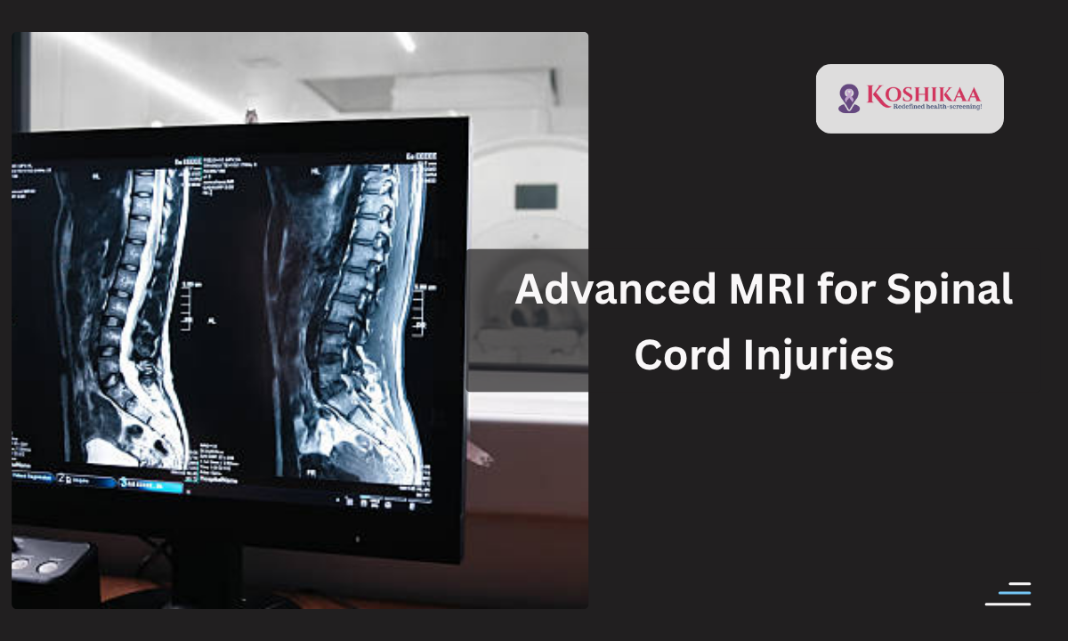
Spinal Cord Injuries Complications are often complex and require precise imaging for accurate diagnosis. With advanced MRI techniques, we can detect structural changes, assess the extent of damage, and guide treatment effectively. 

At our best health screening centre in Bangalore, we use advanced MRI technology to help patients and doctors understand these complications. 

Curious about how an MRI can uncover hidden problems in spinal cord injuries? Let’s explore.

Understanding Spinal Cord Injuries

A spinal cord trauma occurs when the spinal cord is damaged due to accidents, falls, or other injuries. The severity can range from minor nerve irritation to permanent paralysis.

Common spinal cord injury causes include:

  • Road traffic accidents
  • Sports injuries
  • Falls from height
  • Occupational hazards

Spinal cord injury symptoms can vary depending on the location and severity of the injury. Common symptoms include:

  • Weakness or paralysis in limbs
  • Loss of sensation
  • Loss of bladder or bowel control
  • Severe pain or pressure in the neck or back

Why Advanced MRI is Essential

MRI scan

Imaging techniques like MRI are indispensable for evaluating spinal trauma management. Unlike X-rays or CT scans, MRI provides:

  • Detailed soft tissue imaging
  • Clear visualization of nerves and the spinal cord
  • Early detection of secondary complications

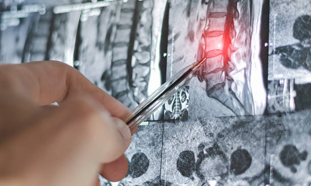
Tip: MRI is particularly useful for assessing edema, hemorrhage, or ligament injuries that are not visible in CT scans.

Example: A patient with neck trauma had persistent numbness. While CT showed no fractures, MRI revealed subtle spinal cord edema, guiding proper rehabilitation.

What MRI Can Detect in Spinal Cord Injuries

ComplicationDetection through Advanced MRI
Spinal cord edemaShows swelling and inflammation of nerve tissues
HemorrhageIdentifies bleeding within or around the cord
Ligament or disc injuryVisualizes soft tissue tears or compression
Nerve compressionDetects pressure from vertebrae or disc herniation
Scar tissue formationTracks post-injury healing complications

Tip: Regular follow-ups with MRI can help monitor progression and guide spinal cord injury management plans.

MRI vs CT for Spinal Cord Injuries

Many patients ask, “Is CT or MRI better for the spinal cord?”

  • CT scans are excellent for detecting fractures and bone abnormalities.
  • MRI scans are superior for soft tissue evaluation, including spinal cord edema, ligament tears, and nerve compression.

At our best MRI scan in Bangalore, we often combine imaging results with clinical assessments to ensure comprehensive evaluation.

Most Severe and Common Complications

The most severe complication of a spinal cord injury is permanent paralysis, which can affect mobility, bladder, bowel, and respiratory functions.

The most frequently observed complication is partial loss of sensation or motor weakness, which often requires long-term therapy and rehabilitation.

Tip: Early detection using advanced MRI significantly reduces risks of permanent damage by guiding timely intervention.

Duration and Safety of MRI

  • Typically, a spinal MRI takes 30 to 60 minutes, depending on the area being scanned and whether contrast is used.
  • The CT scan procedure is faster but may miss soft tissue complications.

Safety Note: MRI is non-invasive, radiation-free, and safe for most patients. Patients with metal implants should inform the technician.

Contrast in Spinal MRI

Sometimes, a contrast MRI is recommended to highlight areas of inflammation or abnormal tissue growth.

Tip: Drinking plenty of water before and after the scan helps flush contrast agents from the body.

How to Understand Your MRI Report

A CT scan report or MRI report includes details on:

  • Spinal cord integrity
  • Presence of edema or hemorrhage
  • Disc herniation
  • Nerve compression

If MRI results are normal, the report usually mentions “spinal cord signal normal” and “no evidence of hemorrhage, edema, or compression detected”.

Tip: Discuss every finding with your doctor or radiologist for proper interpretation.

Spinal Cord Injury Management

Spinal trauma management involves:

  • Immediate medical care after trauma
  • Surgery for stabilization if required
  • Physical rehabilitation and occupational therapy
  • Regular monitoring with advanced MRI

Example: A patient with partial paralysis underwent MRI-guided therapy. The scan revealed scar tissue formation that was addressed through physiotherapy, improving mobility within months.

Tip: Combining imaging with clinical evaluation ensures the best outcomes.

Choosing the Best MRI Scan in Bangalore

Patients seeking spinal imaging often ask, “What is the best scan for spinal cord injuries?”

We recommend advanced MRI because it provides:

  • High-resolution images of spinal cord tissue
  • Detection of complications that CT cannot reveal
  • Guidance for surgical and non-surgical treatment

At our best health screening centre in Bangalore, we combine advanced MRI machines with expert radiologists to deliver accurate and timely results.

Wrapping Up

Spinal Cord Injuries Complications can be complex, but early detection and precise imaging using advanced MRI are critical for effective management. Choosing the best health screening centre in Bangalore ensures accurate scans and professional interpretation.

We, at Koshikaa, combine advanced MRI technology with patient-centred care to guide recovery, monitor complications, and provide peace of mind. For those dealing with spinal cord trauma, proactive imaging is key. Trust expert care to track, manage, and improve outcomes while keeping patients informed and safe.

FAQ Section

1. What is the most severe complication of a spinal cord injury?

Permanent paralysis affecting mobility, bladder, bowel, or respiratory function is the most severe complication, potentially requiring lifelong support and rehabilitation.

2. Is CT or MRI better for the spinal cord?

MRI is superior for evaluating soft tissue, nerve damage, edema, or hemorrhage, whereas CT scans are better for detecting fractures and bone abnormalities.

3. What is the most frequently observed complication of spinal cord injury?

Partial loss of sensation, motor weakness, or limited mobility are the most commonly observed complications, often requiring long-term therapy and rehabilitation.

4. How long does a spinal MRI last?

A spinal MRI usually lasts 30–60 minutes, depending on the area scanned and whether contrast is used, and is non-invasive and radiation-free.

5. When to worry about MRI results?

Abnormal signals, spinal cord compression, or persistent neurological symptoms after trauma indicate the need for urgent medical consultation and follow-up scans.

MRI of the uterus is primarily used for diagnosing and evaluating conditions such as uterine fibroids, adenomyosis, and malignancies. It is particularly useful in the planning of surgical procedures and assessing the extent of gynecological diseases. This imaging test is valuable for its ability to deliver detailed cross-sectional images of the uterus and surrounding structures.
An MRI of the spleen can help diagnose various disorders affecting the spleen including splenomegaly (enlarged spleen), tumors, and other abnormalities. It provides clear, detailed images that are crucial for assessing the health of the spleen and planning potential surgical interventions.
Prostate MRI is a non-invasive imaging technique that provides detailed pictures of the prostate gland and surrounding tissues. It helps in the detection, staging, and monitoring of prostate cancer. Prostate MRIs are increasingly used to guide biopsy procedures and assist in the planning of treatment strategies for prostate health.
An MRI of the pancreas is used to identify issues such as pancreatitis, pancreatic cancer, and other pancreatic disorders. It offers high-resolution images that help in detecting small lesions and assessing the pancreatic ducts. This imaging technique is valuable for guiding biopsies and planning surgeries involving the pancreas.
MRI scans of the liver, kidney, and bile ducts provide detailed images that help diagnose various conditions, such as tumors, cysts, and blockages in the bile ducts. They are crucial for assessing organ damage, staging cancer, and planning liver or kidney-related surgeries. This type of MRI can also evaluate the health and function of these organs comprehensively.
Breast MRI is highly sensitive for detecting breast cancer, especially in individuals with dense breast tissue or a high risk of cancer. It is often used in conjunction with mammography to provide a more comprehensive evaluation. Breast MRIs can also monitor the response to breast cancer treatments and provide detailed pre-surgical mapping.
Brain and spine MRIs are critical tools for diagnosing and monitoring neurological conditions, including tumors, spinal cord injuries, and multiple sclerosis. These MRIs can reveal abnormalities in brain tissue and spinal cord structure that might not be visible with other imaging methods. They are essential for planning surgeries and assessing the effectiveness of treatments in neurology.
PET-CT scans of transplant organs like the liver, kidney, or pancreas are performed to assess metabolic activity, detect complications, and monitor post-transplant function. These scans play a vital role in the early detection of issues such as organ rejection, recurrence of disease, or infection, guiding timely interventions and optimizing transplant outcomes for patients.
PET-CT examinations of organs such as the liver, kidney, or prostate are crucial for assessing metabolic activity, detecting abnormalities, and monitoring disease progression. These scans help diagnose conditions such as liver metastases, renal cell carcinoma, or prostate cancer, guiding treatment decisions and monitoring response to therapy over time.
PET-CT scans of soft-tissue masses are utilized to assess metabolic activity and detect malignant tumors or other abnormalities. By combining PET imaging with CT imaging, these scans provide detailed information about tumor location, size, and metabolic characteristics, aiding in accurate diagnosis and treatment planning for soft-tissue cancers.
PET-CT scans are valuable for detecting cysts or fluid collections within the body, including cysts in the liver, kidneys, or other organs. These scans help characterize the nature and metabolic activity of cysts, guiding diagnosis and treatment planning for conditions such as cystic liver disease, renal cysts, or pancreatic cysts.
PET-CT scans of muscles are performed to assess metabolic activity and detect abnormalities such as inflammation, infection, or tumors. These scans provide valuable information about muscle function and pathology, aiding in the diagnosis and management of conditions like myositis, muscular dystrophy, or soft-tissue sarcomas.
PET-CT scans of the brain are utilized to evaluate brain metabolism and detect abnormalities such as tumors, epilepsy, or neurodegenerative diseases. By combining functional PET imaging with anatomical CT imaging, these scans offer comprehensive information about brain function and structure, guiding neurosurgical interventions and treatment decisions for neurological disorders
PET-CT scans of bones are commonly used to detect bone metastases in cancer patients, assess bone health in conditions like osteoporosis, and evaluate response to treatment. By combining PET and CT imaging, these scans provide detailed information about metabolic activity and anatomical changes in bone structures, aiding in accurate diagnosis and treatment planning for various bone-related conditions.
CT scans of the spine are conducted to assess the vertebrae, spinal cord, and surrounding soft tissues for conditions like herniated discs, spinal fractures, or spinal stenosis, aiding in the diagnosis and management of spinal disorders with detailed imaging essential for surgical planning and intervention.
CT scans of the pelvis are utilized to visualize the pelvic bones, reproductive organs, bladder, and surrounding structures for conditions such as fractures, tumors, or pelvic inflammatory disease, guiding treatment plans and surgical interventions with precise anatomical information.
CT scans of the neck are performed to evaluate structures such as the throat, thyroid gland, lymph nodes, and blood vessels for abnormalities like tumors, infections, or injuries, assisting in diagnosis and management of head and neck disorders with detailed imaging.
CT scans of the extremities are employed to assess bones, joints, and soft tissues in the arms or legs, aiding in the diagnosis of fractures, arthritis, or other musculoskeletal conditions, guiding orthopedic treatment and rehabilitation plans for optimal recovery.
CT scans of the chest are utilized to examine the lungs, heart, blood vessels, and surrounding structures for conditions such as lung nodules, pneumonia, or pulmonary embolism, facilitating accurate diagnosis and management of respiratory and cardiovascular diseases.
CT scans of the brain are conducted to assess brain structures, identify abnormalities like tumors, hemorrhages, or strokes, and aid in diagnosing neurological conditions, guiding neurosurgical interventions and treatment strategies with precise imaging of the brain’s anatomy.
CT scans of the abdomen are performed to visualize and evaluate the internal organs such as the liver, kidneys, pancreas, and intestines for conditions like tumors, infections, or injuries, providing detailed images essential for diagnosis and treatment planning.
Transplant organ ultrasound scans, including those of the liver, kidney, or pancreas, are performed to assess organ viability, detect complications, and monitor post-transplant function. These scans play a vital role in the early detection of issues such as organ rejection, vascular complications, or fluid collections, guiding timely interventions and optimizing transplant outcomes for patients.
Ultrasound examinations of organs such as the liver, kidney, or prostate are crucial for assessing organ structure, detecting abnormalities, and monitoring disease progression. These scans help diagnose conditions such as liver cirrhosis, kidney stones, or prostate enlargement, guiding treatment decisions and monitoring response to therapy over time.
Soft-tissue masses can be evaluated using ultrasound to determine their size, location, and characteristics. Ultrasound imaging helps differentiate between benign and malignant masses, guiding further diagnostic evaluation and treatment planning. By providing real-time imaging, ultrasound assists in the precise localization of soft-tissue masses for biopsy or surgical removal if needed.
Ultrasound scans are valuable for detecting cysts or fluid collections within the body, including cysts in the liver, kidneys, or other organs. These scans help characterize the nature and size of cysts, aiding in diagnosis and treatment planning for conditions such as cystic liver disease or renal cysts, and guiding interventions such as cyst drainage or sclerotherapy.
Musculoskeletal ultrasound scans are used to assess the health and integrity of muscles, identifying issues such as strains, tears, or muscle inflammation. These scans provide detailed images of muscle structures, allowing healthcare providers to diagnose muscle injuries accurately and develop personalized rehabilitation plans to promote recovery and restore function.
Ultrasound examinations of joints are essential for evaluating joint health, detecting abnormalities such as inflammation, fluid accumulation, or cartilage damage. By visualizing the joint structures in real-time, ultrasound helps diagnose conditions like arthritis, bursitis, or ligament injuries, guiding treatment decisions and monitoring disease progression over time.
Ultrasound scans of tendons are performed to assess for injuries, inflammation, or tears, aiding in the diagnosis and management of conditions such as tendonitis or tendon tears. These scans provide detailed images of tendon structures, helping healthcare providers determine the extent of damage and plan appropriate treatment strategies, including physical therapy or surgical intervention if necessary.
Pelvis X-rays play a crucial role in diagnosing injuries, diseases, and developmental conditions affecting the pelvic region. They provide detailed images of the hip bones, sacrum, and coccyx, aiding in the assessment of fractures, dislocations, or signs of arthritis. Pelvic X-rays are instrumental in guiding surgical interventions, monitoring healing progress, and evaluating conditions such as osteoporosis or tumors. By providing precise anatomical information, these imaging studies enable healthcare professionals to deliver personalized care and improve patient outcomes.
Teeth X-rays, known as dental radiographs, are indispensable tools for dentists to assess oral health. They aid in diagnosing dental decay, gum disease, and abnormalities in tooth structure or alignment. These images provide valuable insights into hidden dental issues, allowing for early intervention and prevention of complications. Additionally, dental X-rays are essential for planning treatments such as orthodontics, root canals, or dental implants, ensuring optimal outcomes and patient satisfaction.
Spine X-rays are pivotal in diagnosing spinal fractures, degenerative conditions, and spinal misalignments, providing critical information for treatment planning and monitoring spinal health. They offer detailed images of the vertebrae and surrounding structures, enabling healthcare professionals to identify issues such as scoliosis, disc herniation, or spinal stenosis
Skull X-rays are valuable for detecting fractures, assessing head trauma, and evaluating the alignment of facial bones, crucial in diagnosing conditions like fractures, tumors, or developmental abnormalities in the skull. They provide detailed views of the cranial structure, aiding clinicians in determining appropriate treatment strategies and ensuring patient safety.
For the heart, X-ray solutions can aid in the detection of various cardiac problems, ranging from mild to severe, including – Heart failure, Cardiac congestion and other heart-related conditions. In addition to cardiac assessments, X-ray solutions are also used for breast examinations to identify tumors or signs of breast cancer.

Bone X-rays are essential for diagnosing fractures and assessing bone alignment, aiding doctors in developing treatment plans and monitoring healing progress. They provide detailed images of the skeletal system, helping identify abnormalities or injuries that may not be visible through other imaging techniques

Abdominal X-rays are performed when patients experience symptoms such as significant nausea, ongoing vomiting, abdominal pain, flank or back pain, or abdominal swelling. These X-rays can help diagnose various conditions.

Frequently Booked Tests

Frequently Booked Checkups