When it comes to protecting your lungs, low-dose CT scan technology is one of the safest and most advanced tools we have today. This scan helps detect lung diseases, including early-stage lung cancer, while minimizing radiation exposure, making it a preferred choice for both preventive and diagnostic care.
At our Health Screening Centre in Bangalore, we have seen how early chest screening can make all the difference in saving lives.
But what exactly is a low-dose CT, how does it work, and why is it considered safer? Let’s explore all these questions in detail, and one fact that might surprise you by the end.
Understanding the Low-Dose CT Definition
A Low-Dose CT Scan, or Low-Dose Computed Tomography of Thorax, is a specialized type of imaging test designed to capture detailed pictures of your lungs and chest using minimal radiation.
It’s called “low-dose” because the radiation exposure is significantly lower than that of a regular CT scan, about 75–90% less. Yet, it still provides clear, high-resolution images that help doctors detect even the smallest lung nodules or abnormalities.
In simple terms, it’s a safer, faster, and more precise way to screen for lung and chest conditions.
Why Is Chest Screening Important?
Our lungs are constantly exposed to pollutants, smoke, and allergens. Unfortunately, conditions like chronic bronchitis, COPD, and lung cancer often develop silently.
By the time symptoms like breathlessness, coughing, or chest pain appear, the disease may have already advanced. That’s why Computed Tomography Lung scans, especially low-dose CT, play a vital role in preventive screening.
Early detection through a low-dose CT scan of the chest for Lung Cancer Screening can help identify problems before they become serious, allowing timely treatment and better outcomes.
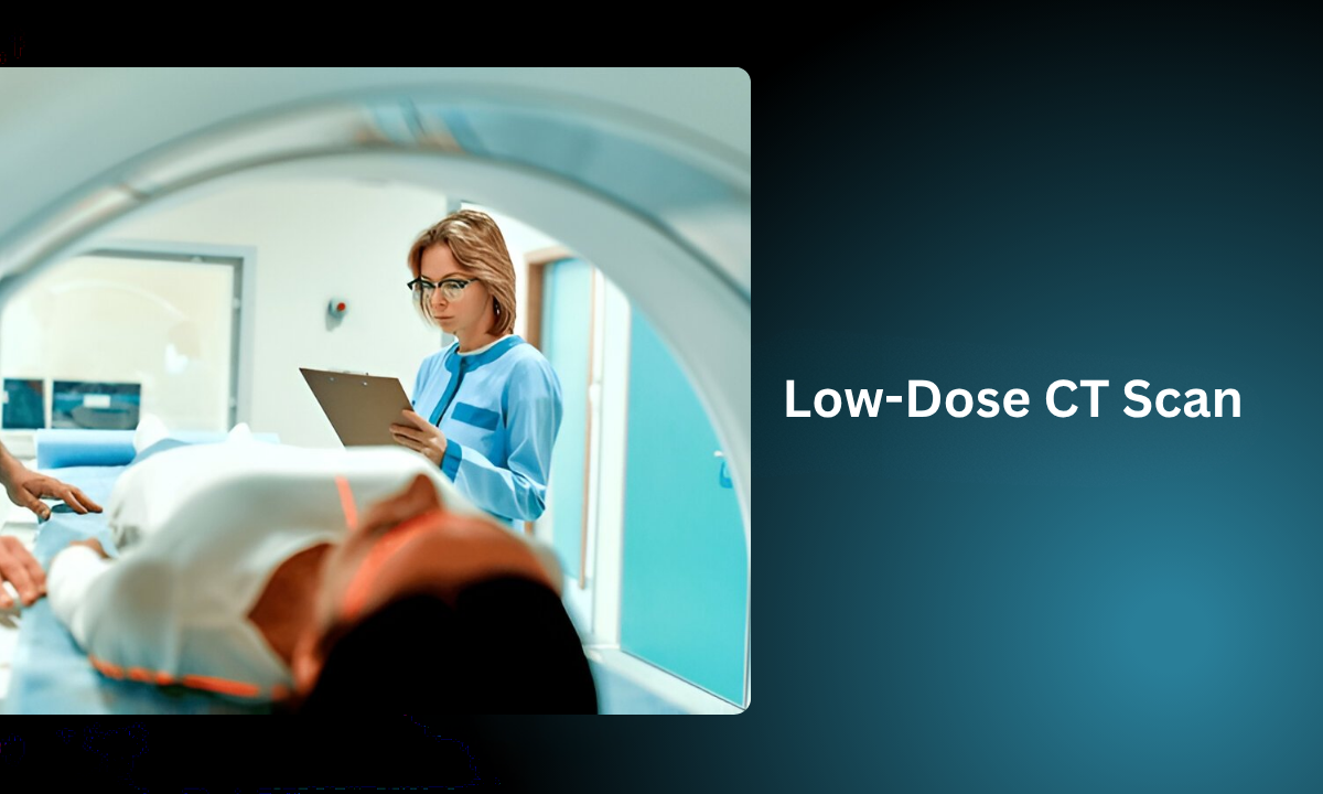
How Does a Low-Dose CT Scan Work?

The process is simple, quick, and completely painless. Here’s what typically happens:
| Step | What Happens |
|---|---|
| 1. Preparation | You may be asked to avoid eating or drinking for a few hours before the scan. |
| 2. Positioning | You lie on a narrow table that slides into the CT scanner. |
| 3. Scanning | The scanner rotates around your chest, taking detailed X-ray images from multiple angles. |
| 4. Image Creation | A computer combines these images to create 3D pictures of your lungs. |
| 5. CT Results | The radiologist interprets the images to detect nodules, infections, or any suspicious growths. |
The entire procedure usually takes less than 10 minutes, with no injections, no pain, and no hospital stay is needed.
Low-Dose CT vs Traditional CT – Key Differences
| Feature | Low-Dose CT Scan | Regular CT Scan |
|---|---|---|
| Radiation Exposure | 75–90% less | Standard dose |
| Purpose | Preventive screening | Diagnostic imaging |
| Common Use | Lung cancer and chest screening | Broad medical evaluations |
| Safety | Highly safe for regular screening | Moderate exposure limits frequency |
This lower exposure makes it ideal for lung cancer CT scan screening, especially for people who may need regular checkups, such as smokers, ex-smokers, or individuals with a family history of lung disease.
Who Should Consider a Low-Dose CT Scan of the Chest?
You should consider a low-dose CT scan of the chest for lung cancer screening if you:
- Are over 40 and have a history of smoking.
- Have a family history of lung or thoracic diseases.
- Experience persistent cough or shortness of breath.
- They are exposed to industrial pollutants or asbestos.
- Want a preventive scan as part of a full-body checkup.
Even if you don’t show symptoms, this scan can act as a proactive health safeguard.
Why Low-Dose CT Is Safer for Your Lungs
Unlike conventional scans, low-dose CT uses advanced detectors that capture detailed lung images with minimal radiation.
It’s particularly useful for repeated monitoring, for example, tracking how your lungs heal after infection, or watching small nodules for any change in size.
Because of its low radiation exposure, it’s a trusted method for annual screening in high-risk individuals.
Computed Tomography Lung Screening Saves Lives
Studies show that early screening with Computed Tomography Lung scans can reduce lung cancer deaths by up to 20%. That’s because it detects small nodules long before symptoms develop.
At our CT scan in Bangalore, many patients discover early-stage issues through this test, which can then be treated with simple medical or surgical interventions instead of complex therapies later.
Interpreting Your CT Results
Your CT results typically include detailed information about your lung structures. Here’s what the report might show:
| Findings | What It Means |
|---|---|
| Normal Lungs | No nodules or abnormal tissue found. |
| Benign Nodules | Small, non-cancerous spots, often due to past infections. |
| Suspicious Nodules | Require further evaluation, possibly with PET-CT or biopsy. |
| Other Conditions | Infections, fibrosis, emphysema, or fluid buildup. |
Your radiologist or pulmonologist will discuss these findings and guide you on the next steps.
Tips Before and After Your Scan
Before the scan:
- Wear loose, comfortable clothing without metal.
- Remove jewelry, eyeglasses, or metal zippers.
- Inform your doctor if you are pregnant or have any allergies.
After the scan:
- You can resume normal activities immediately.
- Drink plenty of water if you received contrast dye.
- Keep a copy of your CT results for future comparison.
The Future of Low-Dose Computed Tomography of the Thorax
Technology continues to evolve rapidly. Today’s low-dose Computed Tomography of Thorax machines use smart algorithms and AI-assisted image reconstruction to improve clarity even further while keeping radiation minimal.
This makes them ideal not just for lung cancer detection, but also for evaluating pneumonia, tuberculosis, or long-term respiratory conditions with unmatched accuracy.
Why Choose a Trusted Health Screening Centre in Bangalore
Finding the right diagnostic partner matters. Our reputed health screening centre in Bangalore ensures your scan is performed using the latest, safest machines and interpreted by experienced radiologists.
This guarantees reliable results, better diagnosis, and peace of mind for patients.
Final Thoughts
A low-dose CT scan is one of the most effective and safest tools for lung and chest screening today. It offers clarity, confidence, and early detection, all while keeping radiation exposure remarkably low.
At our health screening centre in Bangalore, we believe in making advanced imaging both safe and accessible to everyone.
If you or a loved one is considering preventive lung screening, visit Koshikaa, where early detection and expert care come together for healthier living.
FAQs About Low-Dose CT Scans
1. How does lung cancer show on a CT scan?
Lung cancer appears as a distinct nodule, mass, or irregular shadow on the CT images, allowing radiologists to identify suspicious growths for further diagnosis or biopsy.
2. What does a low-dose lung CT scan show?
A low-dose lung CT scan shows detailed images of the lungs, identifying small nodules, infections, fibrosis, or early signs of lung cancer with minimal radiation exposure.
3. What is the difference between a low-dose CT scan and a regular CT scan?
A low-dose CT scan uses significantly less radiation while maintaining clear imaging, making it ideal for preventive lung screening, unlike regular CT scans used for broader diagnostics.
4. Is a low-dose CT Scan safe for everyone?
Yes. It’s extremely safe due to minimal radiation, but pregnant women should consult their doctor before undergoing the scan.
5. Is the scan available at all diagnostic centres?
It’s available at select advanced centres like Koshikaa, which are equipped with modern low-dose CT technology.