CT Scan of the Abdomen and Pelvis: What You Need to Know

Table of Contents

A CT abdomen and pelvis is one of the non-invasive imaging tests, which utilizes X-ray and computer processing to generate a cross-sectional image of the body. An abdomen and pelvis scan is a test ordered by a physician that leaves many patients wondering what this test is. Simply put, it is a combined examination of the organs, blood vessels, and bones in your chest and the hips to assist in the diagnosis of any pain, injury, infection, or disease.

The current article will take you through the preparation of the patient to the interpretation of results in a simple step-by-step approach.

Understanding These Two Tests

CT scan in Bangalore

Abdomen and Pelvis Scan: What is it?  

Knowing the meaning of an abdomen and pelvis scan may help you relieve anxiety when a doctor prescribes an imaging procedure. Examples of structures that are normally checked using this scan include the liver, spleen, kidneys, bladder, intestines, reproductive organs, and surrounding blood vessels. In contrast to an ultrasound that involves sound waves or an MRI image used with magnetic fields, this examination takes advantage of computed tomography in high-resolution detail.

Key Components of the Scan 

  • Contrast Material: A dye can be injected in order to show the blood vessels and particular tissues.  
  • Scan range: The machine rotates around your midsection, starting a bit over the diaphragm and going to mid-thigh.  
  • Image Quality: Slices of information, which are but a few millimeters thick, will be merged into 3-dimensional figures.

Prepare Yourself For Scan

Proper preparation will ensure proper images and proper results. The following is a simple checklist on patient preparation for a CT scan:

1. Fasting: You might be requested to fast or go without intake of food or water for 4-6 hours.  

2. Medication Review: Remember to state any medicines you take, both prescription and over the counter, and any allergies to the provider.  

3. Accessories (dressing, etc.): Remove all metals and metal jewelry and eyewear, and wear loose, comfortable clothes.  

To get a better outcome, patient preparation for a CT scan is likely to include the consumption of oral contrast or wearing a gown. Concise directions facilitate the reduction of delays and duplication of the scans. 

Having a CT scan is usually frightening. It is helpful to know what steps to take.

Step 1: Arrival and Registration

  • He or she will complete forms and chat about your history.  
  • When contrast is employed, a small IV line is inserted into your arm.

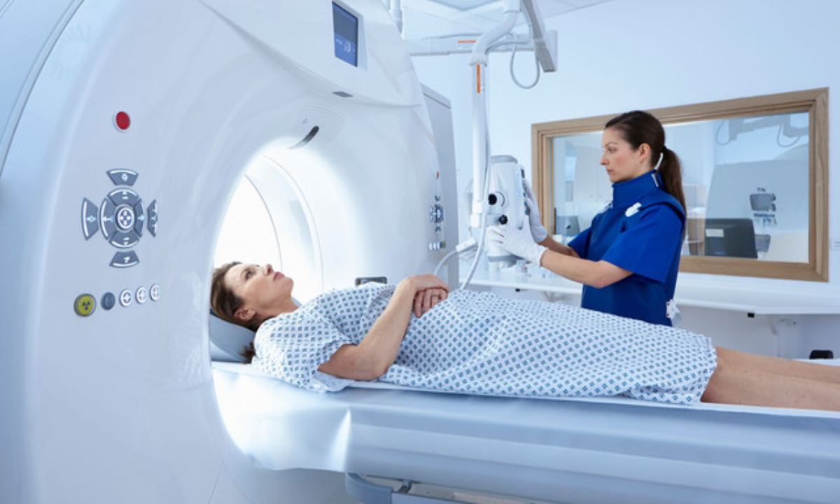
Step 2: CT scan abdomen procedure

  • You are placed on a long, thin table, which slides into the CT machine.  
  • The technologist can also give you some advice to hold your breath briefly so as to avoid the blurring of the scanner.

Step 3 CT scan pelvis procedure  

  • To attack your lower pelvis, the table is set up.  
  • More contrasted/sequential images can be taken as and when required.

Time acquisition and CT scan duration 

The entire protocol lasts 15 -30 minutes to set up the device and carry out follow-up checks after the scan. The X-ray certification takes only seconds, but positioning and contrast injections add time.

Difference between abdomen CT and CT pelvis  

CT abdomen  

  • Examines organs above the hip bones (such as the liver, pancreas, and spleen).  
  • Typical of pain, cancer/tumors, or inter-bleeding.

CT pelvis  

  • Concentrates on the bladder, reproductive organs, lower intestines, and the pelvic bones.  
  • As with any pelvic pain, urinary trauma or issues.
  • Having both scans simultaneously captures the entire picture and the possibilities of other tests being unnecessary hasten the process of diagnosis.

What is diagnosed by a CT scan? 

Upon a CT scan, it may:  

  • Determine the malignant and benign masses as well as tumors.  
  • See spot inflammation and infection (appendicitis, diverticulitis, or abscesses).  
  • Determine vascular complications (aneurysms, blood thrombosis, or vessel obstruction). 

The question most patients frequently ask is what CT scans can detect other than their tumor. They can display fluid masses, organ swelling, and structural deviation.

Safety Considerations  

1. Radiation We are exposed to Radiation; doses are regulated and exceed random X-rays.  

2. Contrast Reactions: There is a small likelihood of an allergic reaction to dye, but in the case of this rarity, your care team is ready to react.  

3. Pregnancy: Pregnant women need to talk to the provider to find out risks and options.

Better where to get/ scanned  

There are locations with different access to quality imaging. When considering choices on CT scan in Bangalore, the following are some elements to consider:  

• Accreditation: Search out those centers that are nationally accredited in imaging services.  

• Technologist Skills: Experienced workers are required to offer a proper technique, as well as comfort to the patient.  

• Turnaround Time: Effective delivery of reports within a brief period can accelerate the process in making the right decision in terms of treatment.

Numerous hospitals in India are providing CT scan in Bangalore with advanced machines that have low-dose protocols and quick image reconstruction. Coverage and prior-authorization needs should never be in doubt.

Tips for Hassle-Free Experience  

1. Be on Time: Doing the forms carefully will do away with stress.  

2. Hydration: The passage of IV contrast can be alleviated and dye flushed by hydration unless one is fasting.  

3. Ask Questions: Clarify any doubts within your mind regarding the method of CT scan abdomen or pelvis scanning process before you go.

Improving Your Diagnostic Journey 

Once you finish your CT abdomen and pelvis scan, it is important to have knowledge of further steps in order to make informed decisions. Talking with your physician about your results as soon as possible will ensure that something out of the ordinary is handled immediately. There are quite a number of centers where you have the option of digital access to photos and reports, and thus you can review them at home.  

Hydrating well post-scan assists in flushing any residual contrast dye. If you experience unusual symptoms like rash or breathing difficulty, notify staff immediately. Inquire about follow-up imaging schedules to monitor conditions or treatment progress. Keep a copy of your scan in a secure file for future reference and comparison. Store a copy of your scan(in a safe file) to compare in the future. 

Conclusion  

Having a scan of the tummy and hips might appear complicated, but knowing what to expect in each step, like preparing the patient before a CT scan, and the detection capacity of CT scans, could calm your nerves. If you require a CT pelvis to assess a question of injury to a bone or a CT abdomen to look into abdominal pains, this examination provides quick and accurate information.

In case you want to find a CT scan in Bangalore or any other area, you should focus on a place or center that has well-trained and competent personnel with modern machines. With this knowledge, you will feel confident with your appointment.  

To individuals who may require a CT scan in Bangalore, Koshikaa Screening Center offers customized facilities and radiology services to facilitate the accomplishment of accurate and exact results. Necessary procedures should also be followed to ensure insurance coverage and preauthorization requirements are verified before scheduling. Ask yourself questions on technology, report turnaround, and radiation-dose optimization to get as many benefits out of a scan as possible, including sedation options, should you wish.

MRI of the uterus is primarily used for diagnosing and evaluating conditions such as uterine fibroids, adenomyosis, and malignancies. It is particularly useful in the planning of surgical procedures and assessing the extent of gynecological diseases. This imaging test is valuable for its ability to deliver detailed cross-sectional images of the uterus and surrounding structures.
An MRI of the spleen can help diagnose various disorders affecting the spleen including splenomegaly (enlarged spleen), tumors, and other abnormalities. It provides clear, detailed images that are crucial for assessing the health of the spleen and planning potential surgical interventions.
Prostate MRI is a non-invasive imaging technique that provides detailed pictures of the prostate gland and surrounding tissues. It helps in the detection, staging, and monitoring of prostate cancer. Prostate MRIs are increasingly used to guide biopsy procedures and assist in the planning of treatment strategies for prostate health.
An MRI of the pancreas is used to identify issues such as pancreatitis, pancreatic cancer, and other pancreatic disorders. It offers high-resolution images that help in detecting small lesions and assessing the pancreatic ducts. This imaging technique is valuable for guiding biopsies and planning surgeries involving the pancreas.
MRI scans of the liver, kidney, and bile ducts provide detailed images that help diagnose various conditions, such as tumors, cysts, and blockages in the bile ducts. They are crucial for assessing organ damage, staging cancer, and planning liver or kidney-related surgeries. This type of MRI can also evaluate the health and function of these organs comprehensively.
Breast MRI is highly sensitive for detecting breast cancer, especially in individuals with dense breast tissue or a high risk of cancer. It is often used in conjunction with mammography to provide a more comprehensive evaluation. Breast MRIs can also monitor the response to breast cancer treatments and provide detailed pre-surgical mapping.
Brain and spine MRIs are critical tools for diagnosing and monitoring neurological conditions, including tumors, spinal cord injuries, and multiple sclerosis. These MRIs can reveal abnormalities in brain tissue and spinal cord structure that might not be visible with other imaging methods. They are essential for planning surgeries and assessing the effectiveness of treatments in neurology.
PET-CT scans of transplant organs like the liver, kidney, or pancreas are performed to assess metabolic activity, detect complications, and monitor post-transplant function. These scans play a vital role in the early detection of issues such as organ rejection, recurrence of disease, or infection, guiding timely interventions and optimizing transplant outcomes for patients.
PET-CT examinations of organs such as the liver, kidney, or prostate are crucial for assessing metabolic activity, detecting abnormalities, and monitoring disease progression. These scans help diagnose conditions such as liver metastases, renal cell carcinoma, or prostate cancer, guiding treatment decisions and monitoring response to therapy over time.
PET-CT scans of soft-tissue masses are utilized to assess metabolic activity and detect malignant tumors or other abnormalities. By combining PET imaging with CT imaging, these scans provide detailed information about tumor location, size, and metabolic characteristics, aiding in accurate diagnosis and treatment planning for soft-tissue cancers.
PET-CT scans are valuable for detecting cysts or fluid collections within the body, including cysts in the liver, kidneys, or other organs. These scans help characterize the nature and metabolic activity of cysts, guiding diagnosis and treatment planning for conditions such as cystic liver disease, renal cysts, or pancreatic cysts.
PET-CT scans of muscles are performed to assess metabolic activity and detect abnormalities such as inflammation, infection, or tumors. These scans provide valuable information about muscle function and pathology, aiding in the diagnosis and management of conditions like myositis, muscular dystrophy, or soft-tissue sarcomas.
PET-CT scans of the brain are utilized to evaluate brain metabolism and detect abnormalities such as tumors, epilepsy, or neurodegenerative diseases. By combining functional PET imaging with anatomical CT imaging, these scans offer comprehensive information about brain function and structure, guiding neurosurgical interventions and treatment decisions for neurological disorders
PET-CT scans of bones are commonly used to detect bone metastases in cancer patients, assess bone health in conditions like osteoporosis, and evaluate response to treatment. By combining PET and CT imaging, these scans provide detailed information about metabolic activity and anatomical changes in bone structures, aiding in accurate diagnosis and treatment planning for various bone-related conditions.
CT scans of the spine are conducted to assess the vertebrae, spinal cord, and surrounding soft tissues for conditions like herniated discs, spinal fractures, or spinal stenosis, aiding in the diagnosis and management of spinal disorders with detailed imaging essential for surgical planning and intervention.
CT scans of the pelvis are utilized to visualize the pelvic bones, reproductive organs, bladder, and surrounding structures for conditions such as fractures, tumors, or pelvic inflammatory disease, guiding treatment plans and surgical interventions with precise anatomical information.
CT scans of the neck are performed to evaluate structures such as the throat, thyroid gland, lymph nodes, and blood vessels for abnormalities like tumors, infections, or injuries, assisting in diagnosis and management of head and neck disorders with detailed imaging.
CT scans of the extremities are employed to assess bones, joints, and soft tissues in the arms or legs, aiding in the diagnosis of fractures, arthritis, or other musculoskeletal conditions, guiding orthopedic treatment and rehabilitation plans for optimal recovery.
CT scans of the chest are utilized to examine the lungs, heart, blood vessels, and surrounding structures for conditions such as lung nodules, pneumonia, or pulmonary embolism, facilitating accurate diagnosis and management of respiratory and cardiovascular diseases.
CT scans of the brain are conducted to assess brain structures, identify abnormalities like tumors, hemorrhages, or strokes, and aid in diagnosing neurological conditions, guiding neurosurgical interventions and treatment strategies with precise imaging of the brain’s anatomy.
CT scans of the abdomen are performed to visualize and evaluate the internal organs such as the liver, kidneys, pancreas, and intestines for conditions like tumors, infections, or injuries, providing detailed images essential for diagnosis and treatment planning.
Transplant organ ultrasound scans, including those of the liver, kidney, or pancreas, are performed to assess organ viability, detect complications, and monitor post-transplant function. These scans play a vital role in the early detection of issues such as organ rejection, vascular complications, or fluid collections, guiding timely interventions and optimizing transplant outcomes for patients.
Ultrasound examinations of organs such as the liver, kidney, or prostate are crucial for assessing organ structure, detecting abnormalities, and monitoring disease progression. These scans help diagnose conditions such as liver cirrhosis, kidney stones, or prostate enlargement, guiding treatment decisions and monitoring response to therapy over time.
Soft-tissue masses can be evaluated using ultrasound to determine their size, location, and characteristics. Ultrasound imaging helps differentiate between benign and malignant masses, guiding further diagnostic evaluation and treatment planning. By providing real-time imaging, ultrasound assists in the precise localization of soft-tissue masses for biopsy or surgical removal if needed.
Ultrasound scans are valuable for detecting cysts or fluid collections within the body, including cysts in the liver, kidneys, or other organs. These scans help characterize the nature and size of cysts, aiding in diagnosis and treatment planning for conditions such as cystic liver disease or renal cysts, and guiding interventions such as cyst drainage or sclerotherapy.
Musculoskeletal ultrasound scans are used to assess the health and integrity of muscles, identifying issues such as strains, tears, or muscle inflammation. These scans provide detailed images of muscle structures, allowing healthcare providers to diagnose muscle injuries accurately and develop personalized rehabilitation plans to promote recovery and restore function.
Ultrasound examinations of joints are essential for evaluating joint health, detecting abnormalities such as inflammation, fluid accumulation, or cartilage damage. By visualizing the joint structures in real-time, ultrasound helps diagnose conditions like arthritis, bursitis, or ligament injuries, guiding treatment decisions and monitoring disease progression over time.
Ultrasound scans of tendons are performed to assess for injuries, inflammation, or tears, aiding in the diagnosis and management of conditions such as tendonitis or tendon tears. These scans provide detailed images of tendon structures, helping healthcare providers determine the extent of damage and plan appropriate treatment strategies, including physical therapy or surgical intervention if necessary.
Pelvis X-rays play a crucial role in diagnosing injuries, diseases, and developmental conditions affecting the pelvic region. They provide detailed images of the hip bones, sacrum, and coccyx, aiding in the assessment of fractures, dislocations, or signs of arthritis. Pelvic X-rays are instrumental in guiding surgical interventions, monitoring healing progress, and evaluating conditions such as osteoporosis or tumors. By providing precise anatomical information, these imaging studies enable healthcare professionals to deliver personalized care and improve patient outcomes.
Teeth X-rays, known as dental radiographs, are indispensable tools for dentists to assess oral health. They aid in diagnosing dental decay, gum disease, and abnormalities in tooth structure or alignment. These images provide valuable insights into hidden dental issues, allowing for early intervention and prevention of complications. Additionally, dental X-rays are essential for planning treatments such as orthodontics, root canals, or dental implants, ensuring optimal outcomes and patient satisfaction.
Spine X-rays are pivotal in diagnosing spinal fractures, degenerative conditions, and spinal misalignments, providing critical information for treatment planning and monitoring spinal health. They offer detailed images of the vertebrae and surrounding structures, enabling healthcare professionals to identify issues such as scoliosis, disc herniation, or spinal stenosis
Skull X-rays are valuable for detecting fractures, assessing head trauma, and evaluating the alignment of facial bones, crucial in diagnosing conditions like fractures, tumors, or developmental abnormalities in the skull. They provide detailed views of the cranial structure, aiding clinicians in determining appropriate treatment strategies and ensuring patient safety.
For the heart, X-ray solutions can aid in the detection of various cardiac problems, ranging from mild to severe, including – Heart failure, Cardiac congestion and other heart-related conditions. In addition to cardiac assessments, X-ray solutions are also used for breast examinations to identify tumors or signs of breast cancer.

Bone X-rays are essential for diagnosing fractures and assessing bone alignment, aiding doctors in developing treatment plans and monitoring healing progress. They provide detailed images of the skeletal system, helping identify abnormalities or injuries that may not be visible through other imaging techniques

Abdominal X-rays are performed when patients experience symptoms such as significant nausea, ongoing vomiting, abdominal pain, flank or back pain, or abdominal swelling. These X-rays can help diagnose various conditions.

Frequently Booked Tests

Frequently Booked Checkups