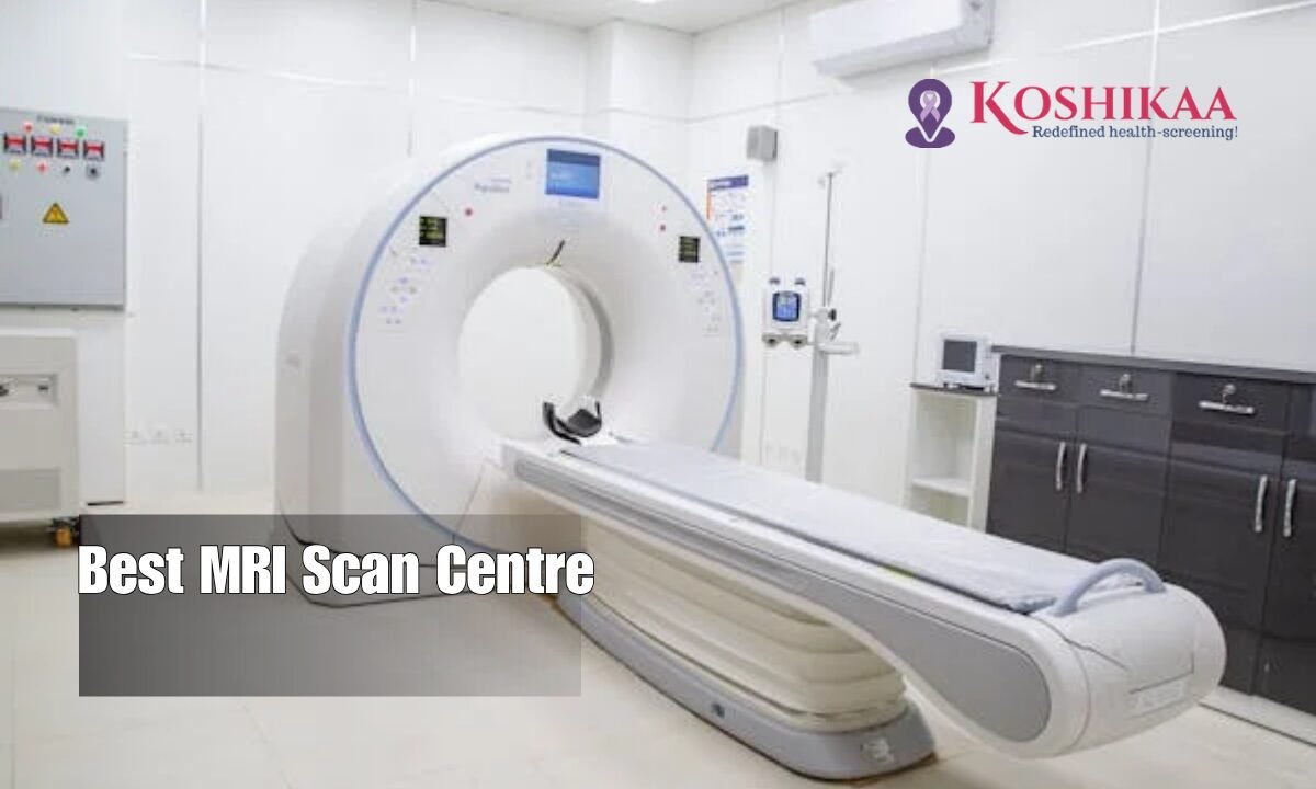
Your Ultimate Guide to Choosing the Best MRI Scan Centre in Bangalore: 7 Insider Tips for Smart Healthcare Decisions

X-ray in Bangalore

Table of Contents

Introduction

In a city like Bangalore, where medical options are plentiful and time is always ticking, choosing the right MRI scan centre is more than a health decision—it’s a strategic one. With the rise in demand for advanced diagnostics, finding a reliable and accurate MRI scan facility can directly impact the accuracy of your diagnosis, your peace of mind, and even your treatment plan.

This guide offers a step-by-step breakdown of everything you need to know when selecting the best MRI scan centre in Bangalore—from technology and staff expertise to pricing, safety standards, and turnaround times. Whether you’re a first-timer or seeking a better alternative, this blog will ensure your next diagnostic decision is a confident one.

Why It’s Crucial to Choose the Right MRI Scan Centre

MRI scan in Bangalore

MRI (Magnetic Resonance Imaging) is one of the most advanced diagnostic tools in modern medicine. It captures high-resolution images of internal organs, soft tissues, and bones, often identifying problems that other scans might miss.

But here’s the catch: not all MRI centres are created equal.

The clarity of your scan, the interpretation of your results, and even your safety depend on the quality of the center you choose. A wrong choice could lead to misdiagnosis, delayed treatment, or unnecessary costs.

Frequently Asked Questions About Choosing an MRI Scan Centre in Bangalore

Q1: What should I look for in a good MRI centre?

  • Accreditation and certifications
  • Latest MRI technology (1.5T or 3T machines)
  • Experienced radiologists and technicians
  • Fast and reliable report delivery
  • Transparent pricing
  • Clean, safe, and patient-friendly environment

Q2: How much does an MRI scan typically cost in Bangalore?

The average MRI scan cost in Bangalore ranges between ₹3,500 to ₹12,000, depending on the body part and contrast usage.

Q3: Can I trust online reviews?

Yes, but always cross-verify with Google ratings, patient testimonials, and whether the centre has a dedicated support team for inquiries.

7 Key Factors to Help You Choose the Best MRI Scan Centre in Bangalore

1. Technology & Equipment

Look for centres equipped with 3 Tesla (3T) MRI machines. They offer higher resolution images and faster scan times. This matters significantly in complex cases like neurological or cancer diagnostics.

2. Qualified Radiologists & Support Team

An advanced machine is only as good as the professional operating it. Ensure the centre has certified radiologists with specialization in your condition area—neurology, orthopedics, cardiology, etc.

3. Turnaround Time

In critical cases, delays in reporting can affect your treatment. Choose a centre that offers report delivery within 12-24 hours, especially for urgent scans.

4. Transparency in Pricing

No one likes surprise bills. Opt for centres with clear, itemized billing, and if possible, ones that offer packages or combo scans for savings. Also, check whether insurance or corporate health cards are accepted.

5. Location and Accessibility

Traffic in Bangalore can be daunting. Ensure the centre is easily reachable, has ample parking, and ideally offers online booking and real-time slot availability.

6. Hygiene and Patient Comfort

MRI scans can be uncomfortable, especially for claustrophobic or elderly patients. Look for open MRI options, quiet scan modes, and a patient-first environment with clear pre-scan instructions.

7. Recommendations & Word-of-Mouth

Ask your doctor, friends, or colleagues about their experiences. A centre like Koshikaa, for instance, has built trust in Bangalore with its cutting-edge diagnostics, exceptional care, and consistent patient satisfaction.

Want to explore a top-tier facility? Visit Koshikaa’s MRI Scan in Bangalore to learn more about their advanced services.

Real Scenario: Choosing the Right Saved Time & Health

Take Priya’s story. A 38-year-old working mom, she needed an urgent spinal MRI after weeks of unexplained back pain. Her first scan at a local centre was blurry and inconclusive, leading to a misdiagnosis. She then chose a well-rated center with 3T MRI, experienced radiologists, and got accurate results within 6 hours. Her treatment began immediately, and she was back to work in a week.

Choosing the right centre saved her weeks of pain and confusion.

Red Flags to Avoid

  • Outdated equipment or no mention of machine strength
  • No online presence or very few genuine reviews
  • Unclear pricing or pushy upselling
  • Long wait times and delayed reports
  • Lack of certified professionals on-site

Why Bangalore Residents Need This Guide

With healthcare options growing across the city, it’s easy to get overwhelmed by choice. This guide helps filter quality over advertising. A strong diagnostic foundation ensures accurate treatment and peace of mind—two things Bangalore’s busy lifestyle rarely offers.

And when you find a reliable MRI center, it becomes your go-to for future needs—saving you time, worry, and money in the long run.

Conclusion

Your health deserves more than a random pick from a Google search. By following this guide, you’re not only making an informed choice but also ensuring that the MRI results you receive are clear, reliable, and timely—just what your health journey needs.

Remember: The right MRI scan center in Bangalore isn’t the most expensive or most advertised—it’s the one that puts precision, people, and your peace of mind first.

Don’t wait for symptoms to escalate. Choose smart, safe and the right.

Explore trusted diagnostic excellence with Koshikaa’s MRI Scan in Bangalore today.

MRI of the uterus is primarily used for diagnosing and evaluating conditions such as uterine fibroids, adenomyosis, and malignancies. It is particularly useful in the planning of surgical procedures and assessing the extent of gynecological diseases. This imaging test is valuable for its ability to deliver detailed cross-sectional images of the uterus and surrounding structures.
An MRI of the spleen can help diagnose various disorders affecting the spleen including splenomegaly (enlarged spleen), tumors, and other abnormalities. It provides clear, detailed images that are crucial for assessing the health of the spleen and planning potential surgical interventions.
Prostate MRI is a non-invasive imaging technique that provides detailed pictures of the prostate gland and surrounding tissues. It helps in the detection, staging, and monitoring of prostate cancer. Prostate MRIs are increasingly used to guide biopsy procedures and assist in the planning of treatment strategies for prostate health.
An MRI of the pancreas is used to identify issues such as pancreatitis, pancreatic cancer, and other pancreatic disorders. It offers high-resolution images that help in detecting small lesions and assessing the pancreatic ducts. This imaging technique is valuable for guiding biopsies and planning surgeries involving the pancreas.
MRI scans of the liver, kidney, and bile ducts provide detailed images that help diagnose various conditions, such as tumors, cysts, and blockages in the bile ducts. They are crucial for assessing organ damage, staging cancer, and planning liver or kidney-related surgeries. This type of MRI can also evaluate the health and function of these organs comprehensively.
Breast MRI is highly sensitive for detecting breast cancer, especially in individuals with dense breast tissue or a high risk of cancer. It is often used in conjunction with mammography to provide a more comprehensive evaluation. Breast MRIs can also monitor the response to breast cancer treatments and provide detailed pre-surgical mapping.
Brain and spine MRIs are critical tools for diagnosing and monitoring neurological conditions, including tumors, spinal cord injuries, and multiple sclerosis. These MRIs can reveal abnormalities in brain tissue and spinal cord structure that might not be visible with other imaging methods. They are essential for planning surgeries and assessing the effectiveness of treatments in neurology.
PET-CT scans of transplant organs like the liver, kidney, or pancreas are performed to assess metabolic activity, detect complications, and monitor post-transplant function. These scans play a vital role in the early detection of issues such as organ rejection, recurrence of disease, or infection, guiding timely interventions and optimizing transplant outcomes for patients.
PET-CT examinations of organs such as the liver, kidney, or prostate are crucial for assessing metabolic activity, detecting abnormalities, and monitoring disease progression. These scans help diagnose conditions such as liver metastases, renal cell carcinoma, or prostate cancer, guiding treatment decisions and monitoring response to therapy over time.
PET-CT scans of soft-tissue masses are utilized to assess metabolic activity and detect malignant tumors or other abnormalities. By combining PET imaging with CT imaging, these scans provide detailed information about tumor location, size, and metabolic characteristics, aiding in accurate diagnosis and treatment planning for soft-tissue cancers.
PET-CT scans are valuable for detecting cysts or fluid collections within the body, including cysts in the liver, kidneys, or other organs. These scans help characterize the nature and metabolic activity of cysts, guiding diagnosis and treatment planning for conditions such as cystic liver disease, renal cysts, or pancreatic cysts.
PET-CT scans of muscles are performed to assess metabolic activity and detect abnormalities such as inflammation, infection, or tumors. These scans provide valuable information about muscle function and pathology, aiding in the diagnosis and management of conditions like myositis, muscular dystrophy, or soft-tissue sarcomas.
PET-CT scans of the brain are utilized to evaluate brain metabolism and detect abnormalities such as tumors, epilepsy, or neurodegenerative diseases. By combining functional PET imaging with anatomical CT imaging, these scans offer comprehensive information about brain function and structure, guiding neurosurgical interventions and treatment decisions for neurological disorders
PET-CT scans of bones are commonly used to detect bone metastases in cancer patients, assess bone health in conditions like osteoporosis, and evaluate response to treatment. By combining PET and CT imaging, these scans provide detailed information about metabolic activity and anatomical changes in bone structures, aiding in accurate diagnosis and treatment planning for various bone-related conditions.
CT scans of the spine are conducted to assess the vertebrae, spinal cord, and surrounding soft tissues for conditions like herniated discs, spinal fractures, or spinal stenosis, aiding in the diagnosis and management of spinal disorders with detailed imaging essential for surgical planning and intervention.
CT scans of the pelvis are utilized to visualize the pelvic bones, reproductive organs, bladder, and surrounding structures for conditions such as fractures, tumors, or pelvic inflammatory disease, guiding treatment plans and surgical interventions with precise anatomical information.
CT scans of the neck are performed to evaluate structures such as the throat, thyroid gland, lymph nodes, and blood vessels for abnormalities like tumors, infections, or injuries, assisting in diagnosis and management of head and neck disorders with detailed imaging.
CT scans of the extremities are employed to assess bones, joints, and soft tissues in the arms or legs, aiding in the diagnosis of fractures, arthritis, or other musculoskeletal conditions, guiding orthopedic treatment and rehabilitation plans for optimal recovery.
CT scans of the chest are utilized to examine the lungs, heart, blood vessels, and surrounding structures for conditions such as lung nodules, pneumonia, or pulmonary embolism, facilitating accurate diagnosis and management of respiratory and cardiovascular diseases.
CT scans of the brain are conducted to assess brain structures, identify abnormalities like tumors, hemorrhages, or strokes, and aid in diagnosing neurological conditions, guiding neurosurgical interventions and treatment strategies with precise imaging of the brain’s anatomy.
CT scans of the abdomen are performed to visualize and evaluate the internal organs such as the liver, kidneys, pancreas, and intestines for conditions like tumors, infections, or injuries, providing detailed images essential for diagnosis and treatment planning.
Transplant organ ultrasound scans, including those of the liver, kidney, or pancreas, are performed to assess organ viability, detect complications, and monitor post-transplant function. These scans play a vital role in the early detection of issues such as organ rejection, vascular complications, or fluid collections, guiding timely interventions and optimizing transplant outcomes for patients.
Ultrasound examinations of organs such as the liver, kidney, or prostate are crucial for assessing organ structure, detecting abnormalities, and monitoring disease progression. These scans help diagnose conditions such as liver cirrhosis, kidney stones, or prostate enlargement, guiding treatment decisions and monitoring response to therapy over time.
Soft-tissue masses can be evaluated using ultrasound to determine their size, location, and characteristics. Ultrasound imaging helps differentiate between benign and malignant masses, guiding further diagnostic evaluation and treatment planning. By providing real-time imaging, ultrasound assists in the precise localization of soft-tissue masses for biopsy or surgical removal if needed.
Ultrasound scans are valuable for detecting cysts or fluid collections within the body, including cysts in the liver, kidneys, or other organs. These scans help characterize the nature and size of cysts, aiding in diagnosis and treatment planning for conditions such as cystic liver disease or renal cysts, and guiding interventions such as cyst drainage or sclerotherapy.
Musculoskeletal ultrasound scans are used to assess the health and integrity of muscles, identifying issues such as strains, tears, or muscle inflammation. These scans provide detailed images of muscle structures, allowing healthcare providers to diagnose muscle injuries accurately and develop personalized rehabilitation plans to promote recovery and restore function.
Ultrasound examinations of joints are essential for evaluating joint health, detecting abnormalities such as inflammation, fluid accumulation, or cartilage damage. By visualizing the joint structures in real-time, ultrasound helps diagnose conditions like arthritis, bursitis, or ligament injuries, guiding treatment decisions and monitoring disease progression over time.
Ultrasound scans of tendons are performed to assess for injuries, inflammation, or tears, aiding in the diagnosis and management of conditions such as tendonitis or tendon tears. These scans provide detailed images of tendon structures, helping healthcare providers determine the extent of damage and plan appropriate treatment strategies, including physical therapy or surgical intervention if necessary.
Pelvis X-rays play a crucial role in diagnosing injuries, diseases, and developmental conditions affecting the pelvic region. They provide detailed images of the hip bones, sacrum, and coccyx, aiding in the assessment of fractures, dislocations, or signs of arthritis. Pelvic X-rays are instrumental in guiding surgical interventions, monitoring healing progress, and evaluating conditions such as osteoporosis or tumors. By providing precise anatomical information, these imaging studies enable healthcare professionals to deliver personalized care and improve patient outcomes.
Teeth X-rays, known as dental radiographs, are indispensable tools for dentists to assess oral health. They aid in diagnosing dental decay, gum disease, and abnormalities in tooth structure or alignment. These images provide valuable insights into hidden dental issues, allowing for early intervention and prevention of complications. Additionally, dental X-rays are essential for planning treatments such as orthodontics, root canals, or dental implants, ensuring optimal outcomes and patient satisfaction.
Spine X-rays are pivotal in diagnosing spinal fractures, degenerative conditions, and spinal misalignments, providing critical information for treatment planning and monitoring spinal health. They offer detailed images of the vertebrae and surrounding structures, enabling healthcare professionals to identify issues such as scoliosis, disc herniation, or spinal stenosis
Skull X-rays are valuable for detecting fractures, assessing head trauma, and evaluating the alignment of facial bones, crucial in diagnosing conditions like fractures, tumors, or developmental abnormalities in the skull. They provide detailed views of the cranial structure, aiding clinicians in determining appropriate treatment strategies and ensuring patient safety.
For the heart, X-ray solutions can aid in the detection of various cardiac problems, ranging from mild to severe, including – Heart failure, Cardiac congestion and other heart-related conditions. In addition to cardiac assessments, X-ray solutions are also used for breast examinations to identify tumors or signs of breast cancer.

Bone X-rays are essential for diagnosing fractures and assessing bone alignment, aiding doctors in developing treatment plans and monitoring healing progress. They provide detailed images of the skeletal system, helping identify abnormalities or injuries that may not be visible through other imaging techniques

Abdominal X-rays are performed when patients experience symptoms such as significant nausea, ongoing vomiting, abdominal pain, flank or back pain, or abdominal swelling. These X-rays can help diagnose various conditions.

Frequently Booked Tests

Frequently Booked Checkups