
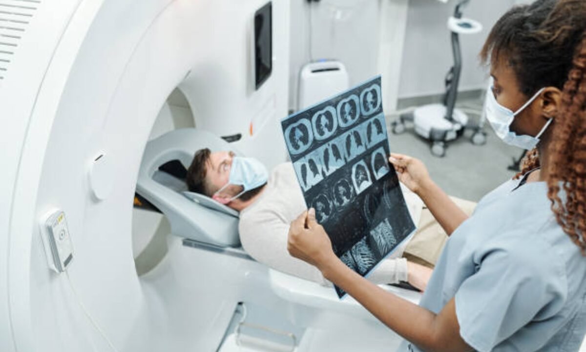
A Chest CT scan may seem scary, but it is a quite common and necessary procedure for modern medicine. CT scan stands for computed tomography scan, which is a form of imaging that allows doctors to see the chest up close (far more detailed than an X-ray).
Many people have questions before their scan, and they want to know what’s involved in the test, what doctors are looking for, how to prepare themselves, and what comes after the test. In this post, everything is explained in easy terms to guide you into feeling knowledgeable and confident.
What is a Chest CT Scan?
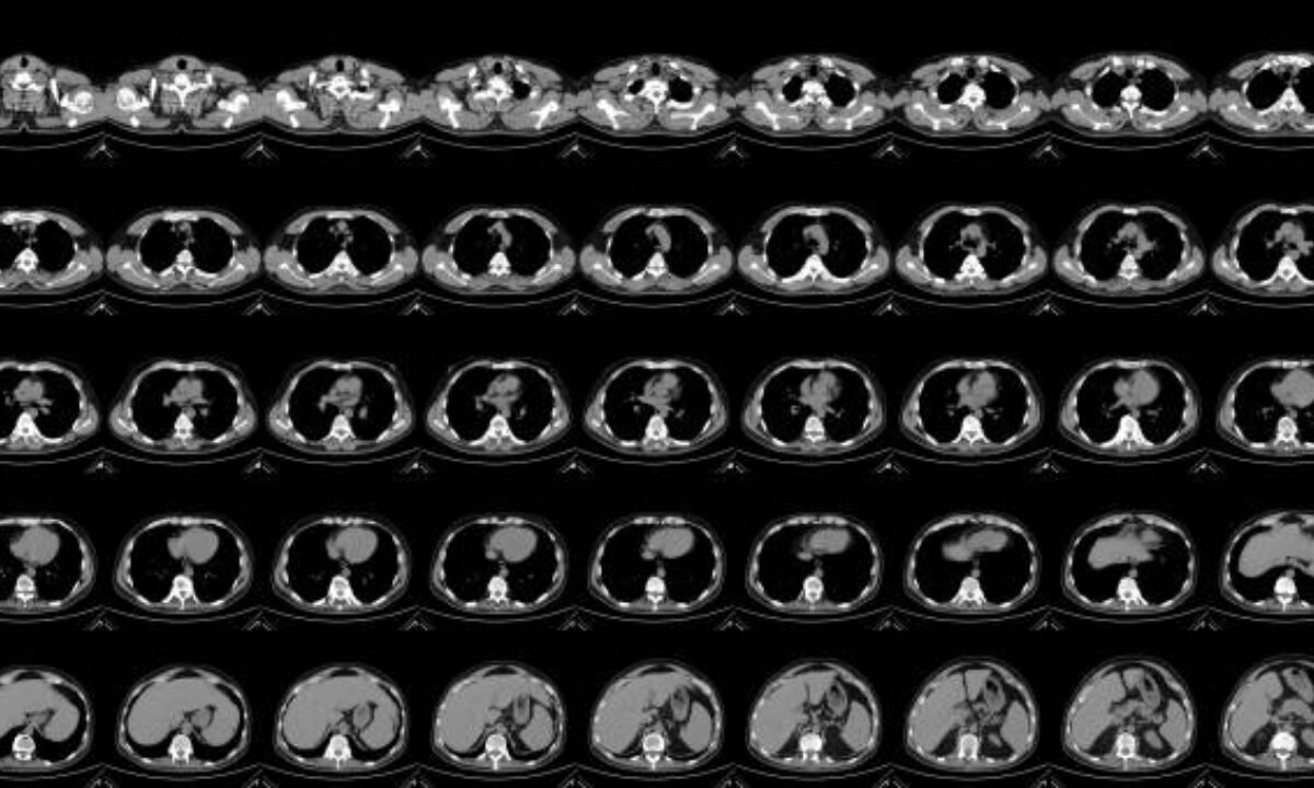
A Chest CT scan is an image test that takes painless X-rays and computer imaging to see the structures, bones, tissues, and blood vessels in the chest. In contrast with the standard X-rays, which only give one “shadowy” picture, the chest CT displays cross-sectional images – just imagine slices or layers – and a doctor would have better vision.
Simple Explanation
A Chest CT scan works like a loaf of bread cut into slices so you can see through each slice and see small details you wouldn’t notice from the outside.
More Detailed Explanation
- There are also improved scanning machines that you lie down on a table that slides in and out of a circular machine and you are scanned there.
- The scanner makes several X-ray images from different angles.
- A computer combines these images into an elaborate view of your chest from all directions.
For what reason do I need a Chest CT Scan?
There are many reasons why doctors request a Chest CT scan. They may prescribe it to you if you have symptoms such as shortness of breath, abnormal chest pain, or a (p) persistent cough, or if something abnormal was found on the X-ray of the whole chest.
Common Reasons Include:
- Looking for infections such as TB or pneumonia.
- detection of tumors, nodules, or lung cancer
- Research on blood clots in the lungs
- Diagnosing heart or blood vessel issues.
- Searching for injuries after an accident
- Monitoring chronic lung conditions
What is a Chest CT Scan looking for?
A chest CT scan is primarily searching for anything abnormal in your chest. This includes:
- Routine and hidden lung diseases.
- Fluid collections
- Swelling of lymphnodes
- Abnormal blood vessels or enlarged heart chambers.
- Cancerous or non-cancerous nodules
Types of Chest CT Scans
There are various chest CT scans. The most common are:
- CT Chest Plain: This is a scan without the use of contrast dye. It provides fundamental but important information about lungs, chest wall, bones and heart.
- CT Chest with Contrast: In this case, a dye is injected to make blood vessels and some tissues stand out. This makes it easier to locate blood clots or tumors.
When is CT Chest Plain Applied?
A CT chest plain is usually the first step. Doctors use it to:
- Eliminate major problems such as big tumors or fractures as soon as possible.
- Look for obvious infections or fluid.
- Give a base scan to compare with future tests.
Preparing for the Scan
Correct preparation will help make your scan smoother and accurate:
- Clothing: Wear comfortable, loose clothes. You might have to put on a hospital gown.
- Jewelry and Metal Objects: Remove these before the scan because they may affect the pictures.
- Eating and Drinking: If your scan requires the use of contrast dye, then you may be advised not to eat for a few hours.
- Medical Conditions: Inform your doctor if you are allergic, have kidney problems, diabetes, or pregnant.
What to Expect during this Process?
1. You will be asked to lie down on a moving table.
2. A technician will assist you in positioning for the best images.
3. Where a contrast dye is needed, this will be injected via a vein in your arm. This may trigger a warm feeling, but it is generally painless.
4. The table slides into the scanner (a big doughnut-shaped machine). You can hear humming or clicking sounds.
5. You will have to hold your breath for a few seconds.
6. The scan typically does not take more than 10 minutes.
After the scan, you can go home and continue with activities unless told otherwise. If you had contrast dye, drink lots of water to flush it out of your system.
Interpreting Your Chest CT Scan Report
After your scan is complete, a radiologist will look at the pictures and write a detailed chest CT scan report. This report is given to your doctor, who will explain the results to you.
Essential Aspects of a Chest CT Scan Report:
- Findings: By noting anything strange, nodules, fluid, or an indication of infection.
- Impression: A summary that will help your doctor to understand the main results quickly.
- Recommendations: Tests if needed, after which.
What is a normal Chest CT Scan?
- No unexpected growths or masses.
- No signs of infection, bleeding, or fluid accumulation.
- Lungs, blood vessels, heart, and chest wall are all healthy.
Common Results: Normal Chest CT Scan vs Abnormal Findings
| Type of Finding | What It Means |
|---|---|
| Normal Chest CT Scan | Everything looks healthy and typical |
| Infection | There may be signs of pneumonia or TB |
| Tumor/Nodule | Possible need for further tests or biopsy |
| Fluid | Could suggest infection or heart failure |
| Clots/Blood Vessel | Indicates possible pulmonary embolism |
If your scan is normal, it’s a good sign—your doctor may look at other causes for your symptoms. If something strange is observed, do not panic; Generally, there is the need for further tests or follow-ups.
CT Scan in Bangalore
Good facilities are essential. When you are looking for a CT scan in Bangalore, research quality hospitals and imaging centers. There are numerous modern CT machines in advanced medical centers in Bangalore. Ask about:
- Time taken to get your chest CT scan report
- Whether they offer CT Chest plain and contrast scans, if necessary
- Cost and whether your insurance covers the scan.
There are numerous credible places that provide CT scan in Bangalore and therefore you will probably receive good care and accurate results.
Risks and Side Effects
CT scans use more radiation than standard X-rays, but the risks are generally low, compared to the benefits. Never fail to inform your doctor if you are pregnant or may be pregnant. The contrast dye is generally safe but may cause:
- Allergic reactions (rare)
- Kidney problems in individuals who already have kidney problems.
If you feel sick after the scan, inform your doctor immediately.
Summary of Key Points
- A Chest CT scan is a safe, fast, and very detailed method of seeing inside your chest.
- It is used to diagnose infections, cancer, heart problems, and so on.
- There are CT Chest plain scans and scans with contrast. Both have specific uses.
- A normal chest CT scan means all is well.
- The chest CT scan report provides your doctor with the detailed answers required.
- CT scan in Bangalore is a good medical center that offers quick and reliable results.
Understanding what is a Chest CT scan looking for and what to expect can make your experience much less stressful. If you have any concerns or questions about your scan, never hesitate to speak to your doctor.
Koshikaa offers the latest imaging services for CT scan in Bangalore with the help of the latest technology and radiologists.