How Contrast Improves CT Scan Results: Insights for Health

Ultrasound scan in Bangalore

Table of Contents

CT scans have advanced to become a fundamental medical instrument which helps physicians make diagnoses. Such imaging methods produce detailed information about the human body which medical professionals need to correctly diagnose illness as well as track patient recovery. This article examines how contrast solutions boost CT scan results and describes their purpose along with highlighting CT scan benefits for the medical sector.

We will also delve into the question “CT scan why is it done” while providing information on CT scan services through Bangalore examples and explaining CT scan result processing durations.

What is a CT Scan?

PET CT Sacan in Bangalore

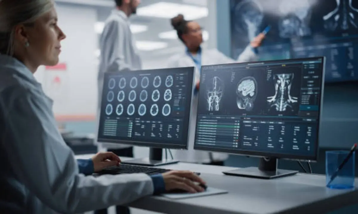
Computed tomography, also known as CT scan, operates as a diagnostic imaging method through which X-ray scans become computer-processed into cross-sectional body images. Operating under these imaging methods enables physician specialists to detect internal body conditions more precisely than X-ray procedures do. The exam process called CT scan allows healthcare providers to study internal structures including organs as well as bones and blood vessels and tissues.

CT Scan: Why Is It Done?

Medical practitioners conduct CT scans to detect various ailments in patients. The medical procedure occurs because of the following major diagnostic needs:

  • Medical analysis uses CT scanners to detect bone fractures in addition to identifying bodily injuries.
  • Doctors use this test to discover tumors along with infections as well as blood clots.
  • Healthcare providers resolve chronic illnesses including cancer through monitoring.
  • CT enables medical personnel to guide biopsy procedures.
  • Doctors investigate the inner compositions of the brain together with the lungs, heart and liver during diagnostic examinations.

The evaluation of “CT scan why is it done” demonstrates how this diagnostic tool precisely diagnoses complex medical problems.

What is the Contrast in CT Scans?

The substance called contrast operates as a specific dye that doctors introduce into bodies to create better visibility of certain areas during CT imaging. CT scan and contrast becomes part of the testing process through oral intake as well as intravenous delivery and rectal administration. The contrast medium serves to illuminate important structures both inside and outside blood vessels and organs so radiologists can identify any potential abnormalities.

Contrast material serves as an essential factor that enhances CT scan image quality.

Medical institutions achieve higher CT scan clarity through the addition of contrast materials. What procedures lead to such enhancements? Let’s break it down:

1. Enhancing Tissue Visibility

Contrast enables specialists to distinguish different tissues which have similar appearances. For example:

  • The contrast agent reveals clearer blood vessel images because it makes it possible for doctors to detect abnormalities and blockages.
  • The distinctive appearance of organs like the liver and pancreas and the kidneys makes cysts or tumor detection possible without difficulties.

2. Detecting Hidden Abnormalities

Diagnosis of small abnormalities including polyps and tiny tumors and minute infections becomes impossible when there is no contrast material present. The contrast technique reveals all details of defective body areas to create a complete visual diagnostic assessment.

3. The diagnosis process for vascular disorders receives better outcomes through contrast utilization.

Contrast functions to improve diagnostic capabilities by revealing spinal fluids arteries and other pathway structures thus aiding poor visibility in aneurysms and blood clots.

CT Scan Benefits: Why Should You Opt for One?

People typically ask themselves about CT scans since multiple imaging alternatives exist. Let’s understand the key of CT scan benefits:

Quick and Clear Imaging

CT scans produce detailed images in a short amount of time despite functioning, unlike traditional X-ray machines. Emergency situations require speedy evaluation, particularly for detecting hidden internal damage after accidents occur.

Non-Invasive Procedure

CT scans perform the examination without causing discomfort to patients and require no invasive procedures. The simple contrast-based examination procedure leads to a small level of discomfort.

Precision and Accuracy

CT scan precision reaches unmatched levels because of both the advanced technology and contrast application thus enabling prompt disease detection, particularly cancer diagnosis.

People easily understand why CT scans have become essential in modern health diagnostics by recognizing these important benefits.

CT Scan and Contrast: Associated Risks and Safety Concerns

CT scans remain generally safe when contrast is used but patients need to understand certain safety precautions and possible risks that may occur.

Allergic Reactions to Contrast

Some patients show mild reactions to contrast material through itching and nausea after receiving the substance. Most people do not experience severe adverse effects following contrast administration although these serious events remain overlooked.

Radiation Exposure

None of the common imaging procedures can reach radiation levels as high as those in CT scans. Your physician should do risk assessments before prescribing repetitive scans because occasional tests do not carry similar safety concerns.

What Happens During a CT Scan with Contrast? 

Being informed about the CT scan and contrast makes the experience less worrying for those who need it. Here’s what you can expect:

Preparation:

  • Your healthcare provider will inform you when to start fasting for several hours before the examination.
  • Before giving you contrast your doctor needs to check whether your allergies and kidney health can safely tolerate it.

Contrast Administration:

  • You will consume a solution containing the dye when receiving oral contrast.
  • The medical staff will administer the intravenous contrast by using a needle to insert it into your vein so that it travels through the bloodstream.
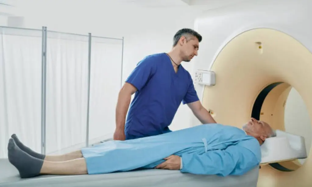
The Scan:

  • The examination table will carry you into the CT scanner when you lie down.
  • During the image acquisition process, you can expect to hear the machine working but the technician will capture clear scans.

Post-Scan:

  • Most individuals can engage in standard post-treatment activities right after the examination.
  • Your body automatically will eliminate the contrast materials through both urine and stool processes. 
  • This simple yet effective process ensures that healthcare professionals get the best possible imaging results.

How Long Do CT Scans Take?

One frequently raised concern is about the CT scan time for results or duration of the Procedure

A CT scan itself usually takes only 10 to 30 minutes. The actual imaging might last just a few minutes, while the rest of the time involves preparation and positioning.

When Will You Receive Results?

While some basic scans might provide immediate results, your doctor usually needs additional time to review detailed images. Your healthcare provider determines how long it takes to get CT scan time for results which may extend over several hours to one complete day.

CT Scan Services in Bangalore: What You Need to Know

CT scanning services in Bangalore can be obtained at different medical facilities that are supported by modern diagnostic technology. Multiple high-end diagnostic facilities and medical centers operating in Bangalore make this city a reliable base for addressing healthcare requirements. 

Before engaging with the “CT scan in Bangalore” service consider these points:

  • Seek medical facilities that have both experienced radiologists together with skilled technicians.
  • Modern scanner systems reduce radiation output which affects the facility quality.
  • People should examine the prices because CT scan rates differ between healthcare facilities in Bangalore.

Why Contrast Enhances the Future of Healthcare

Medical technology innovations will lead to CT scans and contrast becoming more precise and available to patients in the near future. Diagnostic tools prove essential for medical practice because they deliver vital findings necessary for clinical disease recognition.

Understanding CT scan functions becomes possible through answering fundamental questions regarding CT scan procedures as well as benefit analysis and addressing time-related questions and availability queries about CT scan in Bangalore.

Key Takeaways:

  • Clinical images from CT scanners become more precise when contrast substances are applied since they increase image clarity.
  • CT scans work as quick and non-destructive examinations that offer dependable results with or without contrast.
  • Patients can find advanced CT scan services through the facilities located in Bangalore.

The important role of contrast medium in CT scan procedures becomes clearer when people grasp its purpose whether they plan to get scanned or want to understand the medical process better. 

Koshikaa delivers reliable services for CT scan in Bangalore through their advanced imaging system which produces high-quality results and quick report delivery for better patient treatment.

MRI of the uterus is primarily used for diagnosing and evaluating conditions such as uterine fibroids, adenomyosis, and malignancies. It is particularly useful in the planning of surgical procedures and assessing the extent of gynecological diseases. This imaging test is valuable for its ability to deliver detailed cross-sectional images of the uterus and surrounding structures.
An MRI of the spleen can help diagnose various disorders affecting the spleen including splenomegaly (enlarged spleen), tumors, and other abnormalities. It provides clear, detailed images that are crucial for assessing the health of the spleen and planning potential surgical interventions.
Prostate MRI is a non-invasive imaging technique that provides detailed pictures of the prostate gland and surrounding tissues. It helps in the detection, staging, and monitoring of prostate cancer. Prostate MRIs are increasingly used to guide biopsy procedures and assist in the planning of treatment strategies for prostate health.
An MRI of the pancreas is used to identify issues such as pancreatitis, pancreatic cancer, and other pancreatic disorders. It offers high-resolution images that help in detecting small lesions and assessing the pancreatic ducts. This imaging technique is valuable for guiding biopsies and planning surgeries involving the pancreas.
MRI scans of the liver, kidney, and bile ducts provide detailed images that help diagnose various conditions, such as tumors, cysts, and blockages in the bile ducts. They are crucial for assessing organ damage, staging cancer, and planning liver or kidney-related surgeries. This type of MRI can also evaluate the health and function of these organs comprehensively.
Breast MRI is highly sensitive for detecting breast cancer, especially in individuals with dense breast tissue or a high risk of cancer. It is often used in conjunction with mammography to provide a more comprehensive evaluation. Breast MRIs can also monitor the response to breast cancer treatments and provide detailed pre-surgical mapping.
Brain and spine MRIs are critical tools for diagnosing and monitoring neurological conditions, including tumors, spinal cord injuries, and multiple sclerosis. These MRIs can reveal abnormalities in brain tissue and spinal cord structure that might not be visible with other imaging methods. They are essential for planning surgeries and assessing the effectiveness of treatments in neurology.
PET-CT scans of transplant organs like the liver, kidney, or pancreas are performed to assess metabolic activity, detect complications, and monitor post-transplant function. These scans play a vital role in the early detection of issues such as organ rejection, recurrence of disease, or infection, guiding timely interventions and optimizing transplant outcomes for patients.
PET-CT examinations of organs such as the liver, kidney, or prostate are crucial for assessing metabolic activity, detecting abnormalities, and monitoring disease progression. These scans help diagnose conditions such as liver metastases, renal cell carcinoma, or prostate cancer, guiding treatment decisions and monitoring response to therapy over time.
PET-CT scans of soft-tissue masses are utilized to assess metabolic activity and detect malignant tumors or other abnormalities. By combining PET imaging with CT imaging, these scans provide detailed information about tumor location, size, and metabolic characteristics, aiding in accurate diagnosis and treatment planning for soft-tissue cancers.
PET-CT scans are valuable for detecting cysts or fluid collections within the body, including cysts in the liver, kidneys, or other organs. These scans help characterize the nature and metabolic activity of cysts, guiding diagnosis and treatment planning for conditions such as cystic liver disease, renal cysts, or pancreatic cysts.
PET-CT scans of muscles are performed to assess metabolic activity and detect abnormalities such as inflammation, infection, or tumors. These scans provide valuable information about muscle function and pathology, aiding in the diagnosis and management of conditions like myositis, muscular dystrophy, or soft-tissue sarcomas.
PET-CT scans of the brain are utilized to evaluate brain metabolism and detect abnormalities such as tumors, epilepsy, or neurodegenerative diseases. By combining functional PET imaging with anatomical CT imaging, these scans offer comprehensive information about brain function and structure, guiding neurosurgical interventions and treatment decisions for neurological disorders
PET-CT scans of bones are commonly used to detect bone metastases in cancer patients, assess bone health in conditions like osteoporosis, and evaluate response to treatment. By combining PET and CT imaging, these scans provide detailed information about metabolic activity and anatomical changes in bone structures, aiding in accurate diagnosis and treatment planning for various bone-related conditions.
CT scans of the spine are conducted to assess the vertebrae, spinal cord, and surrounding soft tissues for conditions like herniated discs, spinal fractures, or spinal stenosis, aiding in the diagnosis and management of spinal disorders with detailed imaging essential for surgical planning and intervention.
CT scans of the pelvis are utilized to visualize the pelvic bones, reproductive organs, bladder, and surrounding structures for conditions such as fractures, tumors, or pelvic inflammatory disease, guiding treatment plans and surgical interventions with precise anatomical information.
CT scans of the neck are performed to evaluate structures such as the throat, thyroid gland, lymph nodes, and blood vessels for abnormalities like tumors, infections, or injuries, assisting in diagnosis and management of head and neck disorders with detailed imaging.
CT scans of the extremities are employed to assess bones, joints, and soft tissues in the arms or legs, aiding in the diagnosis of fractures, arthritis, or other musculoskeletal conditions, guiding orthopedic treatment and rehabilitation plans for optimal recovery.
CT scans of the chest are utilized to examine the lungs, heart, blood vessels, and surrounding structures for conditions such as lung nodules, pneumonia, or pulmonary embolism, facilitating accurate diagnosis and management of respiratory and cardiovascular diseases.
CT scans of the brain are conducted to assess brain structures, identify abnormalities like tumors, hemorrhages, or strokes, and aid in diagnosing neurological conditions, guiding neurosurgical interventions and treatment strategies with precise imaging of the brain’s anatomy.
CT scans of the abdomen are performed to visualize and evaluate the internal organs such as the liver, kidneys, pancreas, and intestines for conditions like tumors, infections, or injuries, providing detailed images essential for diagnosis and treatment planning.
Transplant organ ultrasound scans, including those of the liver, kidney, or pancreas, are performed to assess organ viability, detect complications, and monitor post-transplant function. These scans play a vital role in the early detection of issues such as organ rejection, vascular complications, or fluid collections, guiding timely interventions and optimizing transplant outcomes for patients.
Ultrasound examinations of organs such as the liver, kidney, or prostate are crucial for assessing organ structure, detecting abnormalities, and monitoring disease progression. These scans help diagnose conditions such as liver cirrhosis, kidney stones, or prostate enlargement, guiding treatment decisions and monitoring response to therapy over time.
Soft-tissue masses can be evaluated using ultrasound to determine their size, location, and characteristics. Ultrasound imaging helps differentiate between benign and malignant masses, guiding further diagnostic evaluation and treatment planning. By providing real-time imaging, ultrasound assists in the precise localization of soft-tissue masses for biopsy or surgical removal if needed.
Ultrasound scans are valuable for detecting cysts or fluid collections within the body, including cysts in the liver, kidneys, or other organs. These scans help characterize the nature and size of cysts, aiding in diagnosis and treatment planning for conditions such as cystic liver disease or renal cysts, and guiding interventions such as cyst drainage or sclerotherapy.
Musculoskeletal ultrasound scans are used to assess the health and integrity of muscles, identifying issues such as strains, tears, or muscle inflammation. These scans provide detailed images of muscle structures, allowing healthcare providers to diagnose muscle injuries accurately and develop personalized rehabilitation plans to promote recovery and restore function.
Ultrasound examinations of joints are essential for evaluating joint health, detecting abnormalities such as inflammation, fluid accumulation, or cartilage damage. By visualizing the joint structures in real-time, ultrasound helps diagnose conditions like arthritis, bursitis, or ligament injuries, guiding treatment decisions and monitoring disease progression over time.
Ultrasound scans of tendons are performed to assess for injuries, inflammation, or tears, aiding in the diagnosis and management of conditions such as tendonitis or tendon tears. These scans provide detailed images of tendon structures, helping healthcare providers determine the extent of damage and plan appropriate treatment strategies, including physical therapy or surgical intervention if necessary.
Pelvis X-rays play a crucial role in diagnosing injuries, diseases, and developmental conditions affecting the pelvic region. They provide detailed images of the hip bones, sacrum, and coccyx, aiding in the assessment of fractures, dislocations, or signs of arthritis. Pelvic X-rays are instrumental in guiding surgical interventions, monitoring healing progress, and evaluating conditions such as osteoporosis or tumors. By providing precise anatomical information, these imaging studies enable healthcare professionals to deliver personalized care and improve patient outcomes.
Teeth X-rays, known as dental radiographs, are indispensable tools for dentists to assess oral health. They aid in diagnosing dental decay, gum disease, and abnormalities in tooth structure or alignment. These images provide valuable insights into hidden dental issues, allowing for early intervention and prevention of complications. Additionally, dental X-rays are essential for planning treatments such as orthodontics, root canals, or dental implants, ensuring optimal outcomes and patient satisfaction.
Spine X-rays are pivotal in diagnosing spinal fractures, degenerative conditions, and spinal misalignments, providing critical information for treatment planning and monitoring spinal health. They offer detailed images of the vertebrae and surrounding structures, enabling healthcare professionals to identify issues such as scoliosis, disc herniation, or spinal stenosis
Skull X-rays are valuable for detecting fractures, assessing head trauma, and evaluating the alignment of facial bones, crucial in diagnosing conditions like fractures, tumors, or developmental abnormalities in the skull. They provide detailed views of the cranial structure, aiding clinicians in determining appropriate treatment strategies and ensuring patient safety.
For the heart, X-ray solutions can aid in the detection of various cardiac problems, ranging from mild to severe, including – Heart failure, Cardiac congestion and other heart-related conditions. In addition to cardiac assessments, X-ray solutions are also used for breast examinations to identify tumors or signs of breast cancer.

Bone X-rays are essential for diagnosing fractures and assessing bone alignment, aiding doctors in developing treatment plans and monitoring healing progress. They provide detailed images of the skeletal system, helping identify abnormalities or injuries that may not be visible through other imaging techniques

Abdominal X-rays are performed when patients experience symptoms such as significant nausea, ongoing vomiting, abdominal pain, flank or back pain, or abdominal swelling. These X-rays can help diagnose various conditions.
Search Bar

Frequently Booked Tests

Frequently Booked Checkups