Understanding PET CT scan: Process, Diagnosis and Results

pet ct scan

Table of Contents

In this article, we will simplify the PET CT scan concept, clarify how it works, discuss its role in diagnosis, and explain what results of the PET CT scan should be interpreted.

If you are someone who is looking to know the significance of a PET CT scan in Bangalore, this article is especially helpful as this technology is growing and becoming accessible in Bangalore. Let’s dive in.

What Is a PET CT Scan?

cost of pet ct scan bangalore

Positron Emission Tomography-Computed Tomography scan is abbreviated as PET CT scan. It is an amalgamation of two imaging techniques, PET (Positron Emission Tomography) and CT (Computed Tomography).

The PET scan measures functional processes in the body. In doing so, it identifies areas of high biochemical activity, which across the board may be linked to diseases, such as cancer, heart issues, neurological disorders, and others. CT Scan is opposite from another hand and uses X-rays to produce detailed images of the body’s internal structures.

How Does a PET CT Scan Work?

It involves three key steps: Imaging, preparation, and assessment. 

1. Preparation for the Scan

Patients are advised to fast for a certain time or avoid food and drink which could give an erroneous result. 

2. The Scanning Process
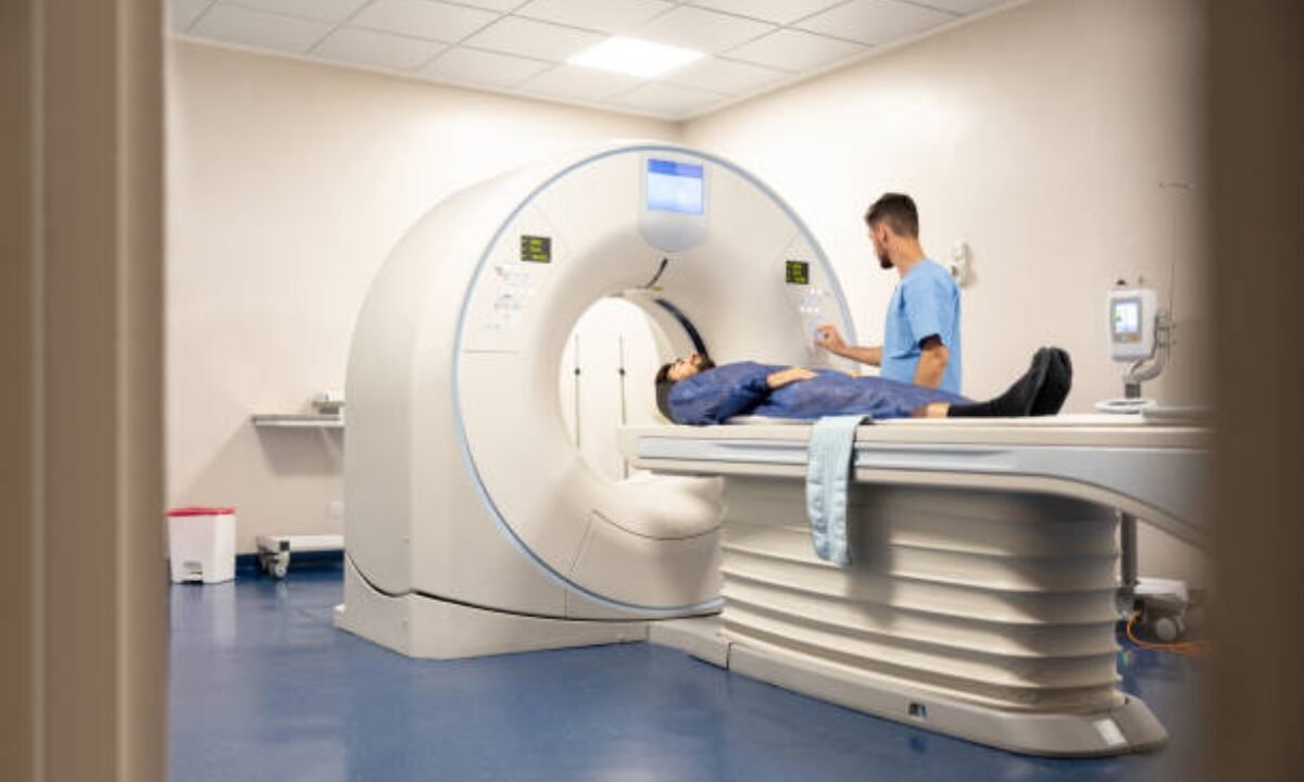
For the actual scan start, you lie on a bed that goes into a donut-shaped scanner where you lie down. 

The reason scans cannot be performed is the need for patients to remain still during the scan. The time it takes is usually 30 to 60 minutes, depending on what kind of imaging is needed. 

3. Post-Scan Evaluation

Following the scan, the patient can resume normal activity. After the results, a radiologist or nuclear medicine specialist interprets them. Such are sent to the patient’s doctor to join in a suitable treatment plan.

Why would you need a PET CT scan?

There are many reasons your doctor may recommend you for a PET CT scan. The most common uses include:

1. Cancer Diagnosis and Monitoring

PET CT scans are the most well-known for detecting and staging different types of cancer. They show the presence of abnormal cells and reveal whether the disease has spread to other areas.  In cities with leading medical services, accessibility to a PET CT scan in Bangalore has made cancer diagnosis considerably faster and more precise.  

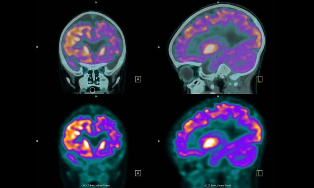
2. Neurological Condition Detection

PET CT scans are also extremely good at spotting neurological diseases like Alzheimer’s disease, epilepsy, and Parkinson’s disease. Doctors also learn about abnormal functions from brain activity analysis.

3. Evaluating Heart Health

In cardiology, it is used to assess blood flow within the heart, to detect damage to the heart tissue after a heart attack, and to assess how effective current treatments are. In such cases, early diagnosis can save one’s life.

4. Monitoring Other Disorders

PET CT scans may also be used, in addition to cancer, heart issues, and brain conditions, where there is unexplained inflammation or infection that may not be seen on other tests.

Why Are PET CT Scans Different Than Other Tests?

Measuring PET and CT together on the same scanner is the primary edge of a PET CT scan in Bangalore. It does not provide either structural or, and in some cases if you are interested in functional information only then MRIs and stand-alone CT scans all provide that, but this does not provide that. PET CT scans combine the parts, making clear images with pinpoint accuracy of the location(s) and function(s) of abnormalities. 

Interpretation of PET CT scan results

Here’s a simplified idea of what the results might indicate:

1. Normal Results

In medical terms, “normal” means the tracer hadn’t any structural abnormalities within the body. This is a good health outcome.

2. Abnormal Results

Any areas of the ear being picked up on the scan can mean disease could be present. For instance, a bright or ‘hot’ spot may indicate an increase in metabolic activity which may be characteristic of cancer. 

3. Follow-Up Steps

Follow-up testing should be performed to obtain a proper diagnosis if abnormal results are achieved. These findings are used to make personalized treatment plans. The growing ease of PET CT scans in Bangalore is making it possible for patients to get accurate, timely testing right in their city.

Is a PET CT Scan Safe?

PET CT scans, however, are generally safe. While they use radiation, it’s low and monitored closely to keep it low and safe for patients. Your doctor will need to know of any pre-existing medical conditions or allergies before you go and have the scan.

Additionally, most of the PET CT scans in Bangalore are run from advanced healthcare centers that are also sticking to certain safety protocols and use the latest technology to reduce exposure and increase diagnostic accuracy.

Finding a PET CT Scan in Bangalore

If you are from Bangalore, you will find several high-quality medical facilities offering PET CT scans. Now the city is a center for advanced diagnostic tools, and both residents and patients from other towns come here. 

If you’re searching for a PET CT scan in Bangalore then it’s ideal to know where to look for centers that give professional care and utilize the most recent scanning technology. 

Final Thoughts

A PET CT scan is a powerful diagnostic tool and provides the accurate information your doctor needs to make informed decisions regarding your health. This imaging technology is instrumental in modern medicine from detecting cancer right up to assessing the brain and heart conditions. With Bangalore being a place that has access to such cutting-edge technology via a PET CT scan in Bangalore, such timely and accurate medical care is ensured to them.

Also remember that our full body checkup in Bangalore is safe, simple, and useful in finding and treating your health problems properly if you are on the verge of scheduling a PET CT scan with Koshikaa.  

MRI of the uterus is primarily used for diagnosing and evaluating conditions such as uterine fibroids, adenomyosis, and malignancies. It is particularly useful in the planning of surgical procedures and assessing the extent of gynecological diseases. This imaging test is valuable for its ability to deliver detailed cross-sectional images of the uterus and surrounding structures.
An MRI of the spleen can help diagnose various disorders affecting the spleen including splenomegaly (enlarged spleen), tumors, and other abnormalities. It provides clear, detailed images that are crucial for assessing the health of the spleen and planning potential surgical interventions.
Prostate MRI is a non-invasive imaging technique that provides detailed pictures of the prostate gland and surrounding tissues. It helps in the detection, staging, and monitoring of prostate cancer. Prostate MRIs are increasingly used to guide biopsy procedures and assist in the planning of treatment strategies for prostate health.
An MRI of the pancreas is used to identify issues such as pancreatitis, pancreatic cancer, and other pancreatic disorders. It offers high-resolution images that help in detecting small lesions and assessing the pancreatic ducts. This imaging technique is valuable for guiding biopsies and planning surgeries involving the pancreas.
MRI scans of the liver, kidney, and bile ducts provide detailed images that help diagnose various conditions, such as tumors, cysts, and blockages in the bile ducts. They are crucial for assessing organ damage, staging cancer, and planning liver or kidney-related surgeries. This type of MRI can also evaluate the health and function of these organs comprehensively.
Breast MRI is highly sensitive for detecting breast cancer, especially in individuals with dense breast tissue or a high risk of cancer. It is often used in conjunction with mammography to provide a more comprehensive evaluation. Breast MRIs can also monitor the response to breast cancer treatments and provide detailed pre-surgical mapping.
Brain and spine MRIs are critical tools for diagnosing and monitoring neurological conditions, including tumors, spinal cord injuries, and multiple sclerosis. These MRIs can reveal abnormalities in brain tissue and spinal cord structure that might not be visible with other imaging methods. They are essential for planning surgeries and assessing the effectiveness of treatments in neurology.
PET-CT scans of transplant organs like the liver, kidney, or pancreas are performed to assess metabolic activity, detect complications, and monitor post-transplant function. These scans play a vital role in the early detection of issues such as organ rejection, recurrence of disease, or infection, guiding timely interventions and optimizing transplant outcomes for patients.
PET-CT examinations of organs such as the liver, kidney, or prostate are crucial for assessing metabolic activity, detecting abnormalities, and monitoring disease progression. These scans help diagnose conditions such as liver metastases, renal cell carcinoma, or prostate cancer, guiding treatment decisions and monitoring response to therapy over time.
PET-CT scans of soft-tissue masses are utilized to assess metabolic activity and detect malignant tumors or other abnormalities. By combining PET imaging with CT imaging, these scans provide detailed information about tumor location, size, and metabolic characteristics, aiding in accurate diagnosis and treatment planning for soft-tissue cancers.
PET-CT scans are valuable for detecting cysts or fluid collections within the body, including cysts in the liver, kidneys, or other organs. These scans help characterize the nature and metabolic activity of cysts, guiding diagnosis and treatment planning for conditions such as cystic liver disease, renal cysts, or pancreatic cysts.
PET-CT scans of muscles are performed to assess metabolic activity and detect abnormalities such as inflammation, infection, or tumors. These scans provide valuable information about muscle function and pathology, aiding in the diagnosis and management of conditions like myositis, muscular dystrophy, or soft-tissue sarcomas.
PET-CT scans of the brain are utilized to evaluate brain metabolism and detect abnormalities such as tumors, epilepsy, or neurodegenerative diseases. By combining functional PET imaging with anatomical CT imaging, these scans offer comprehensive information about brain function and structure, guiding neurosurgical interventions and treatment decisions for neurological disorders
PET-CT scans of bones are commonly used to detect bone metastases in cancer patients, assess bone health in conditions like osteoporosis, and evaluate response to treatment. By combining PET and CT imaging, these scans provide detailed information about metabolic activity and anatomical changes in bone structures, aiding in accurate diagnosis and treatment planning for various bone-related conditions.
CT scans of the spine are conducted to assess the vertebrae, spinal cord, and surrounding soft tissues for conditions like herniated discs, spinal fractures, or spinal stenosis, aiding in the diagnosis and management of spinal disorders with detailed imaging essential for surgical planning and intervention.
CT scans of the pelvis are utilized to visualize the pelvic bones, reproductive organs, bladder, and surrounding structures for conditions such as fractures, tumors, or pelvic inflammatory disease, guiding treatment plans and surgical interventions with precise anatomical information.
CT scans of the neck are performed to evaluate structures such as the throat, thyroid gland, lymph nodes, and blood vessels for abnormalities like tumors, infections, or injuries, assisting in diagnosis and management of head and neck disorders with detailed imaging.
CT scans of the extremities are employed to assess bones, joints, and soft tissues in the arms or legs, aiding in the diagnosis of fractures, arthritis, or other musculoskeletal conditions, guiding orthopedic treatment and rehabilitation plans for optimal recovery.
CT scans of the chest are utilized to examine the lungs, heart, blood vessels, and surrounding structures for conditions such as lung nodules, pneumonia, or pulmonary embolism, facilitating accurate diagnosis and management of respiratory and cardiovascular diseases.
CT scans of the brain are conducted to assess brain structures, identify abnormalities like tumors, hemorrhages, or strokes, and aid in diagnosing neurological conditions, guiding neurosurgical interventions and treatment strategies with precise imaging of the brain’s anatomy.
CT scans of the abdomen are performed to visualize and evaluate the internal organs such as the liver, kidneys, pancreas, and intestines for conditions like tumors, infections, or injuries, providing detailed images essential for diagnosis and treatment planning.
Transplant organ ultrasound scans, including those of the liver, kidney, or pancreas, are performed to assess organ viability, detect complications, and monitor post-transplant function. These scans play a vital role in the early detection of issues such as organ rejection, vascular complications, or fluid collections, guiding timely interventions and optimizing transplant outcomes for patients.
Ultrasound examinations of organs such as the liver, kidney, or prostate are crucial for assessing organ structure, detecting abnormalities, and monitoring disease progression. These scans help diagnose conditions such as liver cirrhosis, kidney stones, or prostate enlargement, guiding treatment decisions and monitoring response to therapy over time.
Soft-tissue masses can be evaluated using ultrasound to determine their size, location, and characteristics. Ultrasound imaging helps differentiate between benign and malignant masses, guiding further diagnostic evaluation and treatment planning. By providing real-time imaging, ultrasound assists in the precise localization of soft-tissue masses for biopsy or surgical removal if needed.
Ultrasound scans are valuable for detecting cysts or fluid collections within the body, including cysts in the liver, kidneys, or other organs. These scans help characterize the nature and size of cysts, aiding in diagnosis and treatment planning for conditions such as cystic liver disease or renal cysts, and guiding interventions such as cyst drainage or sclerotherapy.
Musculoskeletal ultrasound scans are used to assess the health and integrity of muscles, identifying issues such as strains, tears, or muscle inflammation. These scans provide detailed images of muscle structures, allowing healthcare providers to diagnose muscle injuries accurately and develop personalized rehabilitation plans to promote recovery and restore function.
Ultrasound examinations of joints are essential for evaluating joint health, detecting abnormalities such as inflammation, fluid accumulation, or cartilage damage. By visualizing the joint structures in real-time, ultrasound helps diagnose conditions like arthritis, bursitis, or ligament injuries, guiding treatment decisions and monitoring disease progression over time.
Ultrasound scans of tendons are performed to assess for injuries, inflammation, or tears, aiding in the diagnosis and management of conditions such as tendonitis or tendon tears. These scans provide detailed images of tendon structures, helping healthcare providers determine the extent of damage and plan appropriate treatment strategies, including physical therapy or surgical intervention if necessary.
Pelvis X-rays play a crucial role in diagnosing injuries, diseases, and developmental conditions affecting the pelvic region. They provide detailed images of the hip bones, sacrum, and coccyx, aiding in the assessment of fractures, dislocations, or signs of arthritis. Pelvic X-rays are instrumental in guiding surgical interventions, monitoring healing progress, and evaluating conditions such as osteoporosis or tumors. By providing precise anatomical information, these imaging studies enable healthcare professionals to deliver personalized care and improve patient outcomes.
Teeth X-rays, known as dental radiographs, are indispensable tools for dentists to assess oral health. They aid in diagnosing dental decay, gum disease, and abnormalities in tooth structure or alignment. These images provide valuable insights into hidden dental issues, allowing for early intervention and prevention of complications. Additionally, dental X-rays are essential for planning treatments such as orthodontics, root canals, or dental implants, ensuring optimal outcomes and patient satisfaction.
Spine X-rays are pivotal in diagnosing spinal fractures, degenerative conditions, and spinal misalignments, providing critical information for treatment planning and monitoring spinal health. They offer detailed images of the vertebrae and surrounding structures, enabling healthcare professionals to identify issues such as scoliosis, disc herniation, or spinal stenosis
Skull X-rays are valuable for detecting fractures, assessing head trauma, and evaluating the alignment of facial bones, crucial in diagnosing conditions like fractures, tumors, or developmental abnormalities in the skull. They provide detailed views of the cranial structure, aiding clinicians in determining appropriate treatment strategies and ensuring patient safety.
For the heart, X-ray solutions can aid in the detection of various cardiac problems, ranging from mild to severe, including – Heart failure, Cardiac congestion and other heart-related conditions. In addition to cardiac assessments, X-ray solutions are also used for breast examinations to identify tumors or signs of breast cancer.

Bone X-rays are essential for diagnosing fractures and assessing bone alignment, aiding doctors in developing treatment plans and monitoring healing progress. They provide detailed images of the skeletal system, helping identify abnormalities or injuries that may not be visible through other imaging techniques

Abdominal X-rays are performed when patients experience symptoms such as significant nausea, ongoing vomiting, abdominal pain, flank or back pain, or abdominal swelling. These X-rays can help diagnose various conditions.
Search Bar

Frequently Booked Tests

Frequently Booked Checkups