10 Important Facts About PET CT Scans You Should Know  

pet ct scan

Table of Contents

It is normal for you to have questions regarding PET CT scanning since you have been thinking about it or received medical advice to get one. The diagnostic medical imaging method known as PET CT scan has revolutionized both medical examination and diagnostics. This article will present 10 vital points about PET CT scans by explaining their nature along with their importance and the procedures involved. Let’s dive in!

1. What is a PET CT Scan?  

The PET CT scan unites two imaging technologies which integrate Positron Emission Tomography (PET) and Computed Tomography (CT). A CT scan shows detailed anatomical body pictures whereas a PET scan helps detect variations in cellular performance. These two diagnostic methods create an exceptionally precise system to inspect body organs along with their surrounding tissues.  

The medical combination proves beneficial in cancer detection due to its ability to create thorough visual images of atypical cells including both structure and cellular function. Other conditions besides cancer treatment use PET CT scans for neurological examination and heart disease assessments.  

2. Why are PET CT Scans Done?

A PET CT scan serves the main objective of revealing cell functional patterns throughout your entire body. Through PET CT medical staff can track disease development and measure treatment results as well as discover abnormal features including tumours.  

The medical tool serves primarily for oncology applications involving cancer identification but it additionally benefits heart examinations alongside Alzheimer’s disease brain assessments. Early diagnosis relies heavily on this diagnostic method because it reveals the functional status of anatomical components.  

3. Can PET CT Scan Detect Bone Cancer? 

pet ct scan in bangalore

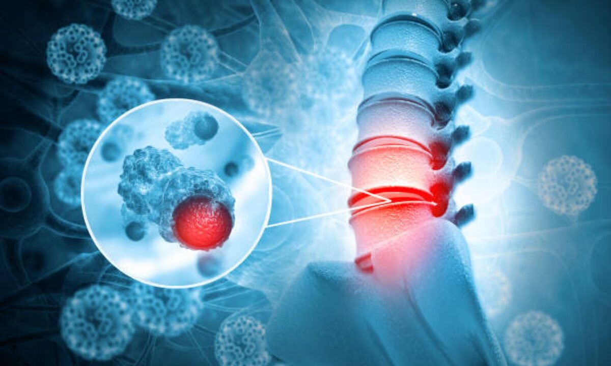
PET CT technology provides professionals with the capability to find bone cancer in patients. The scan tracks irregular metabolic activities occurring within bone tissues to identify potential cancer indications.  

The imaging process points to highly active areas which makes this tool valuable for finding bone cancer while it remains at early stages. Additional testing through biopsies continues to be needed because they serve to validate diagnoses.  

4. What Happens During a PET CT Scan Procedure?

The pet CT scan procedure implements a simple method to reach its final goal. The procedure consists of four main steps as described below.  

1. Medical staff may instruct patients to maintain fasting during the few hours before the scan. Healthcare providers provide particular instructions to patients based on which medical condition requires diagnosis.  

2. A professional staff member administers the special radioactive tracer through your bloodstream when performing the procedure. The tracer designated for this process distinguishes high-activity regions in the human body.  

3. When you participate in the scanning procedure you need to position yourself on a moving table inside the scanning machine. The PET CT scanner records functional and structural scan images which last for under one hour.  

4. The examination process finishes when you need to drink lots of fluids to eliminate the radiotracer from your system.  

People experience minimal discomfort during the painless procedure while accessing excellent information.  

5. Is a PET CT Scan Safe?

The PET CT scan procedure carries an overall safe status for medical patients. Although patients experience slight radiation exposure during the procedure the tracer delivers a very small radiation amount. The examination process creates no adverse reactions which most participants can easily endure without complications.  

PET CT testing must be strongly avoided by expectant mothers who are breastfeeding unless medical necessity confirms the need for the procedure. 

6. How Much Does a PET CT Scan Cost?  

Customers can discover that pet ct scan cost depends on multiple factors when researching scan prices. The test requires a facility, a precise placement, and distinct reasons for scanning as essential factors. For instance, the pet CT scan in Bangalore costs relatively less than what you would pay in Western areas, making it accessible to a wider audience.  

It’s always a good idea to check with your insurance provider to understand what part of the expense, if any, is covered under your plan.

7. How Long Does the Scan Take?  

A standard PET CT scan operates at a time interval ranging from 45 minutes up to 2 hours. The large portion of the scan duration requires patients to wait until the radioactive tracer becomes fully distributed throughout their body during preparation.  

The scanning process extends between 30 to 60 minutes. The complete procedure requires different durations based on the medical condition that healthcare professionals are treating according to their protocols.  

8. How should you prepare before undergoing this scan?  

Accomplished PET CT examinations require proper preparation before the procedure starts. Your test-specific instructions will vary based on the testing procedure you need to complete.  

  • The procedural period requires no food consumption during a 4 to 6-hour window before examination time.  
  • Drink only water before the procedure unless the medical staff provides different instructions.  
  • Before your appointment, you must notify your doctor about all prescriptions under your care.  
  • High-intensity physical exercise must be avoided within 24 hours of the scanning appointment because it will affect test results.  
  • Notify medical staff members about your pregnancy status and breastfeeding choices.  

9. What Are the Benefits of a PET CT Scan?  

The examination provides distinct diagnostic benefits when used as a PET CT scan against alternative imaging technologies.  

  • Metabolic activity evaluation from PET CT scans enables doctors to detect diseases such as cancer at an early stage when no symptoms have manifested yet.  
  • PET CT evaluations enable healthcare professionals to determine treatment effectiveness when using chemotherapy together with radiation therapy.  
  • The device demonstrates versatility through its utility in cancer diagnosis and monitoring of heart problems in addition to brain and nervous system evaluation.  

PET CT offers advanced imaging that results in diagnoses that occur faster and more correctly thereby leading to better patient results.  

10. Where Can You Get a PET CT Scan?  

Various hospitals and advanced diagnostic centres across the world provide PET CT scanning services to their patients. PET CT imaging services exist at distinguished imaging facilities within the Indian cities of Bengaluru and Delhi together with Mumbai. The pet CT scan facilities of Bangalore have earned a solid reputation through their delivery of highly professional services.  

Research about your scan appointment must be finished before you book an appointment. The selection of a diagnostic centre that utilizes radiologists with proper qualifications will produce interpretation results beneficial for doctors.  

Final Thoughts 

PET CT scans represent a top modern medical diagnostic instrument. PET CT scans enable medical practitioners to observe details along with exact results which surpass the capabilities of standard image diagnostic methods when detecting various medical conditions.  

Correct medical assessment in combination with accurate treatment strategies helps reduce costs along with the most essential benefit of saving lives while also conserving time.  

Your understanding of these ten points about PET CT helps you create a better foundation for continued health advancement. Contact your healthcare provider to receive detailed information about the pet CT scan procedure and if you’re wondering can pet CT scan detect bone cancer? Then it will also be answered by your professional.

Koshikaa is a reputed screening centre, providing residents with advanced services of PET CT scan in Bangalore through which they secure accurate results while receiving superior patient support.

MRI of the uterus is primarily used for diagnosing and evaluating conditions such as uterine fibroids, adenomyosis, and malignancies. It is particularly useful in the planning of surgical procedures and assessing the extent of gynecological diseases. This imaging test is valuable for its ability to deliver detailed cross-sectional images of the uterus and surrounding structures.
An MRI of the spleen can help diagnose various disorders affecting the spleen including splenomegaly (enlarged spleen), tumors, and other abnormalities. It provides clear, detailed images that are crucial for assessing the health of the spleen and planning potential surgical interventions.
Prostate MRI is a non-invasive imaging technique that provides detailed pictures of the prostate gland and surrounding tissues. It helps in the detection, staging, and monitoring of prostate cancer. Prostate MRIs are increasingly used to guide biopsy procedures and assist in the planning of treatment strategies for prostate health.
An MRI of the pancreas is used to identify issues such as pancreatitis, pancreatic cancer, and other pancreatic disorders. It offers high-resolution images that help in detecting small lesions and assessing the pancreatic ducts. This imaging technique is valuable for guiding biopsies and planning surgeries involving the pancreas.
MRI scans of the liver, kidney, and bile ducts provide detailed images that help diagnose various conditions, such as tumors, cysts, and blockages in the bile ducts. They are crucial for assessing organ damage, staging cancer, and planning liver or kidney-related surgeries. This type of MRI can also evaluate the health and function of these organs comprehensively.
Breast MRI is highly sensitive for detecting breast cancer, especially in individuals with dense breast tissue or a high risk of cancer. It is often used in conjunction with mammography to provide a more comprehensive evaluation. Breast MRIs can also monitor the response to breast cancer treatments and provide detailed pre-surgical mapping.
Brain and spine MRIs are critical tools for diagnosing and monitoring neurological conditions, including tumors, spinal cord injuries, and multiple sclerosis. These MRIs can reveal abnormalities in brain tissue and spinal cord structure that might not be visible with other imaging methods. They are essential for planning surgeries and assessing the effectiveness of treatments in neurology.
PET-CT scans of transplant organs like the liver, kidney, or pancreas are performed to assess metabolic activity, detect complications, and monitor post-transplant function. These scans play a vital role in the early detection of issues such as organ rejection, recurrence of disease, or infection, guiding timely interventions and optimizing transplant outcomes for patients.
PET-CT examinations of organs such as the liver, kidney, or prostate are crucial for assessing metabolic activity, detecting abnormalities, and monitoring disease progression. These scans help diagnose conditions such as liver metastases, renal cell carcinoma, or prostate cancer, guiding treatment decisions and monitoring response to therapy over time.
PET-CT scans of soft-tissue masses are utilized to assess metabolic activity and detect malignant tumors or other abnormalities. By combining PET imaging with CT imaging, these scans provide detailed information about tumor location, size, and metabolic characteristics, aiding in accurate diagnosis and treatment planning for soft-tissue cancers.
PET-CT scans are valuable for detecting cysts or fluid collections within the body, including cysts in the liver, kidneys, or other organs. These scans help characterize the nature and metabolic activity of cysts, guiding diagnosis and treatment planning for conditions such as cystic liver disease, renal cysts, or pancreatic cysts.
PET-CT scans of muscles are performed to assess metabolic activity and detect abnormalities such as inflammation, infection, or tumors. These scans provide valuable information about muscle function and pathology, aiding in the diagnosis and management of conditions like myositis, muscular dystrophy, or soft-tissue sarcomas.
PET-CT scans of the brain are utilized to evaluate brain metabolism and detect abnormalities such as tumors, epilepsy, or neurodegenerative diseases. By combining functional PET imaging with anatomical CT imaging, these scans offer comprehensive information about brain function and structure, guiding neurosurgical interventions and treatment decisions for neurological disorders
PET-CT scans of bones are commonly used to detect bone metastases in cancer patients, assess bone health in conditions like osteoporosis, and evaluate response to treatment. By combining PET and CT imaging, these scans provide detailed information about metabolic activity and anatomical changes in bone structures, aiding in accurate diagnosis and treatment planning for various bone-related conditions.
CT scans of the spine are conducted to assess the vertebrae, spinal cord, and surrounding soft tissues for conditions like herniated discs, spinal fractures, or spinal stenosis, aiding in the diagnosis and management of spinal disorders with detailed imaging essential for surgical planning and intervention.
CT scans of the pelvis are utilized to visualize the pelvic bones, reproductive organs, bladder, and surrounding structures for conditions such as fractures, tumors, or pelvic inflammatory disease, guiding treatment plans and surgical interventions with precise anatomical information.
CT scans of the neck are performed to evaluate structures such as the throat, thyroid gland, lymph nodes, and blood vessels for abnormalities like tumors, infections, or injuries, assisting in diagnosis and management of head and neck disorders with detailed imaging.
CT scans of the extremities are employed to assess bones, joints, and soft tissues in the arms or legs, aiding in the diagnosis of fractures, arthritis, or other musculoskeletal conditions, guiding orthopedic treatment and rehabilitation plans for optimal recovery.
CT scans of the chest are utilized to examine the lungs, heart, blood vessels, and surrounding structures for conditions such as lung nodules, pneumonia, or pulmonary embolism, facilitating accurate diagnosis and management of respiratory and cardiovascular diseases.
CT scans of the brain are conducted to assess brain structures, identify abnormalities like tumors, hemorrhages, or strokes, and aid in diagnosing neurological conditions, guiding neurosurgical interventions and treatment strategies with precise imaging of the brain’s anatomy.
CT scans of the abdomen are performed to visualize and evaluate the internal organs such as the liver, kidneys, pancreas, and intestines for conditions like tumors, infections, or injuries, providing detailed images essential for diagnosis and treatment planning.
Transplant organ ultrasound scans, including those of the liver, kidney, or pancreas, are performed to assess organ viability, detect complications, and monitor post-transplant function. These scans play a vital role in the early detection of issues such as organ rejection, vascular complications, or fluid collections, guiding timely interventions and optimizing transplant outcomes for patients.
Ultrasound examinations of organs such as the liver, kidney, or prostate are crucial for assessing organ structure, detecting abnormalities, and monitoring disease progression. These scans help diagnose conditions such as liver cirrhosis, kidney stones, or prostate enlargement, guiding treatment decisions and monitoring response to therapy over time.
Soft-tissue masses can be evaluated using ultrasound to determine their size, location, and characteristics. Ultrasound imaging helps differentiate between benign and malignant masses, guiding further diagnostic evaluation and treatment planning. By providing real-time imaging, ultrasound assists in the precise localization of soft-tissue masses for biopsy or surgical removal if needed.
Ultrasound scans are valuable for detecting cysts or fluid collections within the body, including cysts in the liver, kidneys, or other organs. These scans help characterize the nature and size of cysts, aiding in diagnosis and treatment planning for conditions such as cystic liver disease or renal cysts, and guiding interventions such as cyst drainage or sclerotherapy.
Musculoskeletal ultrasound scans are used to assess the health and integrity of muscles, identifying issues such as strains, tears, or muscle inflammation. These scans provide detailed images of muscle structures, allowing healthcare providers to diagnose muscle injuries accurately and develop personalized rehabilitation plans to promote recovery and restore function.
Ultrasound examinations of joints are essential for evaluating joint health, detecting abnormalities such as inflammation, fluid accumulation, or cartilage damage. By visualizing the joint structures in real-time, ultrasound helps diagnose conditions like arthritis, bursitis, or ligament injuries, guiding treatment decisions and monitoring disease progression over time.
Ultrasound scans of tendons are performed to assess for injuries, inflammation, or tears, aiding in the diagnosis and management of conditions such as tendonitis or tendon tears. These scans provide detailed images of tendon structures, helping healthcare providers determine the extent of damage and plan appropriate treatment strategies, including physical therapy or surgical intervention if necessary.
Pelvis X-rays play a crucial role in diagnosing injuries, diseases, and developmental conditions affecting the pelvic region. They provide detailed images of the hip bones, sacrum, and coccyx, aiding in the assessment of fractures, dislocations, or signs of arthritis. Pelvic X-rays are instrumental in guiding surgical interventions, monitoring healing progress, and evaluating conditions such as osteoporosis or tumors. By providing precise anatomical information, these imaging studies enable healthcare professionals to deliver personalized care and improve patient outcomes.
Teeth X-rays, known as dental radiographs, are indispensable tools for dentists to assess oral health. They aid in diagnosing dental decay, gum disease, and abnormalities in tooth structure or alignment. These images provide valuable insights into hidden dental issues, allowing for early intervention and prevention of complications. Additionally, dental X-rays are essential for planning treatments such as orthodontics, root canals, or dental implants, ensuring optimal outcomes and patient satisfaction.
Spine X-rays are pivotal in diagnosing spinal fractures, degenerative conditions, and spinal misalignments, providing critical information for treatment planning and monitoring spinal health. They offer detailed images of the vertebrae and surrounding structures, enabling healthcare professionals to identify issues such as scoliosis, disc herniation, or spinal stenosis
Skull X-rays are valuable for detecting fractures, assessing head trauma, and evaluating the alignment of facial bones, crucial in diagnosing conditions like fractures, tumors, or developmental abnormalities in the skull. They provide detailed views of the cranial structure, aiding clinicians in determining appropriate treatment strategies and ensuring patient safety.
For the heart, X-ray solutions can aid in the detection of various cardiac problems, ranging from mild to severe, including – Heart failure, Cardiac congestion and other heart-related conditions. In addition to cardiac assessments, X-ray solutions are also used for breast examinations to identify tumors or signs of breast cancer.

Bone X-rays are essential for diagnosing fractures and assessing bone alignment, aiding doctors in developing treatment plans and monitoring healing progress. They provide detailed images of the skeletal system, helping identify abnormalities or injuries that may not be visible through other imaging techniques

Abdominal X-rays are performed when patients experience symptoms such as significant nausea, ongoing vomiting, abdominal pain, flank or back pain, or abdominal swelling. These X-rays can help diagnose various conditions.
Search Bar

Frequently Booked Tests

Frequently Booked Checkups