
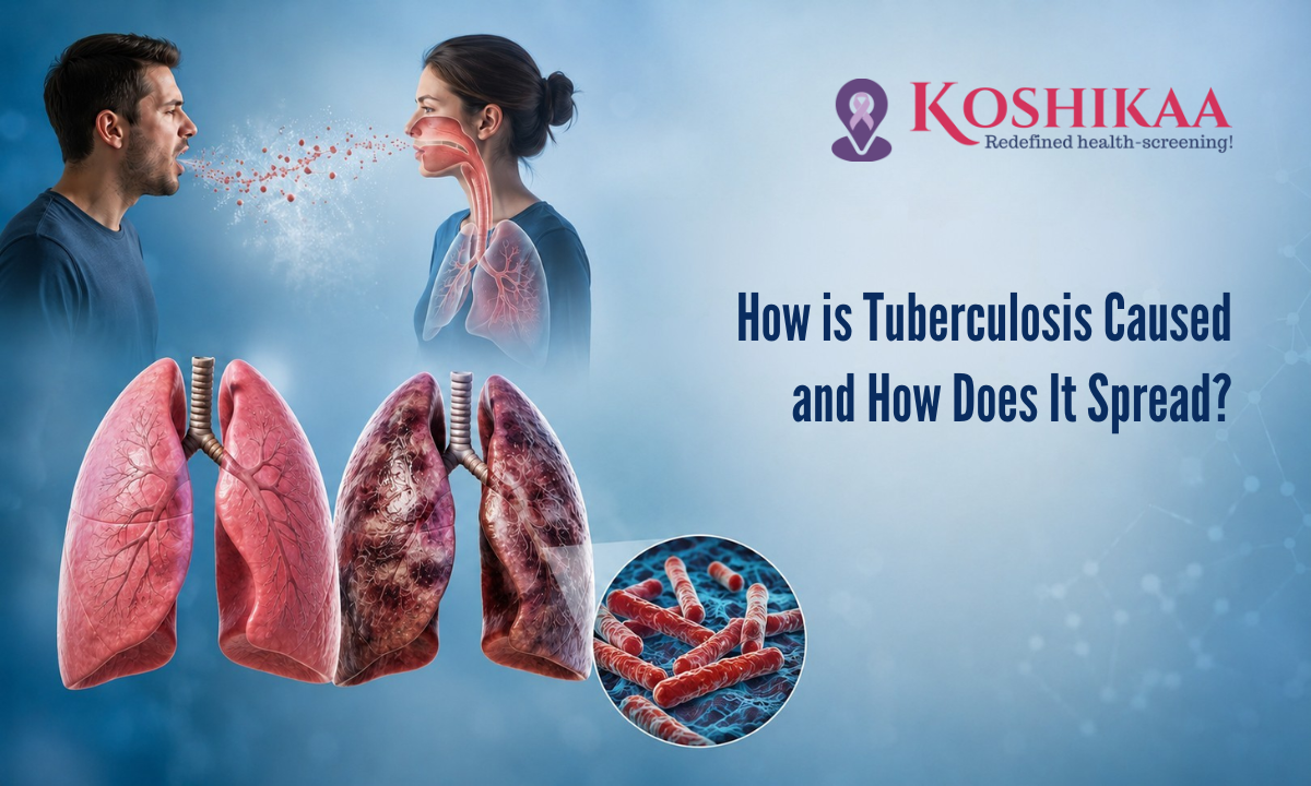
Maintaining respiratory wellness requires a clear understanding of microbial dynamics and proactive cellular evaluation. Educating yourself on how tuberculosis is caused and recognizing precisely how tuberculosis can be spread forms the bedrock of modern public health and personal prevention.
Visiting a specialized Health screening centre in Bangalore transforms clinical anxiety into actionable knowledge, providing individuals with advanced diagnostic insights before early physiological shifts alter their daily lives. By prioritising baseline cellular health, families can confidently navigate respiratory risks.
Historically, public perceptions surrounding lung conditions have been clouded by systemic misconceptions and social isolation.
Modern clinical science, however, views these challenges strictly through the lens of micro-immunology and environmental medicine. When we look at respiratory pathogens from a cellular perspective, we remove the element of fear and replace it with precise, preventative strategies.
Recognizing the early biological warning signs and understanding how external factors interact with internal immune barriers, communities can drastically lower infection baselines and ensure long-term vitality.
Let us take an objective, detailed look at the underlying mechanisms that govern this respiratory condition.
Medical Disclaimer
The biological definitions, cellular breakdowns, and diagnostic matrices provided in this article are intended strictly for educational and health awareness purposes. This informational guide does not serve as a substitute for professional medical advice, direct clinical consultations, official diagnostic interpretations, or personalized therapeutic strategies. Respiratory health conditions are highly complex and can share clinical features with a wide array of viral, fungal, or allergic conditions. Always consult a qualified pulmonologist, infectious disease specialist, or board-certified physician to evaluate your specific physical symptoms or diagnostic laboratory results. Never ignore, delay, or disregard professional medical guidance based on the educational concepts outlined in this document.
The Biological Catalyst for Deconstructing the Cellular Cause
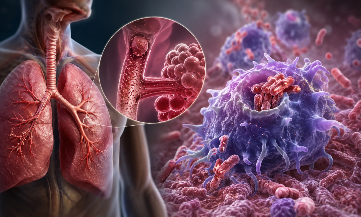
At its fundamental root, this respiratory condition is not a product of environmental exposure to cold weather or generalized fatigue; it is driven by a highly specialized microscopic organism known as Mycobacterium tuberculosis.
This bacterium is an obligate aerobe, meaning it requires high concentrations of oxygen to thrive and replicate, which naturally makes the human lungs its primary biological destination.
What makes this specific pathogen uniquely resilient compared to standard bacteria is its waxy outer cell wall, which is exceptionally rich in mycolic acid. This dense lipid barrier acts as a chemical shield, protecting the organism from drying out in the external environment and defending it against the body’s natural cellular destruction mechanisms.
When these microscopic, rod-shaped bacilli enter the upper respiratory tract, they must first navigate a series of physical defenses designed to trap foreign invaders. The nasal passages, sticky mucus linings, and microscopic hair-like structures called cilia work continuously to sweep out dust and microbes.
However, the particles carrying these bacteria are incredibly small; they can easily bypass these initial mechanical filters. They travel down the trachea and through the branching bronchial tubes, eventually settling deep within the alveoli, the microscopic air sacs responsible for oxygen exchange.
Once the bacteria establish a presence within the alveolar space, the body’s localized immune response is immediately triggered.
Specialized scavenger white blood cells, known as alveolar macrophages, migrate to the site to engulf the foreign cells through a process called phagocytosis. In a routine bacterial infection, the macrophage would successfully digest and eliminate the threat.
However, the unique waxy wall of this pathogen effectively blocks the macrophage’s destructive enzymes. Instead of being destroyed, the bacteria survive and begin to replicate slowly inside the very immune cells that were sent to neutralize them.
This deep cellular compromise marks the official biological onset of tuberculosis disease, shifting the biological landscape from a simple microbial exposure to a complex, long-term cellular standoff.
Aerosol Mechanics: The Reality of Air Transmission

Understanding the precise physics of how particles move through the air is vital to debunking historical stigmas and establishing proper preventative measures. The primary mechanism of tuberculosis transmission relies entirely on airborne pathways, which function quite differently from standard viral infections like the common cold or influenza.
When a person with an active infection forcefully exhales, such as during coughing, sneezing, speaking, or even singing, they release a spectrum of respiratory particles into the surrounding space. These particles are categorized by size, which fundamentally alters how long they remain in the environment and how they interact with nearby individuals.
The Physics of Droplet Nuclei vs. Heavy Droplets
Large Droplets:
These are heavier moisture particles measuring greater than 5 micrometers in diameter. Of their physical mass, gravity pulls them down quickly. They typically travel less than or equal to 1 meter before settling onto floors, tables, or clothing.
As this specific bacillus does not easily infect individuals through surface contact, these large droplets do not serve as the primary vector for spreading the condition.
Droplet Nuclei:
When smaller respiratory droplets are expelled, the liquid layer rapidly evaporates in the air, leaving behind microscopic, dry residues known as droplet nuclei. These tiny particles measure less than or equal to 5 micrometers in diameter.
As they are incredibly light, they easily overcome the gravitational pull, allowing them to remain suspended in the air currents of a room for hours. Anyone sharing that enclosed airspace can inhale these microscopic particles deep into their lungs.
Deconstructing the Contagion Pathway
| Environmental Exposure Type | Biological Interaction Reality | True Vector Status |
|---|---|---|
| Airborne Inhalation | Breathing in suspended droplet nuclei (less than or equal to 5 micrometers) that drift across distances greater than 1 meter. | Primary Vector: Highly effective at delivering bacilli directly to the deep alveolar spaces. |
| Large Droplet Exposure | Droplets greater than 5 micrometers landing on nearby surfaces or skin within a short 1-meter radius. | Negligible Risk: The bacteria cannot cause infection through simple skin contact. |
| Direct Contact | Shaking hands, hugging, or touching the skin of an individual. | Non-Transmission Vector: The pathogen lacks the cellular mechanisms to penetrate intact skin barriers. |
| Indirect Contact | Sharing eating utensils, drinking glasses, bedding, or touching surfaces like doorknobs. | Non-Transmission Vector: The organism cannot survive or replicate productively on inert surfaces to cause respiratory illness. |
The Latent Shield vs. Active Progression: The Cellular Standoff
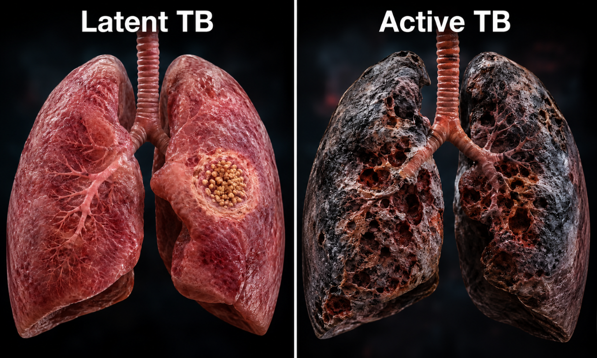
The interaction between the human immune system and the invading bacteria is one of the most complex standoffs in cellular biology. When the bacteria settle deep within the air sacs of the lungs, a healthy immune system recognizes that it cannot easily destroy the waxy outer wall of the microbes. Instead of fighting an endless battle, it deploys a highly effective containment strategy.
Specialized white blood cells travel to the site of infection and tightly pack themselves around the bacteria, building a dense, microscopic wall to wall off the threat. This protective cellular shell is known as a granuloma. By trapping the bacteria inside this biological fortress, the immune system deprives them of the oxygen and nutrients they need to multiply, effectively forcing them into a state of dormancy. This condition is clinically defined as Latent TB.
- Characteristics of the Latent State: During this phase, the bacteria remain alive but completely inactive. The individual feels entirely healthy, shows no clinical signs of illness, and is completely non-contagious. The infection is successfully contained by the body’s cellular shield.
- Characteristics of the Active State: If the body’s immune forces are compromised, the structural integrity of the granuloma begins to erode. The protective cellular wall ruptures, allowing the dormant bacteria to wake up, replicate rapidly, and cause tissue damage within the lungs.
Factors That Disrupt the Cellular Barrier
The transition from a dormant, latent infection to an active, progressive disease is rarely random. It is almost always driven by systemic health shifts, chronic metabolic stress, or lifestyle conditions that exhaust the body’s immune reserves.
When these protective barriers fail, the bacteria break free from their cellular prisons, causing the individual to transition into an active tuberculosis patient who experiences physical symptoms and can now spread the bacteria through the air.
The primary triggers that compromise this protective cellular wall include:
- Chronic Metabolic Stress: Conditions like poorly managed diabetes change the chemical balance of your blood and lower the functional efficiency of scavenger white blood cells, making it much harder for the body to maintain stable granulomas.
- Severe Nutritional Deficiencies: A lack of essential vitamins, proteins, and micronutrients deprives the immune system of the biological building blocks required to repair, maintain, and reinforce the cellular walls surrounding dormant bacteria.
- Immunosuppressive Medical Treatments: Long-term treatments involving high-dose corticosteroids, chemotherapy, or specific biological therapies for autoimmune disorders deliberately lower immune activity, which can inadvertently dissolve the protective walls around contained microbes.
- Natural Age-Related Changes: As the body ages, the immune system naturally undergoes a gradual decline in function. This makes older adults more vulnerable to the reactivation of bacteria that may have been safely trapped for decades.
Clinical Reality Check: Roughly 5 to 10 percent of individuals carrying a latent infection will develop active disease during their lifetime, with the highest risk occurring within the first two years after the initial exposure. This statistical reality highlights why modern medicine values proactive screening.
Catching and managing an infection while it is safely locked behind the latent shield prevents permanent structural changes to your lung tissue and keeps the wider community safe.
Early Detection Framework for Advanced Diagnostic Screening
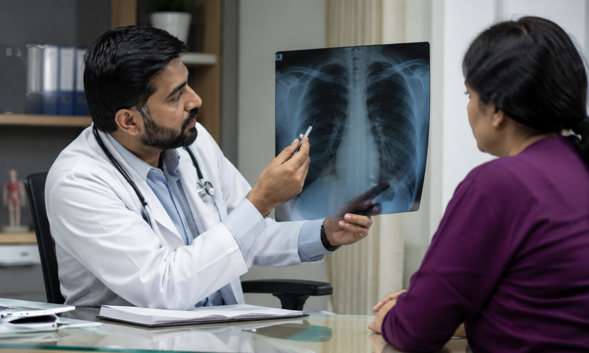
Transitioning from understanding microbial pathways to executing an effective health strategy requires clear, precise diagnostic tracking.
This condition can hide silently behind the body’s immune defenses for months or even years, and relying entirely on the appearance of physical symptoms like a persistent cough or sudden weight loss is a reactive approach that allows structural damage to occur before intervention.
Modern preventive medicine focuses on proactive screening workflows that map both the structural condition of the respiratory system and the specific cellular responses of the immune system.
When an individual chooses to map their respiratory health, clinicians deploy a combined diagnostic approach. This framework combines high-resolution imaging to check for physical tissue changes with sensitive laboratory assays that detect the presence of the pathogen at a microscopic or immunological level.
The Imaging Pillar for Mapping Structural Anomalies
The first line of defense in evaluating active respiratory variations is visual imaging. When the bacteria escape the immune system’s containment shields, they begin to damage the delicate architecture of the lung tissue, typically targeting the oxygen-rich upper lobes.
Securing a high-resolution chest X-ray in Bangalore provides clinical teams with an immediate, detailed view of the internal thoracic space. Radiologists look for specific structural red flags that indicate an active or historical infection:
- Infiltrates: Areas of patchy, dense cloudiness on the film that show where fluid, inflammatory cells, and white blood cells have gathered in the air sacs to fight the bacterial invasion.
- Cavitary Lesions: Distinct, hollowed-out spaces or gas-filled pockets within the lung tissue. These form when the center of a large mass of infected tissue dies and breaks down, leaving a permanent structural mark that requires careful clinical monitoring.
- Fibrosis and Scarring: Rigid strands of scar tissue are left behind as the lungs attempt to heal from localized inflammation, which can restrict breathing capacity if left unmanaged.
Pleural an abnormal buildup of fluid in the narrow space between the outer layers of the lungs and the chest wall, signaling deep tissue irritation.
Cellular and Molecular Assays
While imaging tools are excellent at identifying active structural changes, they cannot reliably detect a dormant, latent infection where the bacteria are safely locked inside microscopic cellular walls. To find these hidden targets, diagnostics must shift from structural imaging to cellular pathology.
Opting for a specialized, highly sensitive diagnostic Blood test in Bangalore allows technicians to measure the body’s immune memory. The most advanced tool in this category is the Interferon-Gamma Release Assay (IGRA).
Unlike older, variable skin tests that require multiple clinic visits and can yield false positives due to historical vaccinations, the IGRA blood test is highly precise. A laboratory technician mixes the patient’s blood sample with specific synthetic proteins that mirror the unique structure of the target bacteria. If the patient’s white blood cells have encountered the pathogen before, they recognize these proteins and instantly release a chemical defense signal called interferon-gamma.
By measuring the exact concentration of this chemical marker, the test determines if an infection is present, providing clear results even when a chest X-ray appears completely normal.
The Comprehensive Diagnostic Matrix
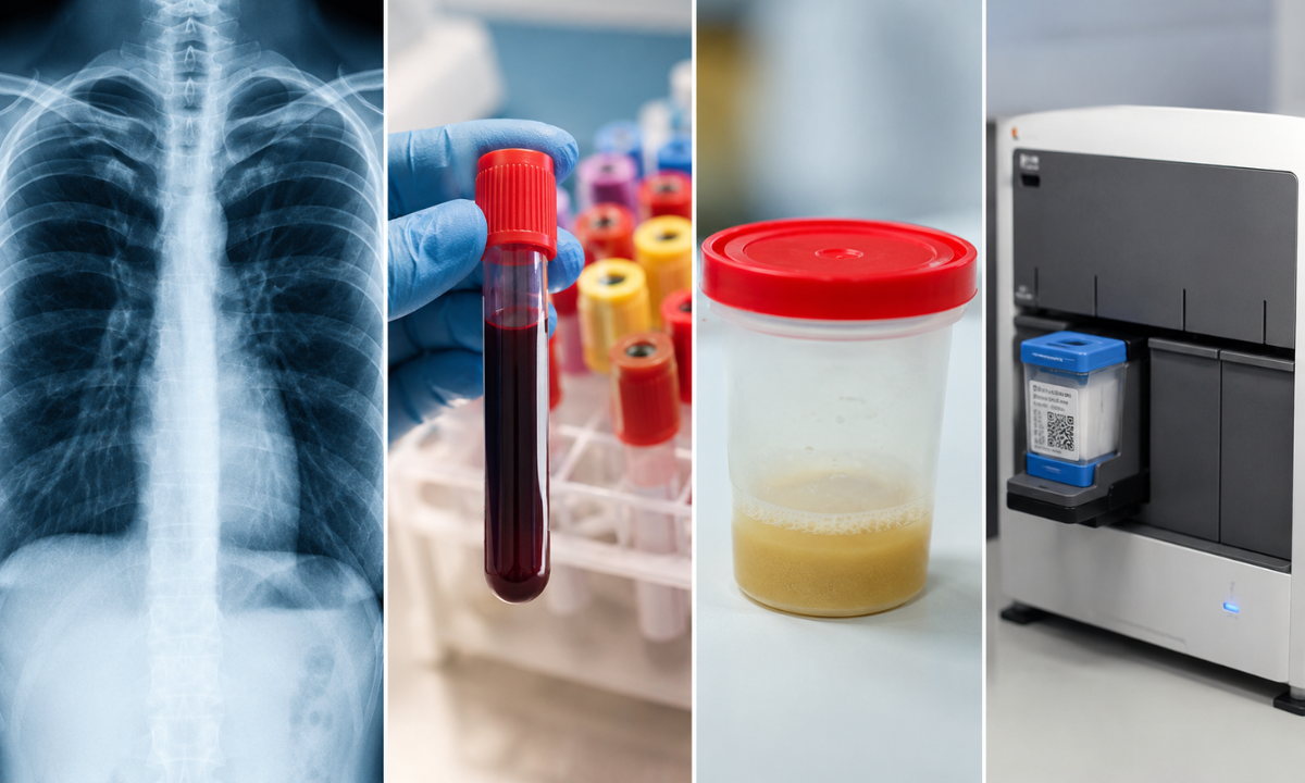
| Diagnostic Tool Category | Core Biological Objective | Clinical Tracking Capability | Primary Preventive Utility |
|---|---|---|---|
| Chest Radiography | Identifies macro-structural changes and tissue density variations within the lungs. | Detects active lung damage, cavitary lesions, and inflammatory fluid accumulation. | Essential for confirming or ruling out active, transmissible respiratory conditions. |
| Interferon-Gamma Assays (IGRA) | Measures the immune system’s cellular response to specific bacterial proteins. | Confirms the presence of the pathogen at a cellular level, whether active or dormant. | The gold standard for identifying hidden, latent infections before they cause tissue damage. |
| Sputum Acid-Fast Bacilli (AFB) Smear | Directly visualizes the physical bacteria under a high-powered microscope using special stains. | Confirms the presence of active, multiplying microbes in coughed-up mucus. | Used to quickly gauge how contagious an individual is during an active phase. |
| Molecular PCR Testing (GeneXpert) | Amplifies and detects the specific DNA sequences of the target pathogen. | Delivers rapid genetic confirmation of the bacteria and checks for drug resistance markers. | Provides high-speed diagnostic accuracy to streamline and personalize treatment planning. |
Why Choose Koshikaa for Preventive Cellular Screening?
Prioritizing your respiratory health requires a partnership with a diagnostic team that understands health at its most foundational level. At Koshikaa, our preventive screening philosophy is built around the deep study of cellular integrity. We believe that true medical empowerment means identifying internal physiological variations long before they manifest as active wellness challenges. Our advanced facility in Banashankari provides individuals and families with a precise, seamless environment to evaluate their health baselines with absolute confidence.
Our preventative care framework stands apart through several dedicated service standards:
- Tailored Age-Wise Screening Models: We recognize that your physiological risks and diagnostic needs evolve. Instead of offering generic, uniform checkups, we design highly personalized screening protocols based on your distinct life stage, lifestyle choices, and genetic family history.
- Advanced Multi-Modal Diagnostics: Our center integrates automated clinical laboratory processing with advanced imaging technology. This dual approach ensures that your cellular immune responses and physical tissue structures are analyzed simultaneously, providing a high-resolution map of your internal health.
- Proactive Focus on Cellular Health: Guided by our name derived from the fundamental building block of life, the cell, our clinical pathways are focused entirely on early identification. We catch subtle biological red flags early, allowing for simple, highly effective lifestyle adjustments and medical guidance.
We understand that visiting a diagnostic center can feel overwhelming. Our facility is deliberately designed to provide a calm, reassuring, and highly supportive atmosphere, transforming essential health checks into a stress-free component of your annual wellness routine.
Conclusion
Maintaining clear, healthy respiratory function relies on proactive tracking rather than waiting for physical discomfort to appear. Understanding the clear dividing line between a dormant, contained microbial presence and an active condition allows individuals to manage their lifestyle risks with complete clarity. By keeping a close watch on your immune system’s baselines and respecting the physics of environmental air transmission, you protect your home, your workplace, and your long-term vitality.
True prevention means acting when your body is at its strongest. Partnering with dedicated specialists to build a documented record of your cellular health ensures that minor internal shifts are identified and managed swiftly. Invest in your peace of mind by scheduling your comprehensive diagnostic screening today, securing a vibrant, uncompromised future for yourself and those who rely on you.