
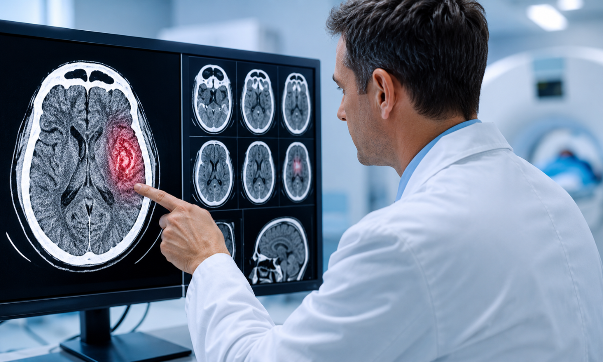
A blood clot in the brain is one of the most serious medical emergencies a person can face. When blood flow to any part of the brain gets blocked, brain cells begin to die within minutes, and the window for effective treatment is painfully short. This is why doctors around the world rely on a Blood Clot in Brain CT Scan as the very first imaging test in suspected stroke cases.
It is fast, painless, widely available, and gives an accurate picture of what is happening inside the skull within minutes of the patient arriving at the hospital. If your doctor has recommended one, you can book a CT Scan in Bangalore. In this complete guide, we explain what a brain clot actually is, the warning signs you should never ignore, how the scan works, how to prepare for it, and what your results really mean.
What Is a Blood Clot in the Brain?
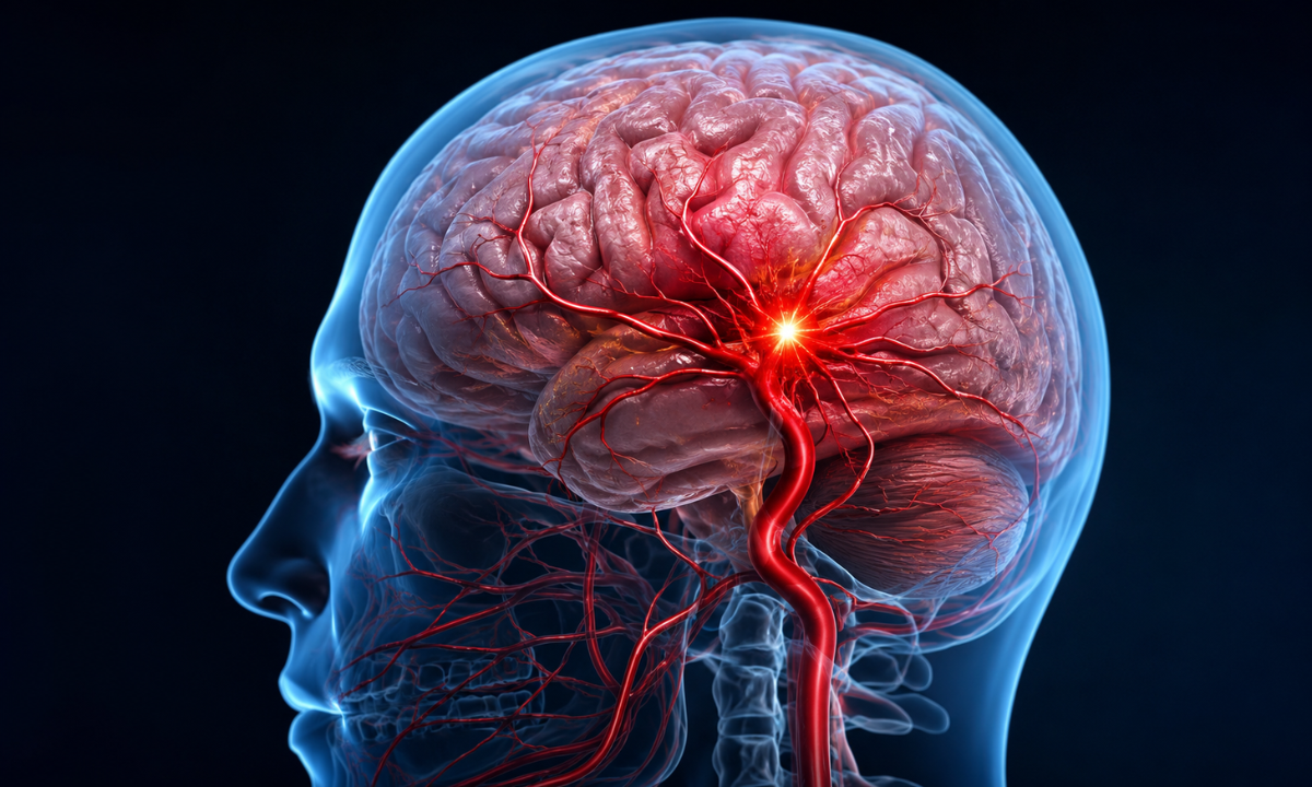
A blood clot in the brain occurs when a clump of blood blocks the normal flow through the arteries or veins that supply the brain. The brain is an incredibly demanding organ. It makes up only about two percent of your body weight, yet consumes nearly twenty percent of your oxygen supply. So when a clot cuts off circulation, the affected brain tissue starts dying almost immediately, leading to what doctors call an ischaemic stroke. This type accounts for the majority of all stroke cases seen in Indian hospitals.
Clots reach the brain in two ways. A thrombus forms directly inside a brain artery, usually because fatty deposits called plaque have narrowed the vessel over the years. An embolus, on the other hand, forms elsewhere in the body, most commonly in the heart, and travels through the bloodstream until it gets stuck in a narrower brain vessel. There is also a rarer condition called cerebral venous sinus thrombosis, where the clot forms in the veins that drain blood away from the brain. Whichever type it is, understanding what causes a blood clot in the brain helps you act faster, and a Blood Clot in Brain CT Scan is usually the first step doctors take to confirm the diagnosis.
Symptoms That May Indicate a Blood Clot in the Brain
The tricky part about brain clots is that symptoms appear suddenly and can be easy to dismiss in the first few minutes. Knowing these warning signs and acting on them immediately can genuinely be the difference between full recovery and lifelong disability. Rush to emergency care if you or anyone around you experiences:
- Sudden weakness or numbness on one side of the body, usually affecting the face, arm, or leg
- A drooping face, especially visible when the person tries to smile
- Slurred speech, jumbled words, or difficulty understanding what others are saying
- A sudden, severe headache that feels like the worst headache of one’s life
- Blurred vision, double vision, or sudden loss of sight in one or both eyes
- Dizziness, loss of balance, or unexplained difficulty in walking
- Confusion, memory lapses, or sudden behavioural changes
- Seizures, fainting, or complete loss of consciousness
Doctors and paramedics follow the FAST rule recommended by the American Stroke Association, which stands for Face drooping, Arm weakness, Speech difficulty, and Time to call for emergency help. The reason time features in the acronym is simple. Nearly two million brain cells die every minute during a stroke, so the sooner the patient reaches a hospital, the more brain tissue can be saved. Once the patient arrives, a Blood Clot in Brain CT Scan is typically performed within the first fifteen minutes because no stroke treatment can begin without it.
How Does a Blood Clot in Brain CT Scan Work?
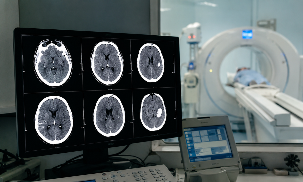
A CT scan, short for computed tomography, uses a rotating ring of X-ray beams that captures images of the head from hundreds of angles. A computer then stitches these together into detailed cross-sectional pictures, allowing radiologists to examine the brain in thin slices, layer by layer, almost like flipping through the pages of a book.
When reviewing the images, the radiologist looks for very specific clues. In the early hours of an ischaemic stroke, the affected brain tissue appears slightly darker than the surrounding areas because it is receiving less blood. Sometimes the blocked artery itself shows up brighter than normal, a finding called the dense artery sign, which points directly to where the clot is sitting. Fresh bleeding, if present, appears as a bright white patch. This last detail is critical because it tells doctors whether they are dealing with a clot or a bleed, and the treatment for each is completely different.
Depending on what the initial images show, doctors may order additional CT-based tests. Here is how the three types compare:
| Type of Scan | What It Shows | When It Is Used |
|---|---|---|
| Non-Contrast CT | Rules out bleeding and shows early tissue changes | The first test in every suspected stroke case |
| CT Angiography | Uses a contrast dye to map blood vessels and locate the exact clot | When doctors need to plan clot removal |
| CT Perfusion | Measures blood flow through different brain regions | To identify brain tissue that can still be saved |
If your doctor has advised any of these tests, you can book a CT Scan Brain in Bangalore, where all three types are available under one roof with expert radiologist reporting.
Why Is a CT Scan the First Choice for Detecting Brain Clots?
You might wonder why hospitals reach for a CT scan rather than an MRI, which is often considered more detailed. The answer comes down to three practical realities of emergency medicine.
Speed comes first. A Blood Clot in Brain CT Scan takes barely five to ten minutes from start to finish, and the images are available for review almost instantly. An MRI of the brain, by comparison, can take thirty to forty-five minutes, which is precious time a stroke patient simply does not have.
Availability comes second. CT scanners run round the clock in most major hospitals and diagnostic centres across India, whereas MRI machines are fewer in number and often have long queues. In an emergency, the nearest working scanner wins.
The third and most important reason is safethe ty of treatment. The main medication for ischaemic stroke is a powerful clot-dissolving drug, but if the patient is actually having a haemorrhagic stroke caused by bleeding, that same drug can make the bleeding catastrophically worse. A CT scan reliably distinguishes between the two within minutes, which is why no doctor will ever start stroke treatment without reviewing the scan images first.
How to Prepare and What to Expect During the Procedure
The good news is that this scan requires almost no preparation, which suits its emergency nature. If your scan is planned in re is what you should know.
You will be asked to remove metal objects such as earrings, hairpins, spectacles, and dentures, since metal interferes with the X-ray images. Wear comfortable clothing without metal zips or buttons near the neck. If contrast dye is planned, you may be asked to avoid food for a few hours before the scan, and it helps to carry any recent kidney function test reports along.
Inside the scan room, you lie on a motorised table that slides gently into the doughnut-shaped scanner. The machine does not touch you, and the scan itself is completely painless. The technician will ask you to stay perfectly still for a few minutes, since even small movements can blur the images. If contrast dye is used, it is injected through a vein in your arm, and a brief warm sensation or a metallic taste in the mouth is completely normal. Always inform the technician in advance about allergies, kidney problems, or pregnancy.
The whole visit, including preparation, rarely takes more than twenty minutes, and there is no recovery time. Choosing an experienced Health Screening Centre in Bangalore ensures the entire process is handled smoothly, from scanning to same-day reporting by qualified radiologists.
Understanding Your Blood Clot in Brain CT Scan Results
Once the imaging is complete, a radiologist studies the slices carefully and prepares a detailed report. A normal result means no visible clot, bleeding, or damaged tissue was found. That said, it is worth knowing that a very early ischaemic stroke does not always show up on the first scan, because visible tissue changes can take a few hours to develop. In such situations, doctors may repeat the Blood Clot in Brain CT Scan after some hours or recommend an MRI for confirmation, especially if symptoms strongly suggest a stroke.
An abnormal result can reveal several things. It may show the exact location and size of the clot, how much brain tissue has been affected, whether there is swelling pressing on surrounding structures, and even evidence of older, silent strokes the patient never knew about. Based on these findings, the medical team decides on treatment for a blood clot in the brain, which may include clot-dissolving injections given within the golden window, mechanical removal of the clot through a catheter, blood thinners to prevent new clots, or supportive care and rehabilitation.
Never hesitate to sit with your doctor and go through the report line by line. Understanding your own scan findings helps you take recovery and prevention far more seriously.
Who Is at Risk and When Should You Get Screened?
Brain clots rarely come out of nowhere. In most cases, the groundwork is laid over years by a combination of lifestyle and medical factors. You carry a higher risk if you have high blood pressure, diabetes, or high cholesterol, if you smoke, if you are overweight, or if you have an irregular heartbeat condition called atrial fibrillation. A family history of stroke, long periods of immobility such as extended bed rest or long flights, and certain clotting disorders add to the risk. Age matters too, with risk climbing sharply after 55, although doctors are now seeing strokes in people in their thirties and forties far more often, largely due to stress, poor sleep, and sedentary routines.
If several of these apply to you, an annual health screening test is one of the smartest health investments you can make. Regular monitoring of blood pressure, blood sugar, cholesterol, and heart rhythm can flag trouble long before it becomes an emergency. A comprehensive preventive package at a trusted Health Screening Centre in Bangalore can assess your overall stroke risk and give you a clear, personalised prevention plan. And if you have already experienced warning episodes like recurring headaches, unexplained dizziness, or brief numbness that passed on its own, do not brush them aside. These can be mini-strokes signalling a bigger event ahead, and booking a CT Scan in Bangalore promptly ensures you get clear answers before it is too late.
Why Choose Koshikaa for Your Brain CT Scan?
When it comes to something as critical as brain imaging, where you get scanned matters just as much as when. Koshikaa is a trusted health screening centre in Bangalore equipped with advanced CT technology that delivers high-resolution images with minimal radiation exposure. Every scan is reviewed by experienced radiologists who specialise in reading brain imaging, so subtle early signs of a clot are far less likely to be missed.
Reports are delivered promptly, often on the same day, because in matters of brain health, waiting days for a result is simply not an option. The pricing is transparent with no hidden charges, the staff guide you through every step of the process, and the centre maintains strict quality standards across all its imaging services. Whether it is an urgent scan prescribed by your doctor or a preventive health check you have been putting off, Koshikaa combines accuracy, speed, and care under one roof.
Conclusion
A Blood Clot in Brain CT Scan is often the deciding factor between timely treatment and irreversible damage. It is quick, painless, affordable, and available round the clock, and it gives doctors the one piece of information no stroke treatment can begin without. Knowing the FAST warning signs, understanding your personal risk factors, and acting within minutes rather than hours can quite literally save a life or protect someone from lifelong disability.
At Koshikaa, advanced CT imaging is paired with experienced radiologist reporting, so you receive accurate answers exactly when they matter most. If stroke risk runs in your family or your lifestyle ticks too many risk boxes, do not wait for an emergency to think about your brain health. Speak to your doctor about preventive screening today.
FAQ’s
Can A CT Scan Detect All Blood Clots In The Brain?
It detects most clots, especially with CT angiography, where contrast dye makes the blocked vessel clearly visible. Very small or very early clots may occasionally need a repeat scan or an MRI for confirmation.
How Long Does The Scan Take?
The scan itself takes five to ten minutes. With registration and preparation, the entire visit is done in about twenty to thirty minutes, and there is no recovery time afterwards.
Is The Contrast Dye Safe?
Yes, it is safe for most people. You may feel a brief warm flush or a metallic taste that fades in seconds. Inform your doctor in advance about kidney issues, allergies, or pregnancy so precautions can be taken.
What Is The Cost Of A Brain CT Scan In Bangalore?
It depends on the type of scan. A plain CT is the most affordable, while CT angiography costs more due to contrast usage. At Koshikaa, you get transparent pricing along with expert radiologist reporting.
Can A Blood Clot In The Brain Dissolve On Its Own?
Very small clots sometimes dissolve naturally, as seen in mini-strokes where symptoms vanish quickly. But a mini-stroke is often a warning before a major one, so always get imaging done immediately instead of waiting.