
Evaluating metabolic stability strictly requires examining the biological response to nutritional intake. While many individuals prioritize fasting metrics, securing a comprehensive Health Screening Test in Bangalore is the only method to accurately assess total glycemic control.
A standard Blood Test in Bengaluru provides a critical baseline, but measuring postprandial glucose is the absolute priority for identifying early-stage insulin resistance.
This specific metric quantifies exactly how the human body processes glucose within the circulatory system following a meal. Understanding these post-meal fluctuations is essential for preventing chronic metabolic dysfunction and maintaining long-term physiological health.
In this clinical guide, we provide the evidence-based data necessary to manage your metabolic profile effectively. This article will thoroughly detail the following critical clinical areas:
- The precise biological definition and the physiological significance of monitoring blood sugar levels after a meal.
- The exact standardized protocols utilized by medical professionals to conduct diagnostic testing.
- The specific clinical ranges utilized to differentiate between optimal health and metabolic impairment.
Strictly adhering to these clinical parameters, patients can secure a complete understanding of their internal metabolic environment. Recognizing the importance of post-meal data allows for highly targeted medical and nutritional interventions that protect the primary vascular system from severe oxidative stress.
Defining the Metric of What is Postprandial Glucose?

To provide a precise biological definition, what is postprandial glucose strictly refers to the concentration of sugar present in the circulatory system following the ingestion of a meal. This physiological state represents the peak of the metabolic digestive cycle.
During the ingestion of macronutrients, the digestive system physically and chemically breaks down complex carbohydrates into simple glucose molecules. These molecules are then absorbed through the intestinal mucosa and enter the bloodstream, causing a transient rise in systemic glucose levels.
The human body utilizes a highly regulated endocrine response to manage this influx of energy. In a healthy physiological state, the pancreas detects the rising glucose concentration and immediately secretes insulin.
This hormone acts as a critical biological signaling agent, facilitating the transport of glucose from the blood into the primary muscular and hepatic cells for immediate energy utilization or localized storage. The efficiency of this transition determines the stability of the post-meal metabolic profile.
Medical professionals categorize the stages of this metabolic response based on the precise chronological timeline of digestion and insulin activity.
Physiological Stages of the Postprandial State
| Metabolic Phase | Biological Mechanism | Impact on Blood Chemistry |
|---|---|---|
| Ingestion and Digestion | Salivary and gastric enzymes physically break down carbohydrates into simple monosaccharides. | An initial slow rise in blood glucose concentration as absorption initiates within the upper digestive tract. |
| Absorption Peak | Glucose molecules rapidly traverse the intestinal barrier directly into the portal circulation. | Blood sugar levels reach their maximum concentration, typically occurring sixty to ninety minutes after ingestion. |
| Endocrine Response | The pancreatic beta cells release insulin to manage the systemic glycemic load. | Insulin facilitates the rapid cellular uptake of glucose, initiating a controlled decline in circulatory sugar levels. |
| Homeostatic Return | Excess glucose is converted into glycogen for storage within the liver and muscle tissues. | Blood glucose concentrations return to a baseline state, typically achieved within two to three hours in healthy individuals. |
Identifying the exact peak and decline of these circulatory sugars allows clinicians to assess the functional capacity of the pancreas and the sensitivity of the peripheral tissues to insulin.
When this biological regulation fails, glucose remains in the bloodstream at pathologically high levels for an extended duration. This failure is a primary indicator of metabolic dysfunction. By strictly monitoring these transitions, healthcare providers can identify microscopic errors in carbohydrate metabolism before they manifest as chronic systemic diseases.
Why is postprandial glucose important?
Evaluating metabolic health strictly through fasting blood sugar provides an incomplete diagnostic picture. Clinical endocrinologists emphasize that the post-meal period represents a highly dynamic biological state where microscopic vascular damage frequently initiates.
When patients ask why postprandial glucose is important, medical professionals point directly to the severe physiological consequences of rapid, uncontrolled hyperglycemic spikes. These post-meal fluctuations exert immense oxidative stress on the internal biological structures and actually predict cardiovascular disease mortality more accurately than standard fasting metrics.
While a healthy biological system clears circulatory glucose rapidly, impaired metabolic function leaves toxic levels of sugar directly in the bloodstream. This specific biological failure initiates multiple cascading pathological events.
Pathological Consequences of Postprandial Hyperglycemia
| Biological Pathway | Physiological Mechanism | Systemic Clinical Impact |
|---|---|---|
| Endothelial Dysfunction | Acute post-meal glucose spikes directly impair the inner cellular lining of the vascular system, forcing the biological production of destructive superoxide radicals instead of protective nitric oxide. | Initiates severe microscopic damage directly within the arterial walls, forming the primary biological foundation for chronic cardiovascular disease. |
| Excessive Glycation | High concentrations of circulatory sugar force glucose molecules to permanently bind to structural cellular proteins and hemoglobin. | Creates advanced glycation end products that physically stiffen and damage delicate blood vessel walls throughout the entire circulatory infrastructure. |
| Systemic Inflammation | Repeated daily elevations in blood sugar directly trigger a chronic biological proinflammatory state and the severe overexpression of cellular adhesion molecules. | Accelerates the physical development of atherosclerosis and severely increases the absolute biological risk of experiencing a fatal or nonfatal cardiovascular event. |
| Pancreatic Exhaustion | The continuous physiological demand to secrete massive volumes of insulin to neutralize high post-meal carbohydrate loads physically degrades beta cell function. | Directly drives the biological progression from asymptomatic insulin resistance into full clinical type 2 diabetes. |
Monitoring these exact post-meal responses, specialized healthcare providers can identify these severe cellular disruptions early. Preventing this hidden physiological damage represents the absolute foundation of advanced metabolic disease prevention and long-term biological preservation.
Diagnostic Evaluation of the Postprandial Glucose Test
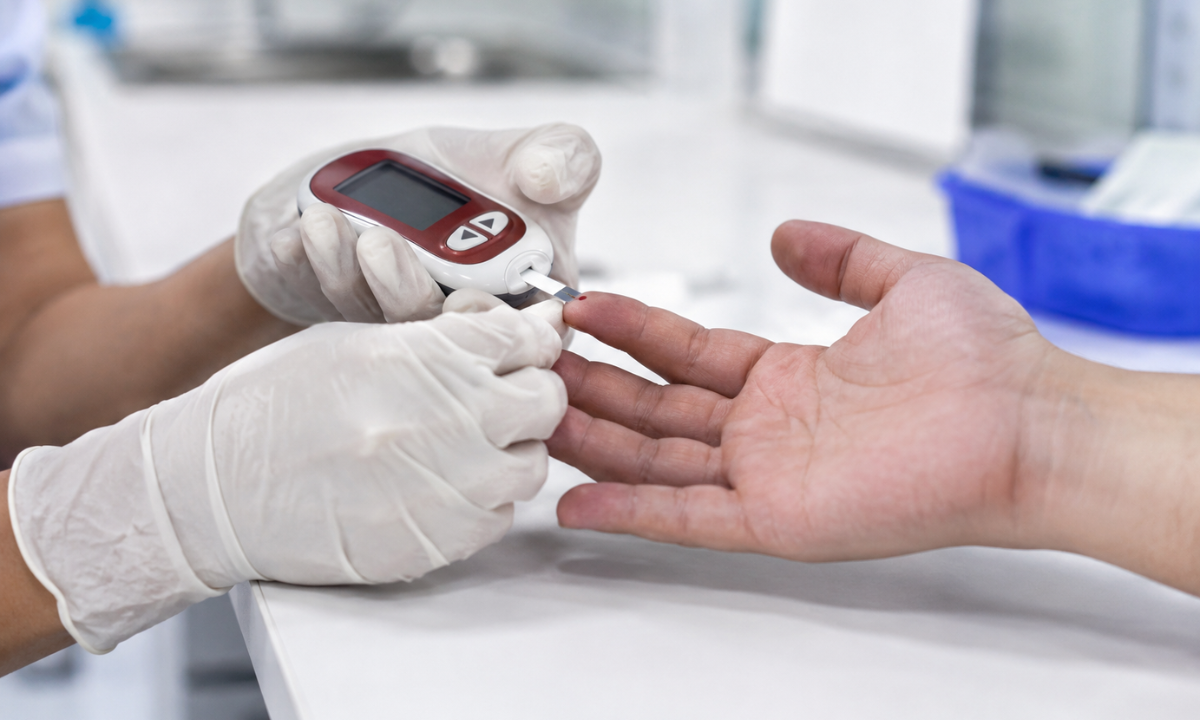
The clinical assessment of post-meal metabolic function strictly requires rigorous adherence to standardized diagnostic protocols. To obtain highly accurate circulatory data, medical professionals administer a specific postprandial glucose test. This precise diagnostic procedure is meticulously timed to capture the exact physiological moment when blood sugar levels should biologically return to a homeostatic baseline.
Executing this evaluation successfully demands complete patient compliance regarding dietary intake and exact chronological timing. Clinical pathologists divide the testing procedure into four strictly regulated phases.
Chronological Clinical Protocol for Postprandial Evaluation
| Procedural Phase | Clinical Action | Diagnostic Purpose |
|---|---|---|
| 1. Baseline Fasting | The patient strictly abstains from all caloric intake for a continuous duration of eight to twelve hours overnight. | Establishes the absolute baseline metabolic state before any dietary carbohydrates are introduced into the biological system. |
| 2. Carbohydrate Administration | The patient consumes a highly specific and measured carbohydrate load within a strict ten-minute window. | Initiates the physiological digestive sequence and triggers the mandatory pancreatic insulin response. |
| 3. Chronological Incubation | The patient remains completely at rest for exactly two hours following the initial carbohydrate consumption. | Allows the biological system the required timeline to process the glucose load and transport the sugar into the cellular infrastructure. |
| 4. Venous Extraction | A specialized phlebotomist extracts a standard venous blood sample precisely at the two-hour marker. | Captures the exact concentration of remaining circulatory glucose to determine the functional efficiency of the metabolic clearing process. |
Strict adherence to this precise chronological timeline completely guarantees the diagnostic validity of the extracted circulatory data.
The absolute precision of this two-hour window is critical. Extracting the blood sample prior to this exact marker provides prematurely elevated data, while delaying the extraction completely misses the critical diagnostic threshold.
Furthermore, the specific type of carbohydrate consumed during the second phase significantly influences the diagnostic results. Medical professionals utilize two primary methods to administer this glucose load based on the specific clinical requirements of the patient.
Clinical Carbohydrate Administration Protocols
- The Standardized Oral Glucose Tolerance Load: The patient consumes exactly 75 grams of anhydrous liquid glucose dissolved entirely in water. This highly concentrated protocol provides an absolute, standardized metabolic stress test, eliminating the variables of protein and fat digestion.
- The Standard Physiological Meal: The patient consumes a typical, balanced nutritional meal containing at least 75 grams of mixed carbohydrates. This protocol evaluates the biological metabolic response under standard daily dietary conditions.
Selecting the appropriate carbohydrate load directly depends on the specific diagnostic objectives established by the attending medical provider. By strictly controlling both the nutritional input and the exact chronological extraction, clinical laboratories provide the highly precise data required to evaluate pancreatic function and biological insulin sensitivity.
Interpreting the Data and Postprandial Glucose Normal Range

After successfully extracting the two-hour metabolic sample, clinical pathologists compare the exact concentration of circulatory sugar against universally established medical thresholds.
The American Diabetes Association provides rigorous diagnostic criteria to accurately define the postprandial glucose normal range and identify exact stages of metabolic failure. Clinical interpretation strictly relies on examining these exact numerical parameters to determine the functional efficiency of the biological insulin response.
Clinical Thresholds for Two-Hour Postprandial Evaluation
| Diagnostic Classification | Circulatory Concentration | Metabolic Interpretation |
|---|---|---|
| Normal Physiological Function | Less than 140 mg/dL | The pancreas secretes adequate insulin, and the cellular structures exhibit optimal biological sensitivity. |
| Prediabetes | 140 mg/dL to 199 mg/dL | The biological system struggles to clear systemic glucose, indicating a highly dangerous progression toward chronic disease. |
| Clinical Diabetes Mellitus | 200 mg/dL or greater | The physiological insulin response has failed, or severe cellular resistance prevents necessary glucose uptake. |
Receiving a diagnostic result that exceeds the optimal parameters immediately necessitates advanced medical intervention. Clinical endocrinologists mandate specific physiological corrections based entirely on the exact diagnostic category presented in the laboratory results.
- Patients diagnosed within the impaired glucose tolerance range strictly require immediate nutritional restructuring. Furthermore, these individuals must implement rigorous mechanical physical activity to reverse the cellular insulin resistance.
- Individuals recording values at or above the 200 mg/dL threshold require rapid clinical evaluation. These patients frequently require pharmacological intervention and continuous metabolic monitoring to prevent acute hyperglycemic crises.
- For patients actively undergoing clinical treatment for established diabetes, the primary medical objective is to strictly maintain post-meal levels below 180 mg/dL.
- Medical professionals strictly advise against ignoring elevated post-meal readings. Prolonged exposure to these toxic glucose concentrations permanently damages the vascular and neurological infrastructure over the long term.
Strictly identifying where your specific metabolic data falls within these established clinical thresholds, you secure the absolute baseline knowledge required to protect your internal organ systems and permanently stabilize your biological health.
Why Choose Koshikaa for Your Health Screening
Executing precise metabolic evaluations strictly requires advanced laboratory infrastructure and highly accurate diagnostic processing.
When individuals select Koshikaa for a comprehensive health screening, they secure immediate access to premier diagnostic facilities conveniently located throughout Bangalore. Our specialized pathology departments operate strictly under rigorous international medical standards to ensure absolute data accuracy for every extracted biological sample.
Our diagnostic facilities provide several definitive clinical advantages for managing your long-term metabolic health:
- Advanced Diagnostic Technology: We strictly utilize highly sophisticated automated chemical analyzers to measure circulatory glucose concentrations with absolute precision. This advanced medical technology eliminates the biological risk of human processing errors and guarantees diagnostic validity.
- Comprehensive Health Packages: Our specialized clinical packages evaluate your complete metabolic profile. We seamlessly combine your exact post-meal glucose data with critical lipid padnels, hepatic indicators, and renal markers to provide your medical provider with a total systemic anatomical assessment.
- Rapid Result Delivery: We guarantee the secure digital delivery of your finalized diagnostic data within twenty-four to forty-eight hours. This highly accelerated processing timeline provides your attending medical provider with the immediate clinical information strictly required to initiate targeted pharmacological or nutritional interventions.
Choosing Koshikaa, patients guarantee that their precise metabolic evaluation receives strictly specialized management utilizing the most advanced diagnostic technology available.
Conclusion
Relying exclusively on fasting diagnostic metrics provides an incomplete and potentially dangerous assessment of your total metabolic health. Understanding exactly how your biological system processes carbohydrates following a meal represents the absolute foundation for preventing severe vascular damage and clinical diabetes. Delaying this critical medical evaluation strictly allows microscopic systemic inflammation and cellular insulin resistance to progress completely unchecked.
Do not wait for advanced physical symptoms of metabolic failure to manifest. Contact the specialized diagnostic coordination team at Koshikaa today to schedule your precise postprandial evaluation, secure your highly accurate metabolic baseline data, and permanently protect your foundational physiological stability.