
Patients experiencing acute abdominal pain or abnormal urinary symptoms frequently require immediate diagnostic imaging. As a premier Health screening centre in Bangalore, Koshikaa provides advanced radiological assessments to accurately diagnose complex conditions of the urinary tract.
When a physician suspects an underlying pathology within the kidneys or bladder, the first line of diagnostic investigation is often a comprehensive Ultrasound scan in Bangalore.
Specifically, doctors rely on a targeted renal ultrasound to safely and efficiently visualize the internal structures of the retroperitoneal organs without exposing the patient to ionizing radiation.
This specific diagnostic modality utilizes high-frequency sound waves to generate real-time, two-dimensional images of the kidneys, ureters, and urinary bladder.
As the kidneys play a critical role in filtering metabolic waste and regulating systemic fluid balance, identifying structural abnormalities promptly is essential for preventing long-term organ damage.
A physician will prescribe this non-invasive imaging study to evaluate the anatomical size, precise location, and overall physical shape of the kidneys, as well as to assess the surrounding blood vessels for proper circulation.
In this comprehensive clinical guide, we will cover the specific biological structures evaluated during this exam, the primary clinical indications for detecting kidney stones, the role of sonography in monitoring chronic kidney disease, the exact step-by-step procedure patients will experience, the general cost expectations for this diagnostic service, and a conclusion highlighting the advanced imaging capabilities available at Koshikaa.
What Does a Renal Ultrasound Evaluate?

A renal ultrasound provides real-time, high-resolution imaging of the primary organs responsible for the human excretory system.
The procedure specifically targets the retroperitoneal space located in the posterior abdominal cavity. By directing high-frequency sound waves through the surrounding tissues, the transducer captures the acoustic echoes reflecting off the internal organs.
This process generates a detailed sonogram that allows radiologists to objectively evaluate the morphological characteristics of the entire renal system.
During this targeted diagnostic examination, the sonographer systematically evaluates three primary anatomical components to construct a complete clinical picture of the patient’s urinary health.
Anatomical Structures and Sonographic Parameters
| Anatomical Structure | Sonographic Evaluation Parameters | Clinical Diagnostic Significance |
|---|---|---|
| Renal Parenchyma (Cortex and Medulla) | Measurement of total organ length, echogenicity (tissue density), and cortical thickness. | Assesses for signs of chronic tissue damage, congenital hypoplasia, or the presence of abnormal solid and cystic growths. |
| Renal Pelvis and Calyces | Observation of the central collecting system for structural dilation or abnormal fluid accumulation. | Identifies hydronephrosis, which indicates a backup of urine caused by downstream physiological blockages. |
| Ureters | Assessment of tubular diameter and overall visibility within the pelvic cavity. | Healthy ureters are rarely visible on a standard sonogram. Their visual presence strongly indicates severe dilation or active obstruction. |
| Urinary Bladder | Measurement of the muscular wall thickness, overall structural capacity, and post-void residual fluid volume. | Evaluates lower urinary tract function, identifies bladder wall thickening, and detects potential bladder outlet obstructions. |
After systematically examining these specific anatomical regions, the interpreting radiologist establishes a comprehensive baseline of the patient’s physical renal health.
This structural assessment provides the referring physician with the exact morphological data required to definitively diagnose acute abnormalities or carefully monitor the progression of chronic conditions affecting the urinary tract.
Kidney Stones and Acute Obstructions
Physicians frequently order a diagnostic sonogram when a patient presents to the emergency department or clinical setting with acute, severe symptoms localized to the urinary tract.
The most common indication for an immediate imaging study is the suspected presence of nephrolithiasis, clinically known as kidney stones. These hard, crystalline mineral deposits form within the renal pelvis and can migrate into the narrow ureters, causing a partial or complete mechanical obstruction of the normal urinary flow.
When evaluating an acute patient, a renal ultrasound for kidney stones is a highly effective diagnostic tool because it relies on the physical density of the calcification.
The high-frequency sound waves emitted by the transducer reflect powerfully off the hard surface of the stone, creating a brightly echogenic focus on the digital monitor.
Furthermore, because the sound waves cannot penetrate the dense mineral mass, the stone casts a distinct, dark acoustic shadow directly behind it. Beyond visualizing the calculus itself, the radiologist utilizes the sonogram to detect secondary complications such as hydronephrosis.
This pathological condition occurs when the mechanical blockage forces urine to back up into the kidney, causing the internal renal pelvis and calyces to physically dilate and swell.
Acute Clinical Symptoms Triggering a Renal Sonogram
| Clinical Symptom | Physiological Presentation | Diagnostic Objective of Ultrasound |
|---|---|---|
| Renal Colic | Acute, severe pain originating in the flank region and radiating downward to the lower abdomen or groin. | To identify the precise location of the obstructing calculus and assess the structural severity of the resulting hydronephrosis. |
| Hematuria | The visible or laboratory confirmed presence of red blood cells circulating within the patient’s urine. | To rule out structural lacerations, obstructive stones causing mucosal trauma, or the presence of solid renal masses. |
| Suspected Pyelonephritis | Sudden onset of high fever, systemic chills, and localized flank tenderness indicate a severe bacterial infection. | To detect the presence of renal abscesses, localized inflammation, or infected fluid collections within the retroperitoneal space. |
| Oliguria or Anuria | A sudden, clinically significant decrease or a complete cessation of daily urine production and output. | To rapidly identify any bilateral physical obstructions preventing the kidneys from successfully draining metabolic waste into the bladder. |
After utilizing sonography to immediately investigate these acute clinical presentations, emergency physicians and urologists can rapidly implement targeted medical interventions to relieve the obstruction, manage the severe pain, and prevent irreversible ischemic damage to the renal tissue.
Diagnosing and Monitoring Chronic Renal Conditions
While acute obstructions demand immediate imaging, physicians also rely heavily on sonography for the long-term management of chronic renal pathologies.
When evaluating a patient for chronic impairment, a renal ultrasound kidney disease protocol provides critical morphological data regarding the progression of organ degradation over time.
Unlike acute conditions that typically cause the kidneys to swell, chronic diseases frequently induce progressive scarring, fibrosis, and a gradual reduction in the overall size of the functional tissue.
Nephrologists utilize regular sonographic examinations to monitor these subtle anatomical changes. By comparing the current ultrasound images against established physiological baselines and the patient’s previous historical scans, the medical team can accurately stage the clinical severity of the disease and adjust therapeutic interventions accordingly.
Chronic Pathologies and Sonographic Findings
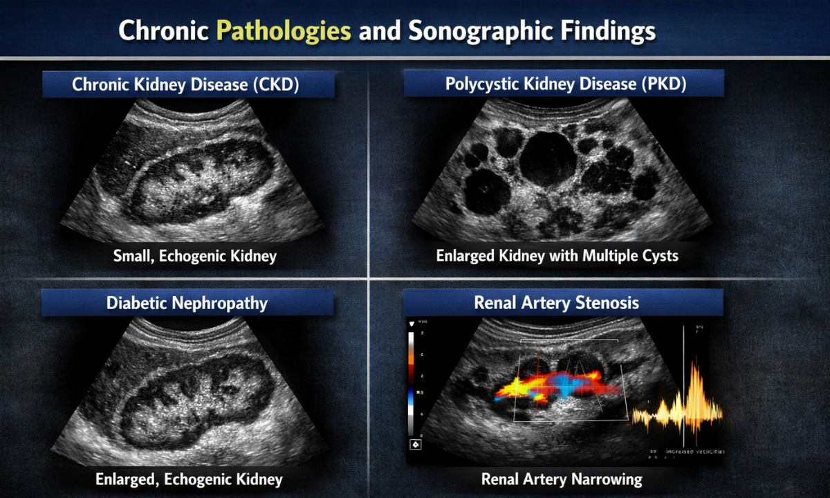
| Chronic Renal Condition | Underlying Pathophysiology | Primary Sonographic Indicators |
|---|---|---|
| Chronic Kidney Disease (CKD) | Progressive loss of renal function due to long-term damage from hypertension or diabetes. | Decreased total organ length, a severely thinned renal cortex, and increased echogenicity, where the tissue appears abnormally bright due to extensive fibrosis. |
| Polycystic Kidney Disease (PKD) | A genetic disorder characterized by the growth of numerous fluid-filled cysts within the renal tissue. | Significantly enlarged kidneys containing multiple anechoic (dark) cysts that progressively destroy the surrounding healthy parenchyma over time. |
| Diabetic Nephropathy | Microvascular damage to the filtering capillaries of the kidneys is caused by prolonged periods of elevated blood glucose. | Initial enlargement of the kidneys during the early stages, followed by a gradual reduction in size and increased cortical density as the vascular damage advances. |
| Renal Artery Stenosis | The physical narrowing of one or both of the primary arteries that supply oxygenated blood to the kidneys. | An abnormal structural discrepancy in total size between the left and right kidneys, prompting the use of a specialized Doppler ultrasound to measure the restricted blood velocity. |
As we continuously track these specific morphological markers, the interpreting radiologist provides the nephrologist with the exact quantitative data required to manage chronic renal failure, potentially delay the clinical necessity for dialysis, and optimize the patient’s long-term physiological outcomes.
How Is the Sonogram Performed?
When patients inquire about how a renal ultrasound is done, radiologists outline a highly standardized, non-invasive clinical protocol.
This specific diagnostic examination requires no ionizing radiation, completely avoids the use of intravenous contrast dyes, and typically concludes within thirty to forty five minutes. The sonographer follows a precise methodology to ensure the highest possible resolution of the retroperitoneal organs.
Step-by-Step Sonographic Protocol
1. Clinical Preparation:
The diagnostic facility typically instructs the patient to consume a specific volume of water approximately one hour before the scheduled examination. The patient must avoid emptying their bladder before the scan begins.
A fully distended urinary bladder acts as a critical acoustic window, allowing the high-frequency sound waves to travel seamlessly through the pelvic cavity to clearly visualize the bladder walls and the distal ureters.
2. Patient Positioning:
The technologist escorts the patient into the darkened examination room and asks them to lie on the clinical table.
To obtain optimal acoustic angles and bypass the dense anatomical shadows cast by the rib cage and intestinal gas, the sonographer will require the patient to assume multiple physical positions. These positional changes frequently include lying supine on the back, prone on the stomach, or in a lateral decubitus position on either side.
3. Application of Acoustic Gel:
The sonographer applies a specialized, water-based conductive gel directly to the patient’s skin over the abdominal and flank areas.
This gel serves a vital physical purpose. It eliminates any microscopic air pockets between the epidermis and the handheld transducer, which guarantees the continuous, uninterrupted transmission of sound waves deep into the internal tissues.
4. Image Acquisition:
The technologist presses the transducer firmly against the gel-coated skin and sweeps it systematically across the target anatomical regions. Because the kidneys naturally move downwards in the abdominal cavity during diaphragmatic inspiration, the sonographer will frequently instruct the patient to take a deep breath and hold it briefly.
This temporary suspension of respiration physically stabilizes the organs, allowing the machine to capture crystal clear, real-time digital images on the adjacent clinical monitor.
Once the radiologist successfully records all necessary anatomical measurements, the technologist wipes the conductive gel from the patient’s skin.
The examination is entirely painless, and the patient experiences no residual physiological effects. Upon completion, the patient may immediately empty their bladder and safely resume all normal daily activities, dietary habits, and prescribed medications without restriction.
Why Choose Koshikaa? Accessibility and Diagnostic Pricing
Patients frequently inquire about the financial aspect of radiological imaging, specifically asking how much a kidney ultrasound cost when prescribed by their referring physician.
Compared to advanced imaging modalities like Magnetic Resonance Imaging or Computed Tomography scans, diagnostic sonography is highly cost-effective and widely accessible. The total cost of a standard renal ultrasound in Bangalore typically ranges between eight hundred and one thousand five hundred Indian Rupees.
This exact price fluctuates based on the specific clinical facility, the potential inclusion of Doppler blood flow analysis, and the overall complexity of the required anatomical measurements.
At Koshikaa, we maintain strict financial transparency for all diagnostic procedures. A targeted kidney ultrasound at our facility is priced highly competitively at eight hundred and fifty rupees. By eliminating hidden fees and providing clear, upfront pricing, we ensure that patients can access critical imaging services without experiencing financial anxiety.
This highly accessible pricing model allows nephrologists and urologists to readily order necessary serial scans to safely monitor chronic renal conditions over an extended period without placing an undue financial burden on the patient.
Conclusion
Early detection of structural renal abnormalities is absolutely critical for preserving long-term kidney function and preventing severe urinary tract complications.
Whether your physician requires immediate imaging to locate an obstructing kidney stone or routine anatomical monitoring for chronic kidney disease, a targeted renal ultrasound provides the exact morphological data needed for highly accurate clinical decision making.
As the procedure is entirely non-invasive and relies exclusively on safe acoustic waves rather than ionizing radiation, it remains the definitive gold standard for initial retroperitoneal evaluations.
As a premier diagnostic facility, Koshikaa is fully equipped to deliver the highest standard of radiological care. Our highly experienced technologists utilize state-of-the-art ultrasound equipment to capture crystal clear, high-resolution images, ensuring that our interpreting radiologists provide your referring physician with a flawless, comprehensive diagnostic report.
Do not delay necessary medical imaging when experiencing acute abdominal pain or abnormal urinary symptoms.
Contact us or visit our dedicated ultrasound centre in Bangalore to schedule your comprehensive renal evaluation and protect your long-term urinary health.