Early Warning Signs That Indicate You Need a Thyroid Test

Table of Contents

We often ignore subtle changes in our bodies, assuming they are due to stress or a busy lifestyle. However, understanding your thyroid test and results at the right time can make a huge difference in identifying hidden health concerns early, especially when access to a reliable blood test in Bangalore is easily available today. 

The thyroid may be small, but it plays a big role in controlling metabolism, energy levels, and hormonal balance. Many of us don’t realise that simple symptoms like fatigue, weight changes, or mood swings could be early signs of a thyroid imbalance. 

So, how do we know when it’s time to take action? Let’s explore the signs your body may already be showing you. By the end of this blog, you might recognise signals you never thought were connected to your thyroid.

Key Takeaways

  • The thyroid controls metabolism, energy, and hormones
  • Early symptoms are often mild but should not be ignored
  • Weight changes, fatigue, and mood swings are common warning signs
  • A thyroid test is simple and usually does not require fasting
  • Timely testing helps prevent long-term complications
  • Imaging, like thyroid ultrasound, may be needed in some cases
  • Regular screening at a trusted health centre ensures peace of mind

What Does the Thyroid Do?

Thyroid Functions Explained

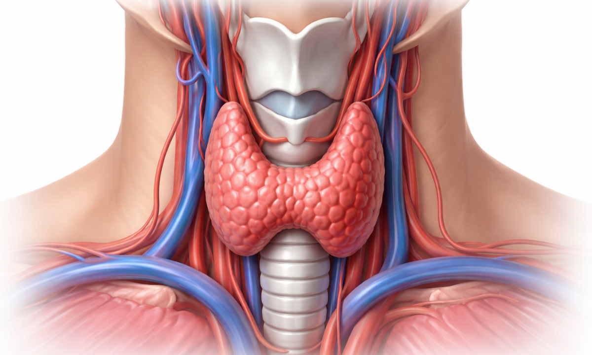
The thyroid is a small, butterfly-shaped gland in the neck that produces hormones controlling how your body uses energy. When it doesn’t function properly, it can lead to hypothyroidism (underactive thyroid) or hyperthyroidism (overactive thyroid).

These conditions may develop slowly, which is why early detection becomes so important.

Early Warning Signs You Should Not Ignore

Sometimes, our body gives us small hints long before a condition becomes serious. We just need to pause and listen carefully to these signals instead of brushing them aside.

1. Constant Fatigue and Low Energy

Low Energy

Do you feel tired even after a full night’s sleep? This could be one of the earliest signs of a thyroid imbalance.

When your thyroid hormones are not balanced, your body’s ability to produce and use energy gets affected. This can leave you feeling drained even after minimal activity.

  • Hypothyroidism often causes sluggishness
  • Hyperthyroidism may lead to restlessness, but poor sleep

If your energy levels don’t match your routine, it’s worth checking.

2. Unexplained Weight Changes

Weight Changes

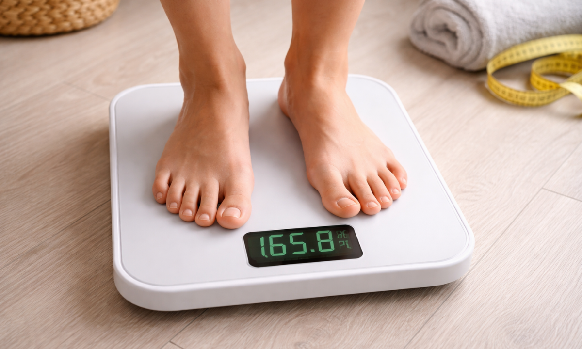
Sudden weight gain or loss without changes in diet or exercise can be linked to thyroid issues.

The thyroid directly controls how fast your metabolism works, which means even small imbalances can affect how your body burns calories. Over time, this can lead to noticeable changes that feel difficult to manage.

ConditionCommon Weight Change
HypothyroidismWeight gain
HyperthyroidismWeight loss

If your weight feels “out of control”, your thyroid might be involved.

3. Hair Thinning and Skin Changes

Hair fall

We often blame hair fall on stress or weather, but thyroid imbalance can also be a cause.

Thyroid hormones play a key role in maintaining healthy skin, hair, and nails by supporting cell renewal. When these hormone levels are off, these visible changes can slowly start to appear.

  • Dry, rough skin
  • Hair thinning or excessive hair fall
  • Brittle nails

These changes happen because thyroid hormones affect cell growth.

4. Mood Swings and Mental Fog

Mood swings

Your thyroid also impacts brain function. If you feel emotionally different, it might not just be stress.

When thyroid hormone levels are imbalanced, they can affect neurotransmitters in the brain, which influence how we think and feel. Over time, this can make even simple daily tasks feel overwhelming or harder to manage.

  • Depression or low mood
  • Anxiety or irritability
  • Difficulty concentrating

These symptoms are often overlooked but are very important.

5. Irregular Periods or Fertility Issues

Irregular Periods

For women, thyroid health plays a major role in reproductive health.

The thyroid helps regulate hormones that control the menstrual cycle and ovulation. When this balance is disturbed, it can lead to noticeable changes in periods and even affect fertility over time.

SymptomPossible Cause
Irregular cyclesHormonal imbalance
Heavy or light periodsThyroid dysfunction
Difficulty conceivingThyroid hormone disruption

If you notice these signs, timely testing can help.

6. Sensitivity to Temperature

Sensitivity

Do you feel unusually cold or hot compared to others?

The thyroid plays a key role in regulating your body’s internal temperature by controlling how energy is produced and used. When hormone levels are imbalanced, your body may struggle to maintain a normal temperature, making you feel uncomfortable in everyday environments.

  • Feeling cold often – Hypothyroidism
  • Feeling too warm – Hyperthyroidism

Your body’s temperature regulation is directly linked to thyroid hormones.

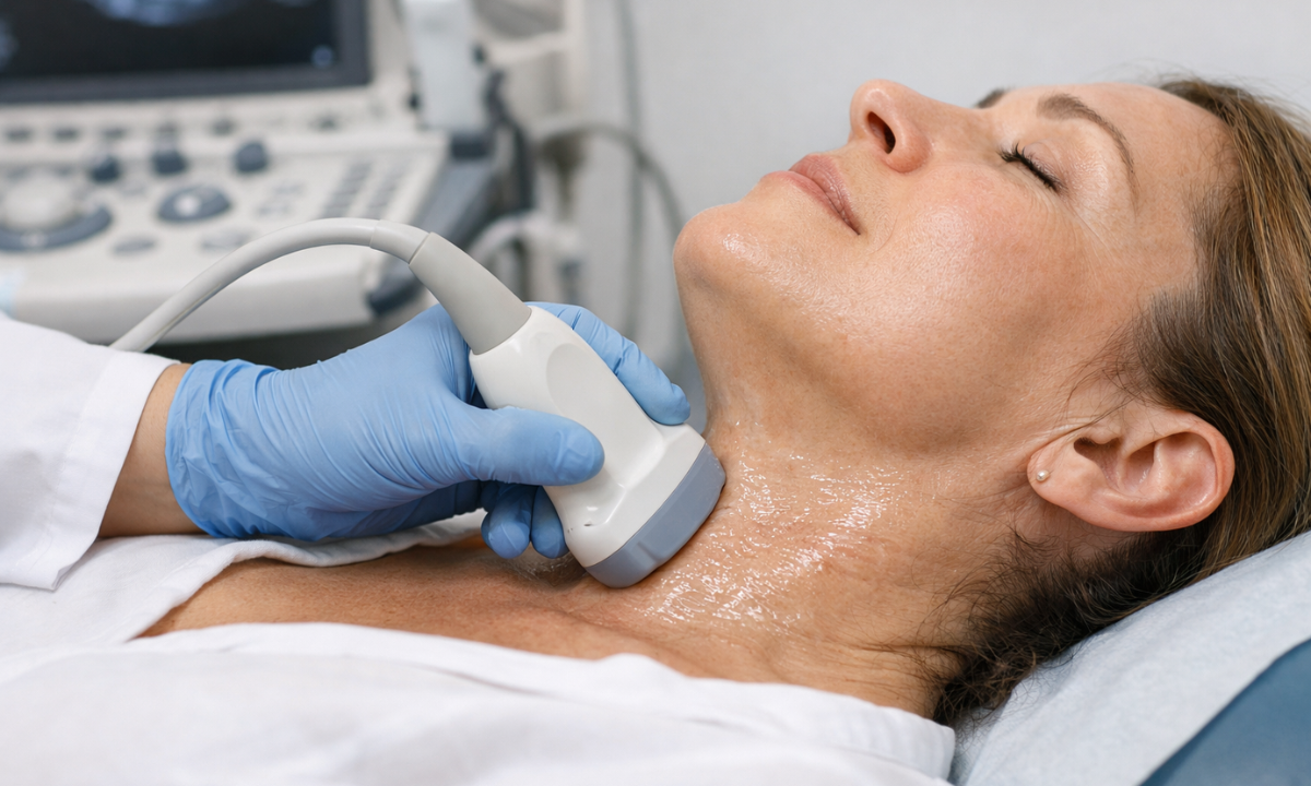
7. Swelling in the Neck

Swelling in the Neck

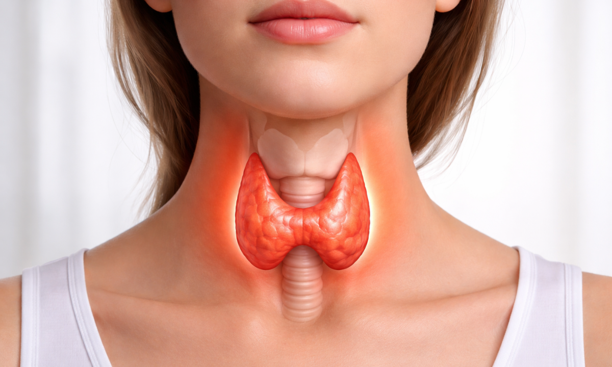
A visible swelling or lump in the neck area may indicate thyroid enlargement (goitre).
This swelling may develop slowly and is often painless, which is why many people ignore it in the early stages. However, it can sometimes affect swallowing or cause a feeling of tightness in the throat if it grows larger.

In such cases, doctors may recommend a thyroid ultrasound to understand the structure of the gland better.

When Should Thyroid Test Be Done?

When to Get a Thyroid Test

Many people ask, when should a thyroid test be done? The answer depends on your symptoms and health history.

In many cases, thyroid issues develop slowly and may not show strong symptoms at first. That’s why knowing the right time to get tested can help detect problems early and prevent them from becoming more serious.

Here are some common situations:

  • If you notice any of the symptoms mentioned above
  • During pregnancy or while planning pregnancy
  • If you have a family history of thyroid disorders
  • As part of a routine health check-up

Early testing helps avoid complications and ensures timely treatment.

How Many Hours of Fasting Is Required for a Thyroid Test?

A very common question is: how many hours of fasting are required for a thyroid test?

Many people worry about test preparation and whether they need to skip meals before getting tested. The good news is that thyroid testing is usually simple and does not require complicated preparation in most cases.

The good news is:

  • Most thyroid blood tests do not require fasting
  • You can usually eat and drink normally
  • However, always follow your doctor’s advice

This makes it easy to schedule your test without much preparation.

Types of Tests for Thyroid Evaluation

Thyroid diagnosis usually involves simple blood tests and sometimes imaging.

1. Blood Tests

Blood test for thyroid scan
Test NamePurpose
TSH (Thyroid Stimulating Hormone)Primary screening test
T3 and T4Measure thyroid hormone levels

These are commonly available at our trusted health screening centre in Bangalore.

2. Imaging Tests

USG scan for thyroid

If required, doctors may suggest imaging tests like:

  • Thyroid ultrasound to detect nodules or swelling
  • Advanced scans if abnormalities are suspected

A good ultrasound scan in Bangalore can help provide detailed insights.

Why Early Detection Matters

Ignoring thyroid symptoms can lead to long-term health issues if left untreated.

Because the thyroid affects multiple systems in the body, even a small imbalance can gradually impact your overall health and quality of life. Identifying the problem early makes it much easier to manage and prevents complications later on.

  • Heart problems
  • Infertility
  • Severe fatigue
  • Weight management issues

Early diagnosis allows simple treatment, often through medication and lifestyle changes.

Simple Tips to Maintain Thyroid Health

We can take small steps to support thyroid health in our daily lives:

  • Eat a balanced diet with iodine and selenium
  • Manage stress through relaxation techniques
  • Exercise regularly
  • Avoid self-medication
  • Go for regular health check-ups

Consistency is key when it comes to hormonal balance.

Final Thoughts

Listening to our body is one of the most important things we can do for our health. If you notice any unusual symptoms, don’t delay getting checked, especially when access to a reliable blood test in Bangalore at a trusted health screening centre in Bangalore makes the process simple and convenient.

At Koshikaa, the focus is on making preventive healthcare easy, accessible, and stress-free for everyone. A small step, like a thyroid test today, can help you avoid bigger health concerns tomorrow.

FAQs

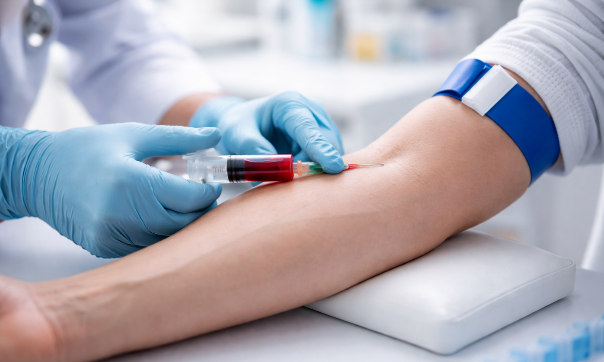
1. Is thyroid testing painful?

No, thyroid testing is a simple blood test and is usually quick and painless. You may feel a slight prick when the needle is inserted, but it lasts only a few seconds. Most people find the process comfortable, and it is completed within minutes without any major discomfort or recovery time.

2. Can thyroid problems be cured?

Many thyroid conditions may not always be completely cured, but they can be effectively managed with the right treatment. With proper medication, lifestyle changes, and regular monitoring, most people are able to lead a normal and healthy life without major complications or disruptions to their daily routine.

3. How often should I get tested?

The frequency of thyroid testing depends on your symptoms, medical history, and whether you already have a diagnosed condition. If everything is normal, testing once a year may be enough. However, if you have thyroid issues, your doctor may suggest more frequent monitoring to keep hormone levels balanced.

4. Is an ultrasound always required?

No, an ultrasound is not always required for thyroid evaluation. A Thyroid ultrasound is usually recommended only when there are signs of swelling, nodules, or structural abnormalities in the neck. In most cases, a blood test is sufficient to assess thyroid function and identify any hormonal imbalance.

MRI of the uterus is primarily used for diagnosing and evaluating conditions such as uterine fibroids, adenomyosis, and malignancies. It is particularly useful in the planning of surgical procedures and assessing the extent of gynecological diseases. This imaging test is valuable for its ability to deliver detailed cross-sectional images of the uterus and surrounding structures.
An MRI of the spleen can help diagnose various disorders affecting the spleen including splenomegaly (enlarged spleen), tumors, and other abnormalities. It provides clear, detailed images that are crucial for assessing the health of the spleen and planning potential surgical interventions.
Prostate MRI is a non-invasive imaging technique that provides detailed pictures of the prostate gland and surrounding tissues. It helps in the detection, staging, and monitoring of prostate cancer. Prostate MRIs are increasingly used to guide biopsy procedures and assist in the planning of treatment strategies for prostate health.
An MRI of the pancreas is used to identify issues such as pancreatitis, pancreatic cancer, and other pancreatic disorders. It offers high-resolution images that help in detecting small lesions and assessing the pancreatic ducts. This imaging technique is valuable for guiding biopsies and planning surgeries involving the pancreas.
MRI scans of the liver, kidney, and bile ducts provide detailed images that help diagnose various conditions, such as tumors, cysts, and blockages in the bile ducts. They are crucial for assessing organ damage, staging cancer, and planning liver or kidney-related surgeries. This type of MRI can also evaluate the health and function of these organs comprehensively.
Breast MRI is highly sensitive for detecting breast cancer, especially in individuals with dense breast tissue or a high risk of cancer. It is often used in conjunction with mammography to provide a more comprehensive evaluation. Breast MRIs can also monitor the response to breast cancer treatments and provide detailed pre-surgical mapping.
Brain and spine MRIs are critical tools for diagnosing and monitoring neurological conditions, including tumors, spinal cord injuries, and multiple sclerosis. These MRIs can reveal abnormalities in brain tissue and spinal cord structure that might not be visible with other imaging methods. They are essential for planning surgeries and assessing the effectiveness of treatments in neurology.
PET-CT scans of transplant organs like the liver, kidney, or pancreas are performed to assess metabolic activity, detect complications, and monitor post-transplant function. These scans play a vital role in the early detection of issues such as organ rejection, recurrence of disease, or infection, guiding timely interventions and optimizing transplant outcomes for patients.
PET-CT examinations of organs such as the liver, kidney, or prostate are crucial for assessing metabolic activity, detecting abnormalities, and monitoring disease progression. These scans help diagnose conditions such as liver metastases, renal cell carcinoma, or prostate cancer, guiding treatment decisions and monitoring response to therapy over time.
PET-CT scans of soft-tissue masses are utilized to assess metabolic activity and detect malignant tumors or other abnormalities. By combining PET imaging with CT imaging, these scans provide detailed information about tumor location, size, and metabolic characteristics, aiding in accurate diagnosis and treatment planning for soft-tissue cancers.
PET-CT scans are valuable for detecting cysts or fluid collections within the body, including cysts in the liver, kidneys, or other organs. These scans help characterize the nature and metabolic activity of cysts, guiding diagnosis and treatment planning for conditions such as cystic liver disease, renal cysts, or pancreatic cysts.
PET-CT scans of muscles are performed to assess metabolic activity and detect abnormalities such as inflammation, infection, or tumors. These scans provide valuable information about muscle function and pathology, aiding in the diagnosis and management of conditions like myositis, muscular dystrophy, or soft-tissue sarcomas.
PET-CT scans of the brain are utilized to evaluate brain metabolism and detect abnormalities such as tumors, epilepsy, or neurodegenerative diseases. By combining functional PET imaging with anatomical CT imaging, these scans offer comprehensive information about brain function and structure, guiding neurosurgical interventions and treatment decisions for neurological disorders
PET-CT scans of bones are commonly used to detect bone metastases in cancer patients, assess bone health in conditions like osteoporosis, and evaluate response to treatment. By combining PET and CT imaging, these scans provide detailed information about metabolic activity and anatomical changes in bone structures, aiding in accurate diagnosis and treatment planning for various bone-related conditions.
CT scans of the spine are conducted to assess the vertebrae, spinal cord, and surrounding soft tissues for conditions like herniated discs, spinal fractures, or spinal stenosis, aiding in the diagnosis and management of spinal disorders with detailed imaging essential for surgical planning and intervention.
CT scans of the pelvis are utilized to visualize the pelvic bones, reproductive organs, bladder, and surrounding structures for conditions such as fractures, tumors, or pelvic inflammatory disease, guiding treatment plans and surgical interventions with precise anatomical information.
CT scans of the neck are performed to evaluate structures such as the throat, thyroid gland, lymph nodes, and blood vessels for abnormalities like tumors, infections, or injuries, assisting in diagnosis and management of head and neck disorders with detailed imaging.
CT scans of the extremities are employed to assess bones, joints, and soft tissues in the arms or legs, aiding in the diagnosis of fractures, arthritis, or other musculoskeletal conditions, guiding orthopedic treatment and rehabilitation plans for optimal recovery.
CT scans of the chest are utilized to examine the lungs, heart, blood vessels, and surrounding structures for conditions such as lung nodules, pneumonia, or pulmonary embolism, facilitating accurate diagnosis and management of respiratory and cardiovascular diseases.
CT scans of the brain are conducted to assess brain structures, identify abnormalities like tumors, hemorrhages, or strokes, and aid in diagnosing neurological conditions, guiding neurosurgical interventions and treatment strategies with precise imaging of the brain’s anatomy.
CT scans of the abdomen are performed to visualize and evaluate the internal organs such as the liver, kidneys, pancreas, and intestines for conditions like tumors, infections, or injuries, providing detailed images essential for diagnosis and treatment planning.
Transplant organ ultrasound scans, including those of the liver, kidney, or pancreas, are performed to assess organ viability, detect complications, and monitor post-transplant function. These scans play a vital role in the early detection of issues such as organ rejection, vascular complications, or fluid collections, guiding timely interventions and optimizing transplant outcomes for patients.
Ultrasound examinations of organs such as the liver, kidney, or prostate are crucial for assessing organ structure, detecting abnormalities, and monitoring disease progression. These scans help diagnose conditions such as liver cirrhosis, kidney stones, or prostate enlargement, guiding treatment decisions and monitoring response to therapy over time.
Soft-tissue masses can be evaluated using ultrasound to determine their size, location, and characteristics. Ultrasound imaging helps differentiate between benign and malignant masses, guiding further diagnostic evaluation and treatment planning. By providing real-time imaging, ultrasound assists in the precise localization of soft-tissue masses for biopsy or surgical removal if needed.
Ultrasound scans are valuable for detecting cysts or fluid collections within the body, including cysts in the liver, kidneys, or other organs. These scans help characterize the nature and size of cysts, aiding in diagnosis and treatment planning for conditions such as cystic liver disease or renal cysts, and guiding interventions such as cyst drainage or sclerotherapy.
Musculoskeletal ultrasound scans are used to assess the health and integrity of muscles, identifying issues such as strains, tears, or muscle inflammation. These scans provide detailed images of muscle structures, allowing healthcare providers to diagnose muscle injuries accurately and develop personalized rehabilitation plans to promote recovery and restore function.
Ultrasound examinations of joints are essential for evaluating joint health, detecting abnormalities such as inflammation, fluid accumulation, or cartilage damage. By visualizing the joint structures in real-time, ultrasound helps diagnose conditions like arthritis, bursitis, or ligament injuries, guiding treatment decisions and monitoring disease progression over time.
Ultrasound scans of tendons are performed to assess for injuries, inflammation, or tears, aiding in the diagnosis and management of conditions such as tendonitis or tendon tears. These scans provide detailed images of tendon structures, helping healthcare providers determine the extent of damage and plan appropriate treatment strategies, including physical therapy or surgical intervention if necessary.
Pelvis X-rays play a crucial role in diagnosing injuries, diseases, and developmental conditions affecting the pelvic region. They provide detailed images of the hip bones, sacrum, and coccyx, aiding in the assessment of fractures, dislocations, or signs of arthritis. Pelvic X-rays are instrumental in guiding surgical interventions, monitoring healing progress, and evaluating conditions such as osteoporosis or tumors. By providing precise anatomical information, these imaging studies enable healthcare professionals to deliver personalized care and improve patient outcomes.
Teeth X-rays, known as dental radiographs, are indispensable tools for dentists to assess oral health. They aid in diagnosing dental decay, gum disease, and abnormalities in tooth structure or alignment. These images provide valuable insights into hidden dental issues, allowing for early intervention and prevention of complications. Additionally, dental X-rays are essential for planning treatments such as orthodontics, root canals, or dental implants, ensuring optimal outcomes and patient satisfaction.
Spine X-rays are pivotal in diagnosing spinal fractures, degenerative conditions, and spinal misalignments, providing critical information for treatment planning and monitoring spinal health. They offer detailed images of the vertebrae and surrounding structures, enabling healthcare professionals to identify issues such as scoliosis, disc herniation, or spinal stenosis
Skull X-rays are valuable for detecting fractures, assessing head trauma, and evaluating the alignment of facial bones, crucial in diagnosing conditions like fractures, tumors, or developmental abnormalities in the skull. They provide detailed views of the cranial structure, aiding clinicians in determining appropriate treatment strategies and ensuring patient safety.
For the heart, X-ray solutions can aid in the detection of various cardiac problems, ranging from mild to severe, including – Heart failure, Cardiac congestion and other heart-related conditions. In addition to cardiac assessments, X-ray solutions are also used for breast examinations to identify tumors or signs of breast cancer.

Bone X-rays are essential for diagnosing fractures and assessing bone alignment, aiding doctors in developing treatment plans and monitoring healing progress. They provide detailed images of the skeletal system, helping identify abnormalities or injuries that may not be visible through other imaging techniques

Abdominal X-rays are performed when patients experience symptoms such as significant nausea, ongoing vomiting, abdominal pain, flank or back pain, or abdominal swelling. These X-rays can help diagnose various conditions.

Frequently Booked Tests

Frequently Booked Checkups