What Is a Blood Clot in the Brain – Causes, Symptoms and Treatments

Table of Contents

Staying aware of serious health conditions can help us act quickly when it matters most. Understanding what a blood clot in the brain is extremely important because it is a life-threatening condition that can lead to stroke or permanent brain damage if not treated on time. 

When diagnosed early through tools like a CT scan in Bangalore, doctors can act quickly and improve outcomes. But how do we recognise the warning signs early enough to actually save a life?

Disclaimer:

This blog is intended for informational and educational purposes only and should not be considered a substitute for professional medical advice, diagnosis, or treatment. A blood clot in the brain is a serious medical emergency that requires immediate attention. If you or someone around you experiences symptoms such as sudden weakness, speech difficulty, or severe headache, please seek urgent medical care or contact emergency services without delay. Always consult a qualified healthcare professional for personalized medical guidance.

Key Takeaways

  • A blood clot in the brain blocks oxygen supply and can cause a stroke
  • Symptoms usually appear suddenly and need immediate attention
  • Early diagnosis through imaging, such as CT or MRI, is critical
  • Lifestyle and medical conditions increase the risk
  • Treatment is most effective during the “golden window”
  • Choosing the right health screening centre in Bangalore improves care quality

What is a Blood Clot in the brain?

Understanding Blood Clots in the Brain

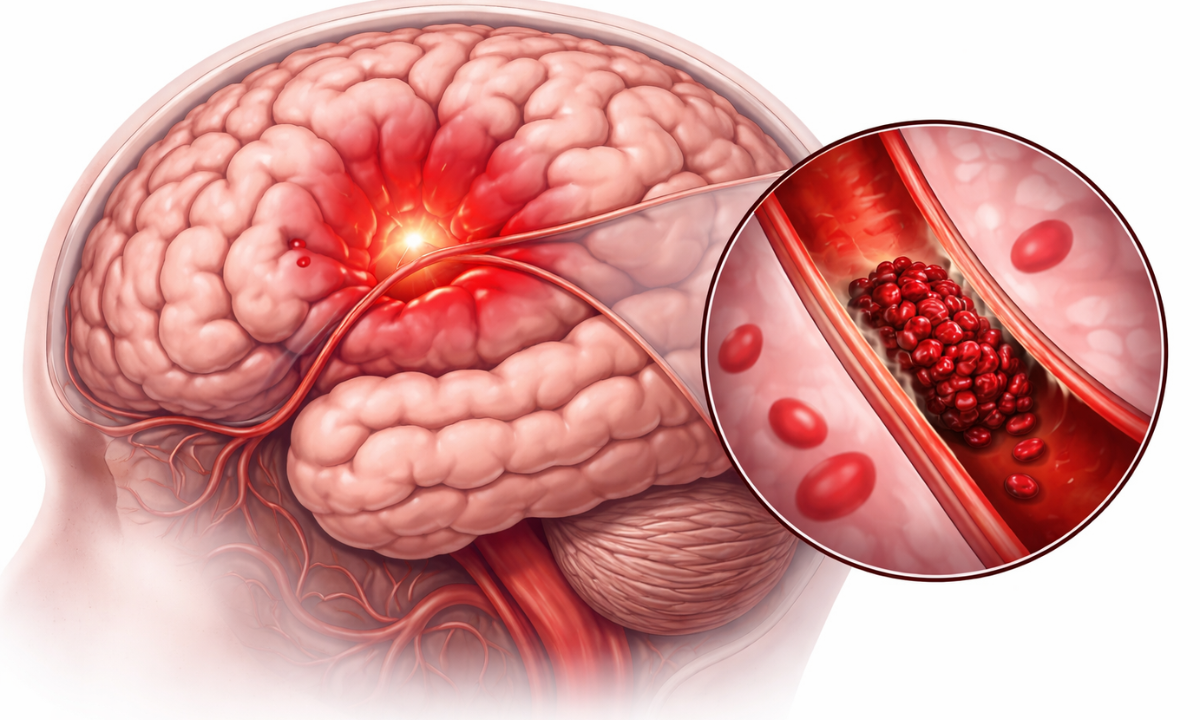
A blood clot in the brain occurs when blood solidifies inside a blood vessel, blocking the normal flow of oxygen-rich blood to the brain. This condition is also known as cerebral thrombosis or embolism.

Without oxygen, brain cells begin to die within minutes, which is why this condition is considered a medical emergency. Immediate diagnosis and treatment are essential to prevent serious complications.

Types of Blood Clots in the Brain

Types of Blood Clots

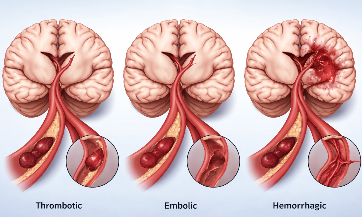
Understanding the types of blood clots in the brain helps us know how and where the blockage occurs.

TypeDescription
Thrombotic StrokeA clot forms directly in the brain artery
Embolic StrokeA clot travels from another part of the body to the brain
Hemorrhagic StrokeCaused by bleeding due to a ruptured vessel

Each type requires a slightly different approach to treatment, making early and accurate diagnosis very important.

Blood Clot in the Brain Causes

Blood clot causes

Many factors contribute to blood clots in the brain, and they are often a combination of medical and lifestyle issues.

Medical Conditions

  • High blood pressure – Damages blood vessels over time, increasing the risk of clot formation.
  • Diabetes – Affects blood circulation and can lead to thickened or damaged blood vessels.
  • High cholesterol – Causes plaque buildup in arteries, which can block blood flow.
  • Atrial fibrillation (irregular heartbeat) – Leads to improper blood flow, increasing the chances of clot formation.
  • Blood clotting disorders – Make the blood more likely to clot even without injury.

Lifestyle Factors

  • Smoking or vaping – Damages blood vessels and increases clotting risk.
  • Obesity – Puts extra strain on the heart and affects blood circulation.
  • Lack of physical activity – Slows blood flow, making clot formation more likely.
  • Dehydration – Thickens the blood, increasing the risk of clots.

Other Risk Factors

  • Pregnancy or hormone therapy – Hormonal changes can increase blood clotting tendency.
  • Long-distance travel – Sitting for long periods reduces circulation and may lead to clots.
  • Head injury or trauma – Can damage blood vessels and trigger clot formation.

Understanding why blood clots in the brain happens helps us take preventive steps early.

Blood Clot in Brain Symptoms

The blood clot in the brain symptoms usually appear suddenly and should never be ignored. Acting fast can save a life.

In many cases, these symptoms may seem mild at first, but they can worsen quickly within minutes or hours. The signs depend on which part of the brain is affected, as different areas control different body functions. Recognising these early warning signals and acting immediately can significantly improve recovery and reduce long-term damage.

SymptomWhat We May Notice
Weakness or numbnessOne side of the body, face drooping
Speech difficultySlurred or confused speech
Vision problemsBlurred or double vision
Severe headacheSuddenly, an intense “thunderclap” headache
DizzinessLoss of balance or coordination

Other Warning Signs

  • Seizures
  • Nausea or vomiting
  • Loss of consciousness

If we notice any of these symptoms, we must seek emergency care immediately.

How Is A Blood Clot Diagnosed?

Diagnosing Blood Clots: How It Works

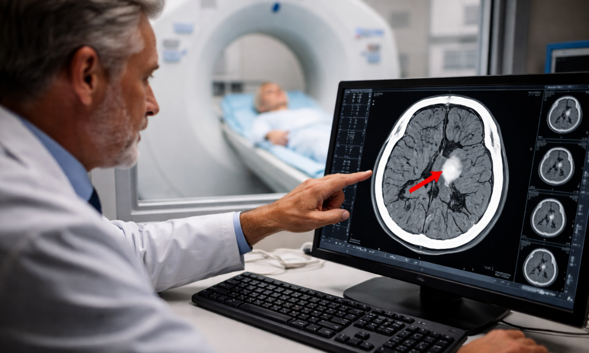
Diagnosis plays a crucial role in saving brain function and life. Doctors use imaging techniques to confirm the presence of a clot.

When a patient arrives with suspected symptoms, doctors act immediately to assess the situation and choose the right tests. Speed is extremely important because even a small delay can affect brain function. These diagnostic tools not only confirm the clot but also help determine its location, size, and severity, which guides the treatment plan.

Common Diagnostic Tests

TestPurpose
CT ScanQuickly detects bleeding or blockage
MRI ScanProvides detailed brain images
Blood TestsIdentify underlying conditions

Both CT scan and MRI scan in Bangalore are widely used for quick and accurate diagnosis. The faster we identify the clot, the better the chances of recovery.

Blood Clot in Brain Treatment

The blood clot in the brain treatment depends on how quickly the patient receives medical care. Time is critical here.

When symptoms appear, immediate hospital care is essential to prevent permanent brain damage. Doctors quickly evaluate the condition and begin treatment based on the type and severity of the clot. The goal is to restore blood flow to the brain as soon as possible and minimise complications or long-term disability.

Emergency Treatments

TreatmentDescription
Clot-busting drugs (tPA)Dissolve the clot and restore blood flow
Mechanical thrombectomyPhysically removes the clot
Blood thinnersPrevent further clot formation
SurgeryRelieves pressure in severe cases

Doctors often refer to the first few hours as the “golden window”, where treatment is most effective.

Why Early Detection Matters

When we delay treatment, the brain suffers irreversible damage. Early detection not only saves lives but also reduces long-term disability.

The brain depends on a constant supply of oxygen, and even a few minutes of blockage can start damaging brain cells. When we act quickly, doctors can restore blood flow and prevent serious complications like paralysis, speech loss, or memory issues. Early diagnosis also increases the chances of faster recovery and a better quality of life after treatment.

Even simple symptoms like sudden weakness or headache should not be ignored. Timely scans and medical attention can make a huge difference.

Tips to Reduce the Risk

Prevention is always better than a cure, especially for serious conditions like this.

  • Maintain healthy blood pressure and sugar levels
  • Stay physically active
  • Avoid smoking and excessive alcohol use
  • Stay hydrated
  • Go for regular health check-ups
  • Manage stress effectively

These small lifestyle changes can significantly reduce the risk of developing a clot.

Choosing the Right Health Screening Centre

Selecting the right health screening centre in Bangalore ensures accurate diagnosis and timely treatment.

Look for:

  • Advanced imaging facilities
  • Experienced medical professionals
  • Quick emergency response
  • Integrated diagnostic services

A reliable centre can make all the difference during critical situations.

Final Thoughts

A blood clot in the brain is not something we can afford to ignore. Quick action, early diagnosis, and the right treatment can save lives and prevent long-term complications. That’s why access to timely imaging, like a CT scan in Bangalore, becomes so important in emergencies.

At Koshikaa, we focus on providing personalised and accurate diagnostic support, helping you make informed decisions about your health with confidence. Because when it comes to your brain, every second truly counts.

Frequently Asked Questions

1. What is a blood clot in the brain?

A blood clot in the brain occurs when a blood vessel becomes blocked, preventing oxygen-rich blood from reaching brain cells. Without oxygen, brain cells begin to get damaged within minutes, which can lead to a stroke or permanent complications. 

A blood clot in the brain is considered a medical emergency, and immediate diagnosis and treatment are essential to prevent serious outcomes, including long-term disability or even life-threatening situations.

2. What are the first signs of a blood clot in the brain?

The first signs usually appear suddenly and can vary depending on the affected area of the brain. Common symptoms include weakness or numbness on one side of the body, slurred speech, confusion, vision problems, and a severe headache.

Recognising these early warning signs and seeking immediate medical attention can greatly improve the chances of survival and reduce the risk of long-term damage.

3. Can a blood clot in the brain be treated?

Yes, a blood clot in the brain can be treated, especially if medical care is provided quickly. Treatment options include clot-dissolving medications, procedures like mechanical thrombectomy to remove the clot, and supportive therapies. 

The success of treatment largely depends on how fast the patient reaches a hospital, as early intervention within the “golden window” significantly improves recovery and reduces complications.

4. How can we prevent blood clots in the brain?

We can lower the risk of blood clots in the brain by adopting a healthy lifestyle and managing existing medical conditions. Controlling blood pressure, diabetes, and cholesterol levels is essential, along with staying physically active and avoiding smoking. 

Regular health check-ups help detect risk factors early, allowing timely intervention and lifestyle changes that can prevent serious complications in the future.

Reference

1. From Google

2. https://my.clevelandclinic.org/health/diseases/24208-ischemic-stroke-clots 

MRI of the uterus is primarily used for diagnosing and evaluating conditions such as uterine fibroids, adenomyosis, and malignancies. It is particularly useful in the planning of surgical procedures and assessing the extent of gynecological diseases. This imaging test is valuable for its ability to deliver detailed cross-sectional images of the uterus and surrounding structures.
An MRI of the spleen can help diagnose various disorders affecting the spleen including splenomegaly (enlarged spleen), tumors, and other abnormalities. It provides clear, detailed images that are crucial for assessing the health of the spleen and planning potential surgical interventions.
Prostate MRI is a non-invasive imaging technique that provides detailed pictures of the prostate gland and surrounding tissues. It helps in the detection, staging, and monitoring of prostate cancer. Prostate MRIs are increasingly used to guide biopsy procedures and assist in the planning of treatment strategies for prostate health.
An MRI of the pancreas is used to identify issues such as pancreatitis, pancreatic cancer, and other pancreatic disorders. It offers high-resolution images that help in detecting small lesions and assessing the pancreatic ducts. This imaging technique is valuable for guiding biopsies and planning surgeries involving the pancreas.
MRI scans of the liver, kidney, and bile ducts provide detailed images that help diagnose various conditions, such as tumors, cysts, and blockages in the bile ducts. They are crucial for assessing organ damage, staging cancer, and planning liver or kidney-related surgeries. This type of MRI can also evaluate the health and function of these organs comprehensively.
Breast MRI is highly sensitive for detecting breast cancer, especially in individuals with dense breast tissue or a high risk of cancer. It is often used in conjunction with mammography to provide a more comprehensive evaluation. Breast MRIs can also monitor the response to breast cancer treatments and provide detailed pre-surgical mapping.
Brain and spine MRIs are critical tools for diagnosing and monitoring neurological conditions, including tumors, spinal cord injuries, and multiple sclerosis. These MRIs can reveal abnormalities in brain tissue and spinal cord structure that might not be visible with other imaging methods. They are essential for planning surgeries and assessing the effectiveness of treatments in neurology.
PET-CT scans of transplant organs like the liver, kidney, or pancreas are performed to assess metabolic activity, detect complications, and monitor post-transplant function. These scans play a vital role in the early detection of issues such as organ rejection, recurrence of disease, or infection, guiding timely interventions and optimizing transplant outcomes for patients.
PET-CT examinations of organs such as the liver, kidney, or prostate are crucial for assessing metabolic activity, detecting abnormalities, and monitoring disease progression. These scans help diagnose conditions such as liver metastases, renal cell carcinoma, or prostate cancer, guiding treatment decisions and monitoring response to therapy over time.
PET-CT scans of soft-tissue masses are utilized to assess metabolic activity and detect malignant tumors or other abnormalities. By combining PET imaging with CT imaging, these scans provide detailed information about tumor location, size, and metabolic characteristics, aiding in accurate diagnosis and treatment planning for soft-tissue cancers.
PET-CT scans are valuable for detecting cysts or fluid collections within the body, including cysts in the liver, kidneys, or other organs. These scans help characterize the nature and metabolic activity of cysts, guiding diagnosis and treatment planning for conditions such as cystic liver disease, renal cysts, or pancreatic cysts.
PET-CT scans of muscles are performed to assess metabolic activity and detect abnormalities such as inflammation, infection, or tumors. These scans provide valuable information about muscle function and pathology, aiding in the diagnosis and management of conditions like myositis, muscular dystrophy, or soft-tissue sarcomas.
PET-CT scans of the brain are utilized to evaluate brain metabolism and detect abnormalities such as tumors, epilepsy, or neurodegenerative diseases. By combining functional PET imaging with anatomical CT imaging, these scans offer comprehensive information about brain function and structure, guiding neurosurgical interventions and treatment decisions for neurological disorders
PET-CT scans of bones are commonly used to detect bone metastases in cancer patients, assess bone health in conditions like osteoporosis, and evaluate response to treatment. By combining PET and CT imaging, these scans provide detailed information about metabolic activity and anatomical changes in bone structures, aiding in accurate diagnosis and treatment planning for various bone-related conditions.
CT scans of the spine are conducted to assess the vertebrae, spinal cord, and surrounding soft tissues for conditions like herniated discs, spinal fractures, or spinal stenosis, aiding in the diagnosis and management of spinal disorders with detailed imaging essential for surgical planning and intervention.
CT scans of the pelvis are utilized to visualize the pelvic bones, reproductive organs, bladder, and surrounding structures for conditions such as fractures, tumors, or pelvic inflammatory disease, guiding treatment plans and surgical interventions with precise anatomical information.
CT scans of the neck are performed to evaluate structures such as the throat, thyroid gland, lymph nodes, and blood vessels for abnormalities like tumors, infections, or injuries, assisting in diagnosis and management of head and neck disorders with detailed imaging.
CT scans of the extremities are employed to assess bones, joints, and soft tissues in the arms or legs, aiding in the diagnosis of fractures, arthritis, or other musculoskeletal conditions, guiding orthopedic treatment and rehabilitation plans for optimal recovery.
CT scans of the chest are utilized to examine the lungs, heart, blood vessels, and surrounding structures for conditions such as lung nodules, pneumonia, or pulmonary embolism, facilitating accurate diagnosis and management of respiratory and cardiovascular diseases.
CT scans of the brain are conducted to assess brain structures, identify abnormalities like tumors, hemorrhages, or strokes, and aid in diagnosing neurological conditions, guiding neurosurgical interventions and treatment strategies with precise imaging of the brain’s anatomy.
CT scans of the abdomen are performed to visualize and evaluate the internal organs such as the liver, kidneys, pancreas, and intestines for conditions like tumors, infections, or injuries, providing detailed images essential for diagnosis and treatment planning.
Transplant organ ultrasound scans, including those of the liver, kidney, or pancreas, are performed to assess organ viability, detect complications, and monitor post-transplant function. These scans play a vital role in the early detection of issues such as organ rejection, vascular complications, or fluid collections, guiding timely interventions and optimizing transplant outcomes for patients.
Ultrasound examinations of organs such as the liver, kidney, or prostate are crucial for assessing organ structure, detecting abnormalities, and monitoring disease progression. These scans help diagnose conditions such as liver cirrhosis, kidney stones, or prostate enlargement, guiding treatment decisions and monitoring response to therapy over time.
Soft-tissue masses can be evaluated using ultrasound to determine their size, location, and characteristics. Ultrasound imaging helps differentiate between benign and malignant masses, guiding further diagnostic evaluation and treatment planning. By providing real-time imaging, ultrasound assists in the precise localization of soft-tissue masses for biopsy or surgical removal if needed.
Ultrasound scans are valuable for detecting cysts or fluid collections within the body, including cysts in the liver, kidneys, or other organs. These scans help characterize the nature and size of cysts, aiding in diagnosis and treatment planning for conditions such as cystic liver disease or renal cysts, and guiding interventions such as cyst drainage or sclerotherapy.
Musculoskeletal ultrasound scans are used to assess the health and integrity of muscles, identifying issues such as strains, tears, or muscle inflammation. These scans provide detailed images of muscle structures, allowing healthcare providers to diagnose muscle injuries accurately and develop personalized rehabilitation plans to promote recovery and restore function.
Ultrasound examinations of joints are essential for evaluating joint health, detecting abnormalities such as inflammation, fluid accumulation, or cartilage damage. By visualizing the joint structures in real-time, ultrasound helps diagnose conditions like arthritis, bursitis, or ligament injuries, guiding treatment decisions and monitoring disease progression over time.
Ultrasound scans of tendons are performed to assess for injuries, inflammation, or tears, aiding in the diagnosis and management of conditions such as tendonitis or tendon tears. These scans provide detailed images of tendon structures, helping healthcare providers determine the extent of damage and plan appropriate treatment strategies, including physical therapy or surgical intervention if necessary.
Pelvis X-rays play a crucial role in diagnosing injuries, diseases, and developmental conditions affecting the pelvic region. They provide detailed images of the hip bones, sacrum, and coccyx, aiding in the assessment of fractures, dislocations, or signs of arthritis. Pelvic X-rays are instrumental in guiding surgical interventions, monitoring healing progress, and evaluating conditions such as osteoporosis or tumors. By providing precise anatomical information, these imaging studies enable healthcare professionals to deliver personalized care and improve patient outcomes.
Teeth X-rays, known as dental radiographs, are indispensable tools for dentists to assess oral health. They aid in diagnosing dental decay, gum disease, and abnormalities in tooth structure or alignment. These images provide valuable insights into hidden dental issues, allowing for early intervention and prevention of complications. Additionally, dental X-rays are essential for planning treatments such as orthodontics, root canals, or dental implants, ensuring optimal outcomes and patient satisfaction.
Spine X-rays are pivotal in diagnosing spinal fractures, degenerative conditions, and spinal misalignments, providing critical information for treatment planning and monitoring spinal health. They offer detailed images of the vertebrae and surrounding structures, enabling healthcare professionals to identify issues such as scoliosis, disc herniation, or spinal stenosis
Skull X-rays are valuable for detecting fractures, assessing head trauma, and evaluating the alignment of facial bones, crucial in diagnosing conditions like fractures, tumors, or developmental abnormalities in the skull. They provide detailed views of the cranial structure, aiding clinicians in determining appropriate treatment strategies and ensuring patient safety.
For the heart, X-ray solutions can aid in the detection of various cardiac problems, ranging from mild to severe, including – Heart failure, Cardiac congestion and other heart-related conditions. In addition to cardiac assessments, X-ray solutions are also used for breast examinations to identify tumors or signs of breast cancer.

Bone X-rays are essential for diagnosing fractures and assessing bone alignment, aiding doctors in developing treatment plans and monitoring healing progress. They provide detailed images of the skeletal system, helping identify abnormalities or injuries that may not be visible through other imaging techniques

Abdominal X-rays are performed when patients experience symptoms such as significant nausea, ongoing vomiting, abdominal pain, flank or back pain, or abdominal swelling. These X-rays can help diagnose various conditions.

Frequently Booked Tests

Frequently Booked Checkups